Assay for rapid detection of human activated protein C and highly specific monoclonal antibody therefor

a technology of activated protein and enzyme capture assay, which is applied in the field of measurement of activated protein c (apc), can solve the problems of inconvenient use of microtiter plates, inability to directly determine the function of apc, etc., and achieves high specificity for binding and high selectivity over protein c.

Inactive Publication Date: 2006-01-24
OKLAHOMA MEDICAL RES FOUND
View PDF2 Cites 130 Cited by
  • Summary
  • Abstract
  • Description
  • Claims
  • Application Information

AI Technical Summary

Benefits of technology

[0010]In another aspect, the invention is a binding fragment of the monoclonal antibody produced by hybridoma cell HAPC 1555 having been deposited under ATCC accession number PTA-2658, the binding segment capable of binding activated protein C with high selectivity over protein C. The binding fragment may comprise an Fab portion or Fc portion of the antibody.
[0011]In another aspect, the invention is an assay for assessing the concentration of activated protein C comprising testing a human plasma sample suspected of containing a quantity of activated protein C for binding to an antibody or portion thereof having high specificity for binding to APC, wherein the antibody is produced by hybridoma cell HAPC 1555 having been deposited under ATCC accession number PTA-2658.
[0012]In another aspect, the invention is an assay for the detection of activated protein C (APC) in a patient plasma sample comprising binding antibody HAPC 1555 or a portion thereof to a solid support to form a surface-bound antibody, contacting the surface-bound antibody with a patient plasma sample in a reaction zone, incubating the reaction zone under conditions effective for binding APC from the patient plasma sample to the surface-bou

Problems solved by technology

Since EPCR and TM are expressed on endothelium, it is not possible to directly determine how well they are functioning without removal of blood vessels.
We have found that in some meningococcemia patients, supplementation with protein C failed to elicit det

Method used


Image

  • Assay for rapid detection of human activated protein C and highly specific monoclonal antibody therefor
  • Assay for rapid detection of human activated protein C and highly specific monoclonal antibody therefor
  • Assay for rapid detection of human activated protein C and highly specific monoclonal antibody therefor

Examples


example 1

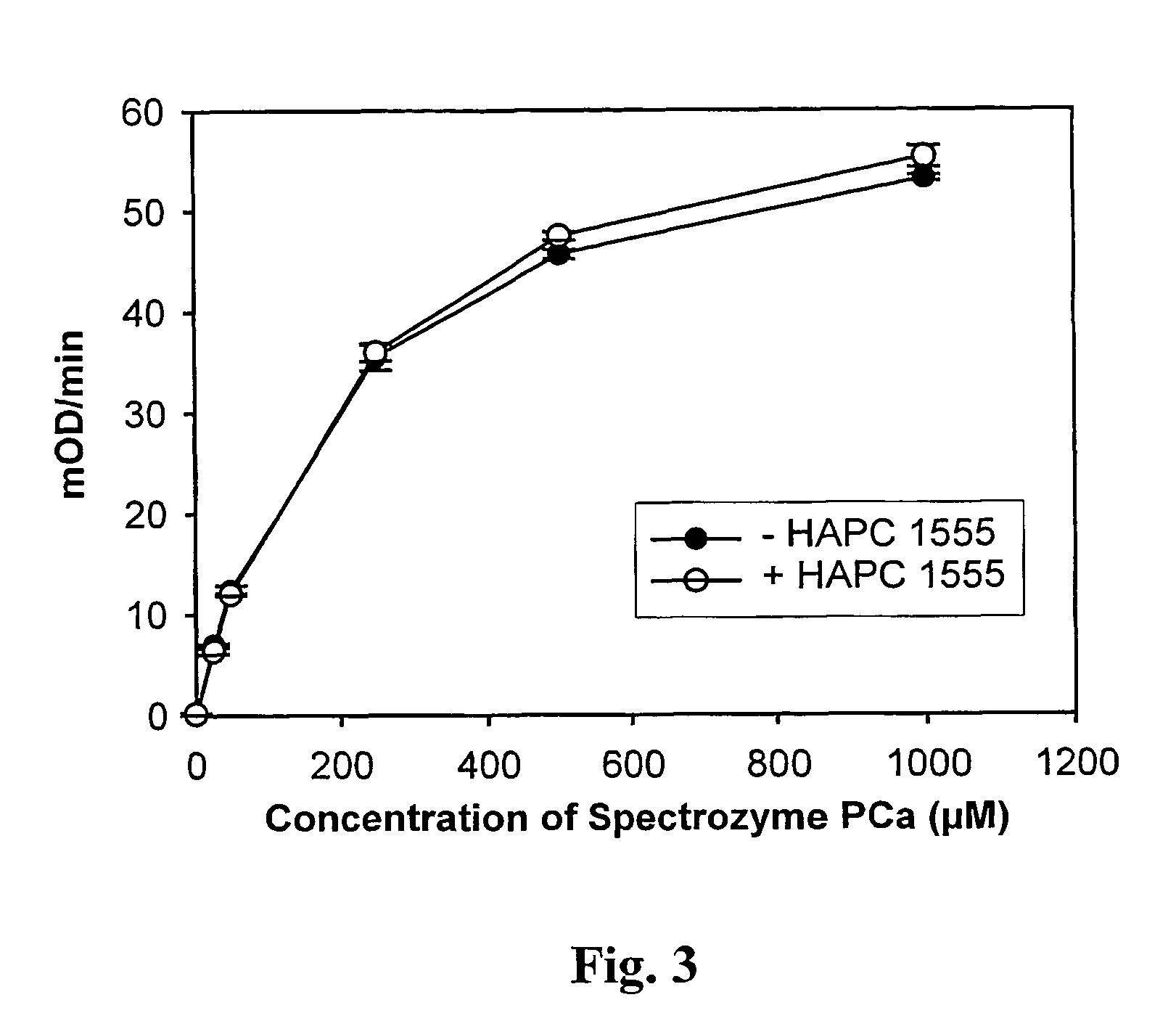
Quantification of APC in Human Plasma Using HAPC 1555

[0032]A. Methods

[0033]Initially, 96-well vinyl microtiter plates (Costar, Cat # 2596) were coated with 100 μl of HAPC 1555 (5 μg / ml) in coating buffer (20 mM Hepes, pH 7.5, 150 mM NaCl, 5 mM CaCl2) for 2 hours at 37° C. As negative controls, microtiter plate wells were coated with either 5 μg / ml HPC4 monoclonal antibody or 1% bovine serum albumin (BSA) in the same buffer. The plates were then blocked for 1 hour at 37° C. or overnight at 4° C. with 200 μl of blocking buffer (coating buffer containing 1% BSA). The solutions were gently removed from the plates by vacuum.

[0034]To generate a standard curve for the quantitative measurement of APC in plasma, increasing amounts of APC (from 0 to 250 ng / ml) were spiked into normal pooled plasma containing 20 mM Hepes, pH 7.5, 2 units / ml heparin, 20 mM benzamidine, and 10 mM CaCl2. The spiked plasma (100 μl) were transferred to the plates and incubated at room temperature for 1 hour. The we...

example 2

Quantification of APC in Human Plasma Specimens

[0038]The procedure of Example 1 can be used to test human plasma for APC. In Example 1, human plasma was spiked with APC, but to assess a clinical specimen for its APC content, a specimen is collected from the patient in standard citrate anticoagulant that also contains 20 mM benzamidine HCl. The blood cells are removed by centrifugation using standard techniques. The plasma is used in the assay after standard preparation (e.g., 20 mM Hepes, pH 7.5, 2 units / ml heparin and 10 mM CaCl2). The plasma sample is then analyzed for APC using an assay such as described in Example 1.

Indications Relating to Deposited Microorganism or Other Biological Material

[0039]A mouse hybridoma cell line useful in the assay of the present invention has been established and given the designation HAPC 1555. The HAPC 1555 cell line was deposited under the provisions of the Budapest Treaty with the American Type Culture Collection (ATCC), 10801 University Bouleva...


PUM

PropertyMeasurementUnit
Molar densityaaaaaaaaaa
Concentrationaaaaaaaaaa

Abstract

A highly specific monoclonal antibody, produced by a hybridoma cell has been found. A new assay, highly specific for activated protein C (APC) has been developed which will permit rapid determination of APC levels in clinical situations.

Description

CROSS-REFERENCE TO RELATED APPLICATIONS[0001]This application claims the benefit of International Application Ser. No. PCT / US01 / 30802, filed Oct. 2, 2001 which claims the benefit of U.S. Ser. No. 60 / 237,481, filed Oct. 2, 2000.TECHNICAL FIELD OF THE INVENTION[0002]This invention relates to measurement of activated protein C (APC) in clinical samples.BACKGROUND[0003]Protein C is the precursor to activated protein C (APC), a potent natural anticoagulant. Protein C is activated by thrombin in complex with thrombomodulin (TM). The activation is augmented by endothelial cell protein C receptor (EPCR). TM and EPCR can be down-regulated due to inflammatory mediators, such as tumor necrosis factor, reviewed by Esmon (Esmon, C. T., “Natural anticoagulants and their pathways,”Handbook of Experimental Pharmacology 132:447–76; G. V. R. Born, Cuatrecasas, P., Ganten, D., Herken, H., Starke, K., and Taylor, P., eds., Springer-Verlag, New York 1999). TM and EPCR have also been found to be reduced ...

Claims


Application Information

Patent Timeline
IPC IPC(8): G01N33/573G01N33/577C07K16/40C12N5/20C12Q1/37
CPCC07K16/40G01N33/573C12Q1/37G01N2333/96461
Inventor ESMON, CHARLES T.LIAW, PATRICIA C. Y.FERRELL, GARY L.
Owner OKLAHOMA MEDICAL RES FOUND
Why Eureka
Try Eureka