
Graphical user interfaces and methods for retrospectively gating a set of images

a graphical user interface and retrospective gating technology, applied in the field of medical imaging, can solve the problems of introducing artifacts into the images, affecting the quality of the images obtained by the ct scanner, and cannot be readily eliminated, so as to improve the calcium scoring of the heart of the patient, improve the imaging of the heart, and improve the effect of the patient's hear

Inactive Publication Date: 2006-02-28
MERATIVE US LP
View PDF40 Cites 59 Cited by
  • Summary
  • Abstract
  • Description
  • Claims
  • Application Information

AI Technical Summary

Benefits of technology

[0009]The present invention provides methods and software for improving the imaging of a patient's heart. In a particular use, the present invention improves calcium scoring of a patient's heart by selecting image slices that were obtained during a specific point (typically diastole) of the patient's cardiac cycle and thereafter calcium scoring the selected image slices.
[0010]Some embodiments of the present invention use a patient's measured ECG signals taken during the acquisition of the image scan to gate an image scan. The ECG signal is a repetitive pattern that reflects the electrical activity of the patient's heart. An ECG signal has a plurality of cardiac cycles (sometimes referred to as R—R cycle), with each cardiac cycle beginning with an R-wave (e.g., highest amplitude peak) during a systole period and ending with a relatively motionless diastole period. Blurring of the images is most likely to occur when imaging during systole. Consequently, it is preferable to use image slices taken during the diastole period so as to reduce the amount of artifacts introduced into the reconstructed image(s).
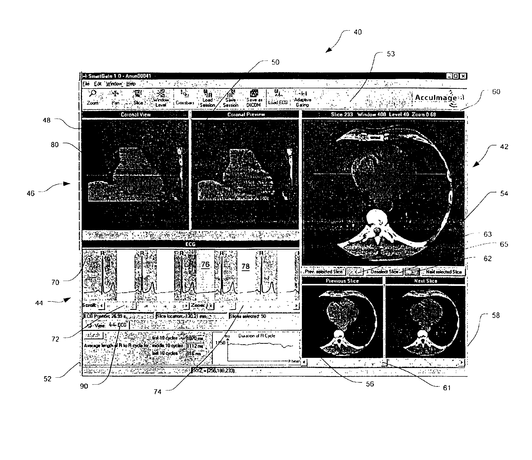
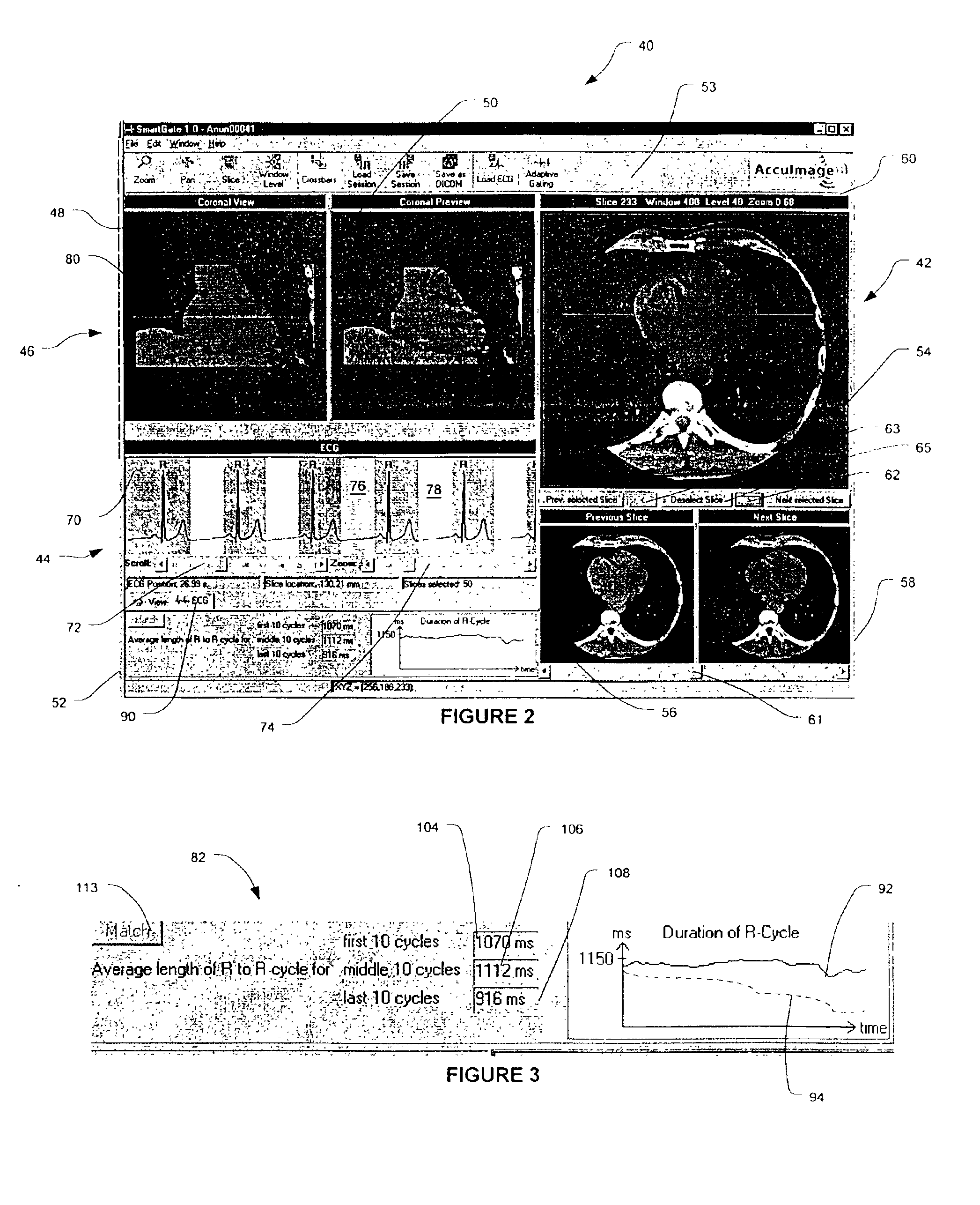
[0015]After the user has selected and applied the global selection criteria, the graphical user interface can display a sagittal or coronal reconstruction projection that incorporates the selected slices. Optionally, the sagittal or coronal reconstruction projection that uses all of the slices can be displayed concurrently with the image that has only the selected slices. This allows the user to see the effect of the chosen slices has on the image quality. The invention of this application is not limited to the use of coronal or sagittal projections. Other projections may be chosen, such as those of the heart's short or long axis.
[0016]The user generally has the option to de-select some of the originally selected slices and replace the de-selected slices with other slices of the image scan. The graphical user interface can display at least one selected slice and the neighboring slice images that are immediately adjacent the selected slice. In other embodiments, the graphical user interface can display two or more slices on each side of the selected slice, if desired. Thus, if the user visually determines that one of the neighboring slice images has better characterisitics than the selected slice image, the user can select the neighboring slice image. Advantageously, the replacement of the slices can be viewed in real-time so that the user can visually determine if the new slices improve the image quality of the coronal / sagittal projection.
[0017]To improve the visualization of the correspondence of the ECG to the slice selection, the graphical interface can display the patient's ECG on the same screen as the selected slice and the coronal / sagittal projection of the selected slices. Typically, the ECG will be highlighted to indicate where the slices were selected relative to the ECG signal.
[0019]Advantageously, in some embodiments the present invention improves the “dose efficiency” of the imaging procedure by increasing the use of information obtained during the image scan. Conventional imaging generally uses only one out of approximately every ten slices to create the image projection. The methods of the present invention generally utilizes three slices out of every ten slices and produces a parsed set of images for calcium scoring. Thus, the present invention effectively uses three times as much information as conventional methods of gating the image scan.

Problems solved by technology

Unfortunately, two body motions can interfere with the quality of the images obtained by the CT scanner: the heart motion and the patient's breathing motion.
The heart motion, on the other hand, cannot be readily eliminated and can lead to blurring, introduction of artifacts into the images, and misregistration.
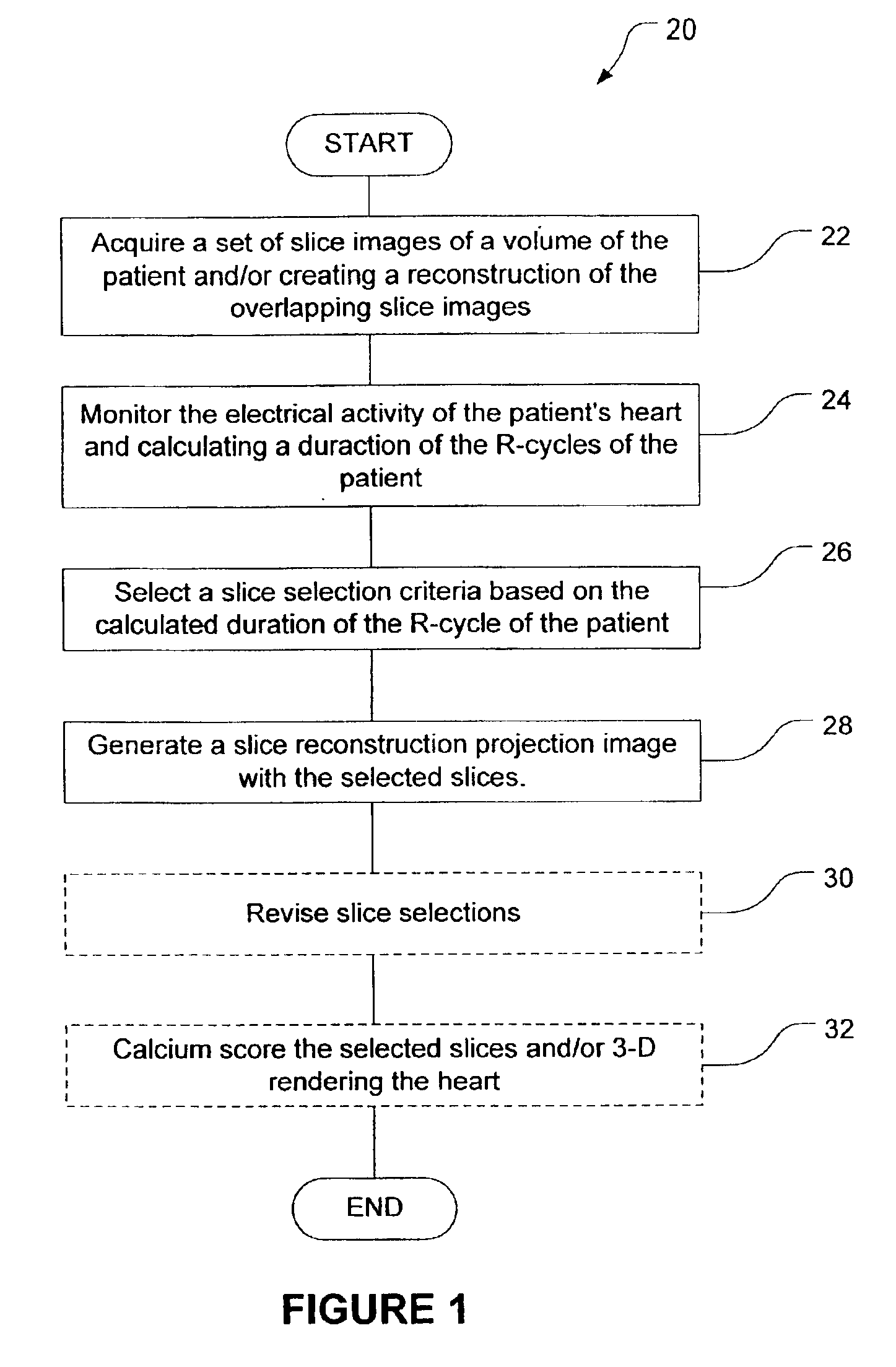
A major issue with prospective gating in subjects with irregular heart beats is that the trigger can only be set to acquire data after the R-wave.
The second problem is that gating from an ECG signal requires the placement of electrodes on the subject and testing to confirm that their placement is adequate.
In a busy screening or diagnostic practice the added steps can decrease utilization and negatively affect the economics of the imaging operation.
Information as to the length of the cardiac cycle during the study, convenient ways to ascertain whether it changed during the study, and measurement of any one cycle are also not readily available.
Except for manually adjusting each slice (there can be 350-500 slices in a study), there is no way to account for changes in the cardiac cycle.

Method used


Image

  • Graphical user interfaces and methods for retrospectively gating a set of images
  • Graphical user interfaces and methods for retrospectively gating a set of images
  • Graphical user interfaces and methods for retrospectively gating a set of images

Examples


Embodiment Construction

[0041]The present invention provides methods and graphical user interfaces for self gating and retrospectively gating a set of image slices (referred to herein as an image scan).

[0042]While the remaining discussion focuses on the gating of an image scan from a CT scanner for use in coronary calcium measurements, it should be appreciated that the methods and devices of the present invention are not limited to such imaging modalities and uses. For example, instead of analyzing the image scan for measuring coronary calcium, the image scan can be used for 3-D reconstructions of the heart, such as those used for CT angiography, or for heart function studies, including dynamic studies.

[0043]In some exemplary embodiments, the present invention uses a patient's measured ECG signals taken during the acquisition of the image scan to gate the image scan. The ECG signal is a repetitive pattern that reflects the electrical activity of the patient's heart. An ECG signal has a plurality of cardiac...


PUM


Abstract

Graphical user interfaces and methods for displaying ECG, slice images and a reconstruction projection image of the slices. In exemplary embodiments, the graphical user interfaces and methods facilitate the selection of slice images that are taken during a quiet part of the patient's heart cycle in which the heart is relatively motionless so as to reduce blurring and the introduction of artifacts into the resultant reconstructed projection image of the heart. In exemplary embodiments, the resultant projection image can be used for coronary calcium detection and scoring or 3-D rendering.

Description

CROSS-REFERENCE TO RELATED APPLICATIONS[0001]The present application claims benefit to U.S. Provisional Patent Application Ser. No. 60 / 306,311 filed Jul. 17, 2001, the complete disclosure of which is incorporated herein by reference.[0002]The present application is also related to U.S. Patent Application entitled “Methods and Software for Retrospectively Gating a Set of Images” filed herewith, and U.S. Patent Application entitled “Methods and Software for Self-Gating a Set of Images,” also filed herewith, the complete disclosures of which are incorporated herein by reference.BACKGROUND OF THE INVENTION[0003]The present invention relates generally to medical imaging. More specifically, the present invention relates to gating of an image scan to improve calcium scoring of a patient's heart and coronary arteries.[0004]CT scanning of the heart is an increasingly common procedure to obtain information about the presence of calcification in the coronary arteries. Unfortunately, two body m...

Claims


Application Information

Patent Timeline
IPC IPC(8): A61B5/05A61B5/044A61B5/352A61B6/03G06T19/00
CPCA61B6/032A61B6/463A61B6/469A61B6/503A61B6/5288A61B8/5276G06T19/00A61B6/467A61B5/0456A61B5/7207A61B5/7289A61B6/03G06T2219/028A61B6/504A61B5/352
Inventor KAUFMAN, LEONGRIESS, FRIEDERIKE
Owner MERATIVE US LP
Learn More