Method for marker-free automatic fusion of 2-D fluoroscopic C-arm images with preoperative 3D images using an intraoperatively obtained 3D data record

a technology of 3d data record and fluoroscopic c-arm, which is applied in the field of superimposing a preoperative 3d image over a 2d image obtained intraoperatively with a carm, can solve the problem that the 2d fluoroscopic image does not adequately display the anatomy of the patien

Active Publication Date: 2006-03-07
SIEMENS HEALTHCARE GMBH
View PDF6 Cites 96 Cited by
  • Summary
  • Abstract
  • Description
  • Claims
  • Application Information

AI Technical Summary

Benefits of technology

[0006]An object of the present invention is to fuse, in a simple manner, 2D fluoroscopic images, which have been obtained in an intraoperative procedure, and which display the medical instrument, with 3D images obtained in the preoperative stage.

Problems solved by technology

From both a medical and a technical viewpoint, a problem is that although during the intervention (operation, examination) the medical instrument (the further text describes a catheter as a non-restrictive example) can be displayed very exactly and with a high resolution in one or more fluoroscopic images (also called 2D fluoroscopic images) using an intraoperative X-ray procedure with a C-arm, the 2D fluoroscopic images insufficiently display the anatomy of the patient.

Method used


Image

  • Method for marker-free automatic fusion of 2-D fluoroscopic C-arm images with preoperative 3D images using an intraoperatively obtained 3D data record
  • Method for marker-free automatic fusion of 2-D fluoroscopic C-arm images with preoperative 3D images using an intraoperatively obtained 3D data record
  • Method for marker-free automatic fusion of 2-D fluoroscopic C-arm images with preoperative 3D images using an intraoperatively obtained 3D data record

Examples


Embodiment Construction

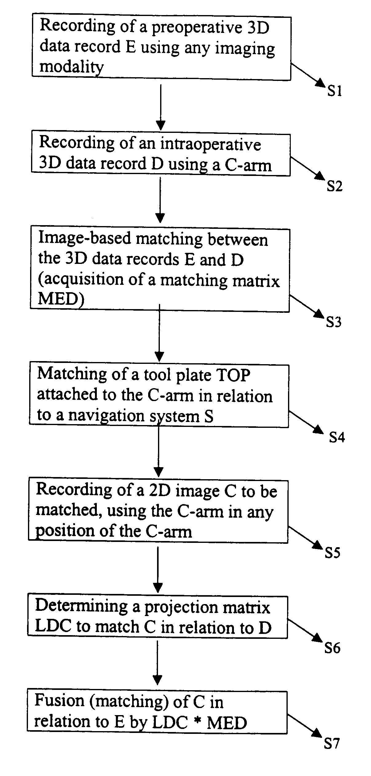
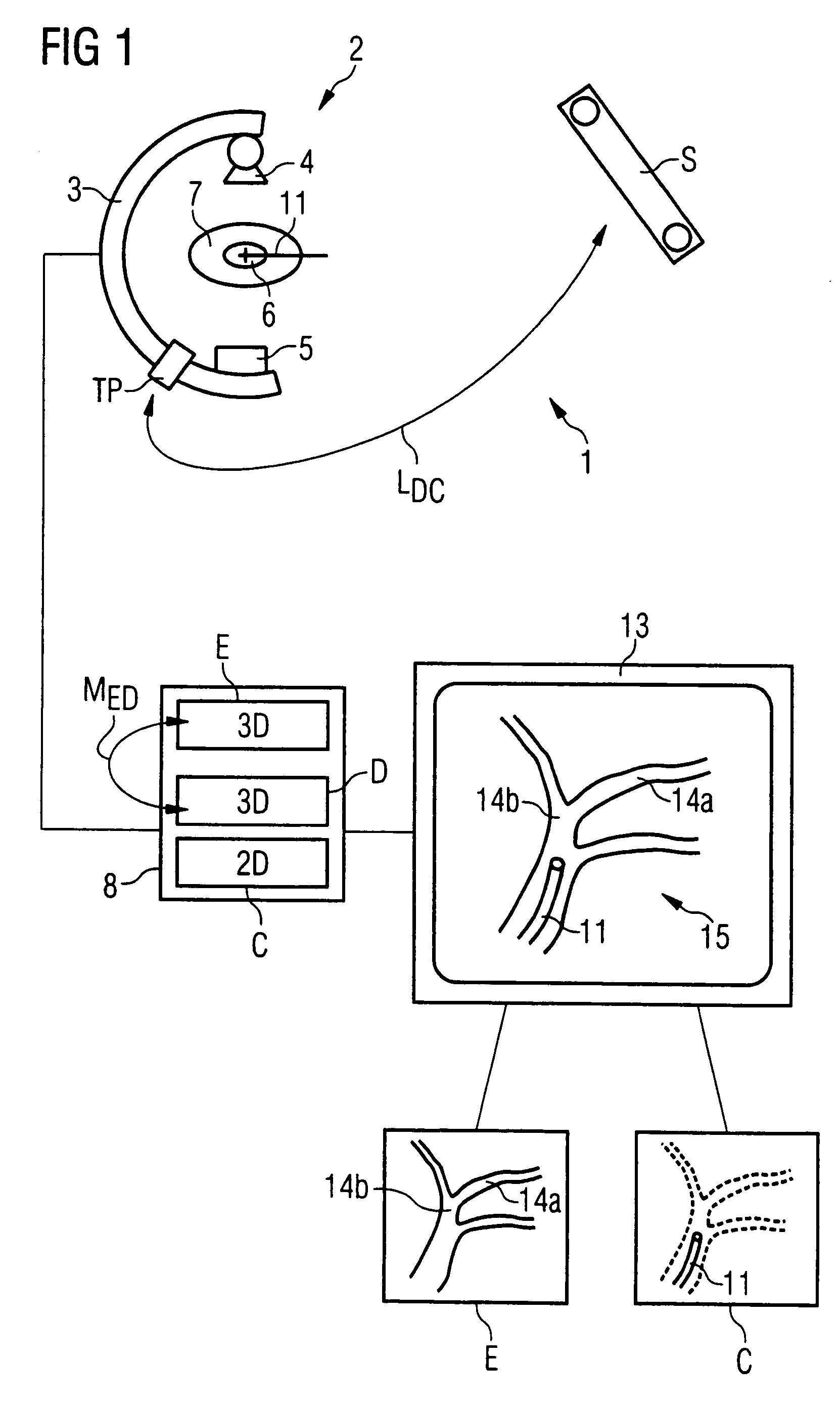
[0016]FIG. 1 is a schematic illustration of a medical examination and / or treatment device 1 according to this invention, wherein only the basic components are shown. The device has a data acquisition device 2 for obtaining two-dimensional X-ray images (2D fluoroscopic images). The data acquisition device has a C-arm 3 on which an X-ray source 4 and a radiation detector 5, for example, a solid-state image detector, are mounted, as well as a tool plate TP. The field of examination 6 of a patient 7 is preferably located in the isocenter of the C-arm 3, so that it is completely visible in the recorded 2D fluoroscopic image.

[0017]In close proximity to the data acquisition device 2 is a navigation sensor S, which detects the actual location of the tool plate TP—and thus the location of the C-arm 3—and the location and position of the medical instrument 11 used for the intervention, as well as the patient. If the locations of the device 2 and the patient are not required, the position of t...


PUM


Abstract

In a method and apparatus for automatic marker-free fusion (matching) of 2D fluoroscopic C-arm images with preoperative 3D images using an intraoperatively acquired 3D data record, an intraoperative 3D image is obtained using a C-arm x-ray system, image-based matching of an existing preoperative 3D image in relation to the intraoperative 3D image is undertaken, which generates a matching matrix of a tool plate attached to the C-arm system is matched in relation to a navigation system, a 2D fluoroscopic image to be matched is obtained, with the C-arm of the C-arm system in any arbitrary location, a projection matrix for matching the 2D fluoroscopic image in relation to the 3D image is obtained, and the 2D fluoroscopic image is fused (matched) with the preoperative 3D image on the basis of the matching matrix and the projection matrix.

Description

BACKGROUND OF THE INVENTION[0001]1. Field of the Invention[0002]The present invention relates to a method for the superimposition of a preoperative 3D image over a 2D image obtained intraoperatively with a C-arm. In particular, the invention relates to the display of a 3D image representing a medical instrument that has been introduced into the area of examination of a patient and is contained within the 2D image.[0003]2. Description of the Prior Art[0004]Increasingly, patients are being examined and treated in a minimally invasive manner, i.e., with as little operative intrusion as possible. Examples of this approach are procedures using endoscopes, laparoscopes, or catheters, which are introduced into the examination or treatment region in the patient through a small body orifice. For example, catheters often are used in cardiology examinations.[0005]From both a medical and a technical viewpoint, a problem is that although during the intervention (operation, examination) the medic...

Claims


Application Information

Patent Timeline
Patent Type & Authority Patents(United States)
IPC IPC(8): A61B6/03A61B6/00A61B6/12G06T7/00
CPCA61B6/12A61B6/4441A61B6/504A61B6/5235A61B6/547G06T7/0038A61B6/5247Y10S378/901G06T2207/30004G06T7/38
Inventor MITSCHKE, MATTHIASRAHN, NORBERTRITTER, DIETER
Owner SIEMENS HEALTHCARE GMBH
Why Eureka
Try Eureka