
Separating motion from cardiac signals using second order derivative of the photo-plethysmogram and fast fourier transforms

a technology of photo-plethysmogram and motion, applied in the field of physiological signals, can solve the problems of substantial disadvantages of existing ppg devices, substantial difficulty in oximeters, and degrade the signal-to-noise ratio

Active Publication Date: 2006-03-28
SPACELABS HEALTHCARE LLC
View PDF28 Cites 255 Cited by
  • Summary
  • Abstract
  • Description
  • Claims
  • Application Information

AI Technical Summary

Benefits of technology

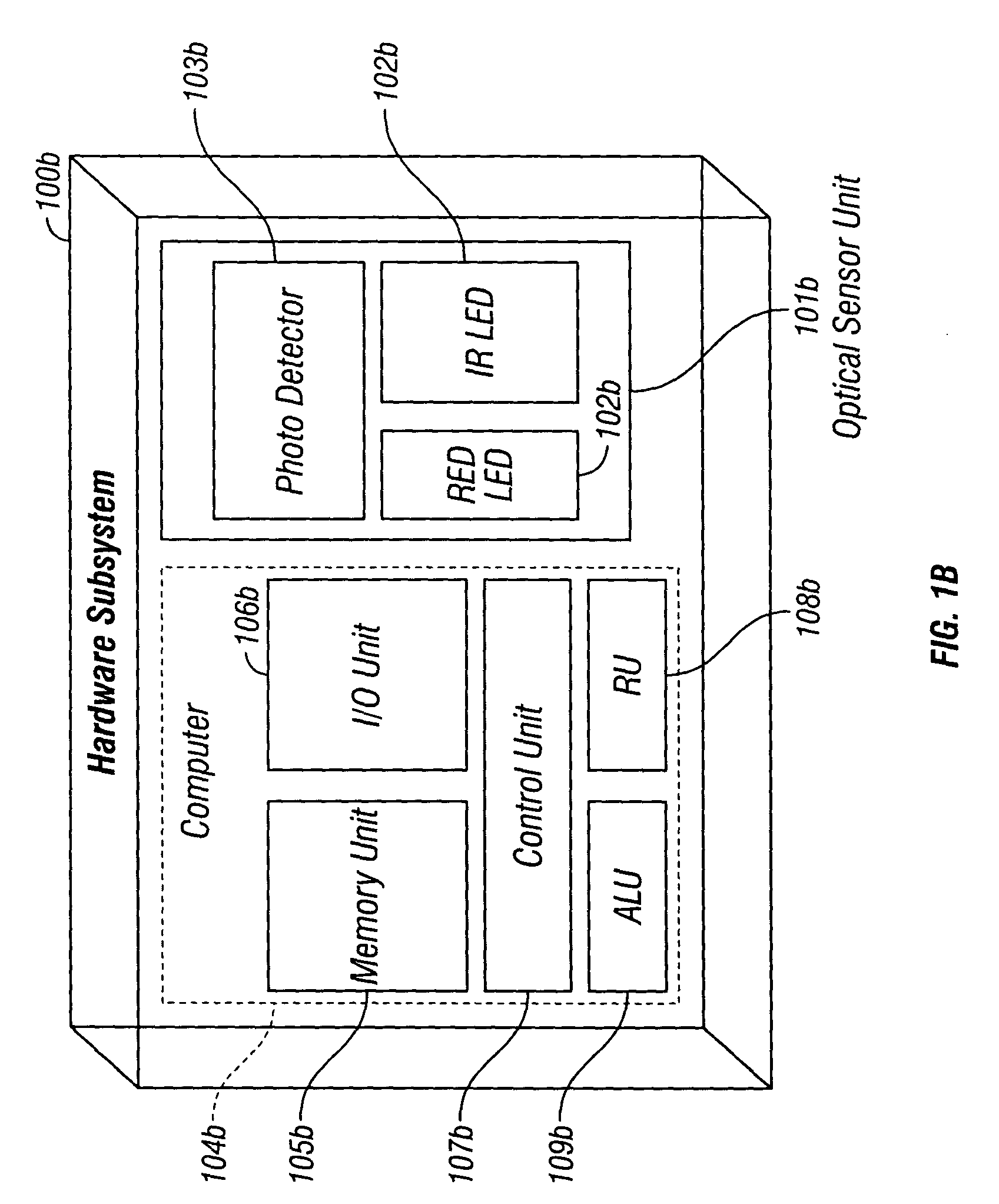
[0019]The hardware subsystem comprises an optical sensor unit for providing the pulse oximetry system with a plurality of photo-plethysmographic data, wherein the sensor may either be transmittance or reflectance type containing at least two light emitting diodes for emitting light of two different wavelengths. The hardware subsystem additionally comprises a photo-detector for detecting the light after being transmi

Problems solved by technology

Existing PPG devices have substantial disadvantages, however.
The PPG device measurements are sensitive to corruption from external dynamics, causing motion artifacts that degrade the signal-to-noise ratio.
For example, variation in the optical coupling between the probe head and the patient, possibly induced by patient movement, may cause an artifact.
Motion artifacts can render it substantially difficult for the oximeter to accurately determine the patient's PPG signal, therefore causing errors in the pulse rate and oxygen saturation outputs.
Another limitation is that PPG relies on AC photo-plethysmogram (PPG) component, which is synchronous with cardiac pulsations but very small compared to the overall DC component, to determine SpO2.
Because the AC component is small relative to the DC component, accurate measurement of the AC component is difficult.
Inaccuracies in AC component measurements cause inaccuracies in SpO2 measurements because oximeters compute SpO2 using the relative magnitudes of the AC components of the different optical photo-plethysmograms.
In addition, in poorly perfused patients, the circulation may not adequately modulate the light beams to the point where it is difficult to distinguish the synchronous cardiac pulsations from system noise.
Thus, the usefulness of most commercial pulse oximeters is limited to situations in which patient is well perfused and subject to minimal motion.
A difficulty with these time-domain algorithms is that the SpO2 is conventionally averaged with several recent values in order to reduce the disruptive effect of corrupted data.
Furthermore, this motion artifact results in correlated red and infrared signals.
A limitation with this algorithm is that when red and infrared signals become corrupted by sources other than moving arterial and venous blood, such as sensor movement relative to skin, the artifact can result in uncorrelated red and infrared signals.
One drawback of this cardiac-based filtering is the assumption that, on average and over several seconds of time, signal noise coming from real patient motion and other sources does not occur at the heart rate.
As a result, the filters associated with this approach assume that noise is chaotic and random and in turn tend to find the underlying cardiac signal that is mixed with noise created by patient movement or other sources of artifact.
In practice, physiological signals such as the PPG are not well behaved and are often modulated with artifacts due to noise, interference, and patient motion.

Method used


Image

  • Separating motion from cardiac signals using second order derivative of the photo-plethysmogram and fast fourier transforms
  • Separating motion from cardiac signals using second order derivative of the photo-plethysmogram and fast fourier transforms
  • Separating motion from cardiac signals using second order derivative of the photo-plethysmogram and fast fourier transforms

Examples


Embodiment Construction

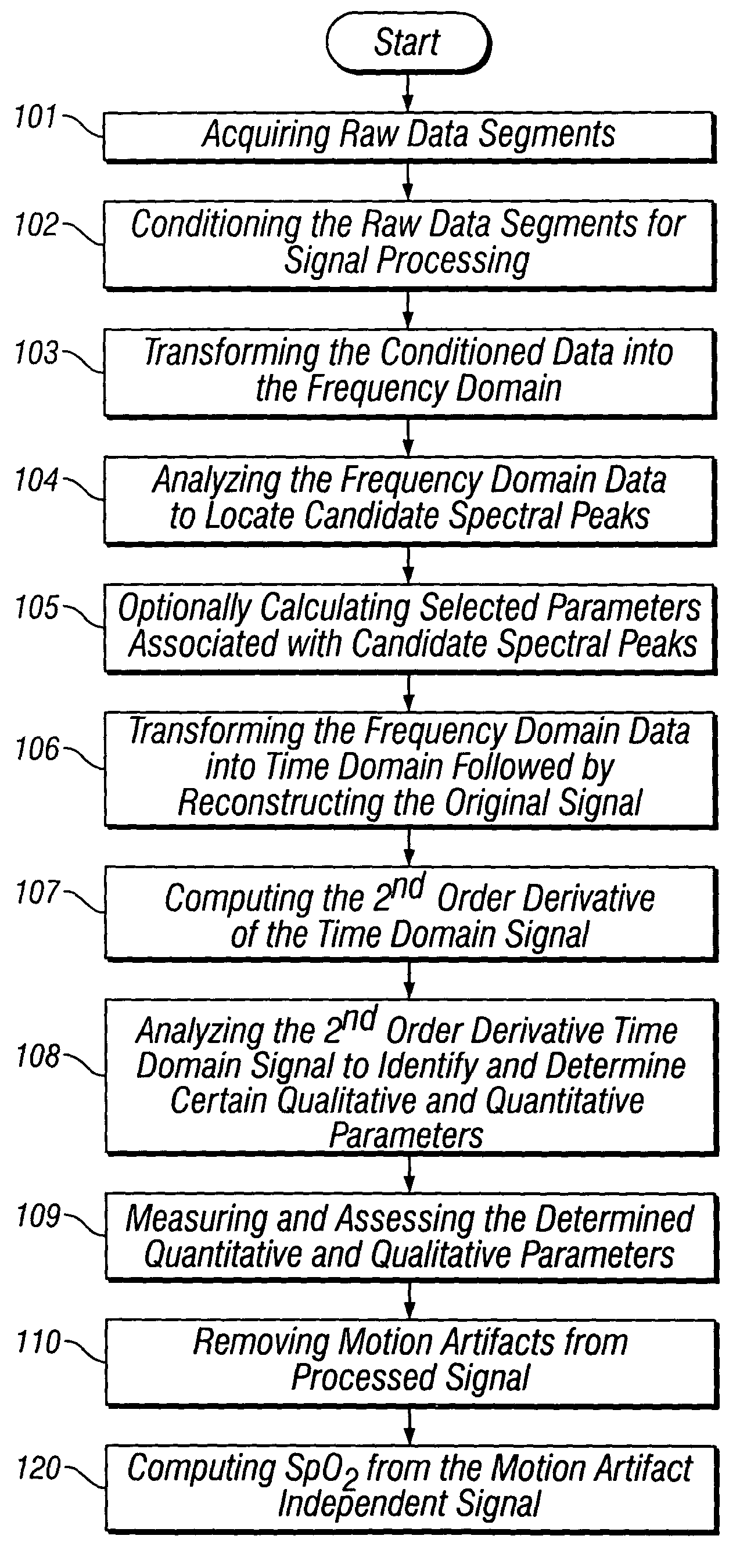
[0037]The present invention discloses methods and systems for removing asynchronous motion artifacts from synchronous cardiac signals by performing second order derivative processing of the measured photo-plethysmographic waveforms. The present invention will be described with reference to the aforementioned drawings. One of ordinary skill in the art would appreciate that the application described herein is an example of how the broader concept can be applied.

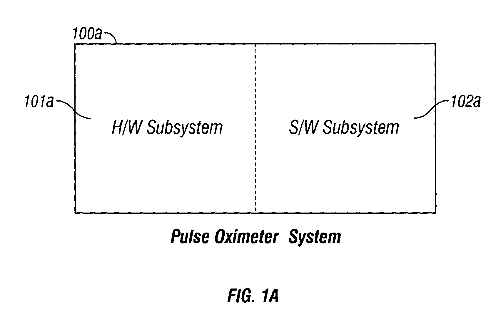
[0038]Referring to FIG. 1a, a system embodiment of the pulse oximeter 100a of the present invention is shown. The pulse oximeter system 100a comprises of a hardware subsystem 101a and an associated software subsystem 102a. The hardware subsystem 101a performs signal acquisition, followed by measurements and optional preconditioning. The software subsystem 102a is responsible for discarding motion artifacts from the acquired, measured and optionally preconditioned signal. Within the software subsystem 102a resides a method embod...


PUM


Abstract

The present invention is directed toward a pulse oximetry system for the determination of a physiological parameter capable of removing motion artifacts from physiological signals comprises a hardware subsystem and a software subsystem. The software subsystem is used in conjunction with the hardware subsystem to perform a method for removing a plurality of motion artifacts from the photo-plethysmographic data and for obtaining a measure of at least one physiological parameter from the data. The method comprises acquiring the raw photo-plethysmographic data, transforming the data into the frequency domain, analyzing the transformed data to locate a series of candidate cardiac spectral peaks (primary plus harmonics), reconstructing a photo-plethysmographic signal in the time domain with only the candidate cardiac spectral peaks (primary plus harmonics), computing the second order derivative of the reconstructed photo-plethysmographic signal, analyzing the candidate second order derivative photo-plethysmographic signal to determine the absence or presence of cardiac physiologic signal characteristics, and finally selecting the best physiologic candidate from the series of potential cardiac spectral peaks (primary plus harmonics) based upon a second derivative scoring system. This scoring system is preferentially based upon second derivative processing analysis, but can be equally applied using the first, third, fourth or other similar derivative processing analysis.

Description

CROSS-REFERENCE TO RELATED APPLICATIONS[0001]This application claims the benefit of U.S. Provisional Application No. 60 / 354,601, filed Jan. 31, 2002, entitled “Separating Motion from Cardiac Signals Using Second Order Derivative of the Photo-plethysmogram and Fast Fourier Transforms”.FIELD OF THE INVENTION[0002]The present invention relates generally to correcting physiological signals, and more specifically to methods and systems for separation of artifacts, generated by undesirable motion, from physiological signals.BACKGROUND OF THE INVENTION[0003]Pulse oximeters are devices used to measure oxygen saturation level (SpO2) in the blood. Conventional pulse oximeters perform photoplethysmography (PPG) to measure SpO2. Photoplethysmography (PPG) is the electro-optic technique of measuring the cardiovascular pulse wave found throughout the human body. This pulse wave is caused by the periodic pulsations of arterial blood volume and is measured by the changing optical absorption. The me...

Claims


Application Information

Patent Timeline
IPC IPC(8): A61B5/00
CPCA61B5/14551A61B5/7207A61B5/7253A61B5/7239A61B5/726A61B5/7257
Inventor SCHARF, JOHN E.SHAH, BHAVIN
Owner SPACELABS HEALTHCARE LLC
Learn More