Medical catheter assembly including a removable inner sleeve and method of using the same

a technology of medical catheters and inner sleeves, which is applied in the field of medical catheters, can solve the problems of unsuitable long-term use, nearly one foot length of tubing that extends externally, and is difficult to conceal, so as to increase the lifetime of the feeding tube and minimize the clogging of the feeding tub

Inactive Publication Date: 2006-05-09
BOSTON SCI SCIMED INC
View PDF36 Cites 363 Cited by
  • Summary
  • Abstract
  • Description
  • Claims
  • Application Information

AI Technical Summary

Benefits of technology

[0021]In a preferred embodiment, the medical catheter assembly is a low profile percutaneous endoscopic gastrostomy (PEG) device comprising a body, a clamp, a feeding tube, a cap and an inner sleeve assembly. The body includes a base portion and a sleeve portion, the base portion being dimensioned to engage the skin of a patient and having a transverse bore, the sleeve portion extending upwardly from the base portion and having a longitudinal slot aligned with the transverse bore and a transverse slot intersecting the longitudinal bore. The clamp, which is slidably mounted on the base portion and across the transverse slot of the sleeve, comprises a plate having a transverse opening. The transverse opening has a wide region and a narrow region, the two regions being alternately alignable with the longitudinal bore to open and to close, respectively, the feeding tube. The feeding tube has a distal end adapted to be anchored to the inside of a patient and a proximal end inserted up through the base portion and the sleeve portion, including the transverse opening of the clamp situated within the sleeve, and then inverted over the top edge of the sleeve. The cap is then mounted on top of the sleeve so as to secure the inverted end of the catheter to the exterior of the sleeve. The cap is provided with an opening through which access to the catheter may be gained. An inner sleeve, sized to engage the inside surface of the feeding tube along its entire non-inverted length, is removably inserted through the cap and the feeding tube, the inner sleeve having a proximal end to which a tubular fitting is secured. Food and / or medications are dispensed to the patient through the fitting and the inner sleeve. In this manner, because the feeding tube is not exposed to the materials being dispensed or to gastric materials from the patient, clogging of the feeding tube is minimized. When, after time, the inner sleeve becomes clogged, it may be removed from the feeding tube and repaired (i.e, cleaned) or replaced. Alternatively, after having been removed, the inner sleeve may not be repaired or replaced, and the feeding tube may simply be used without an inner sleeve. In any event, use of the inner sleeve, for any extent of time, increases the lifetime of the feeding tube.

Problems solved by technology

Certain patients are unable to take food and / or medications transorally due to an inability to swallow.
Although the intravenous administration of food and / or medications to such patients may be a viable short-term approach, it is not well-suited for the longterm.
Although gastrostomy feeding tubes of the type described above work well for their intended purpose, many active patients find the nearly one foot length of tubing that extends externally to be unwieldy, difficult to conceal and susceptible to being inadvertently pulled on.
As can readily be appreciated, these conditions are potential sources of physical and / or psychological trauma to the patient.
Although low-profile replacement PEG devices are less awkward and bulky than initially-implanted gastrostomy tube assemblies, the use of such low-profile replacement PEG devices suffers from its own set of shortcomings.
As can readily be appreciated, such a procedure can be quite painful to the patient and can result in damage to the stoma, thereby delaying when the replacement device can be implanted.
Another shortcoming of many low-profile replacement PEG devices is that such devices typically do not last as long as initially-implanted gastrostomy tube assemblies (most commonly due to failure of their internal anchoring mechanisms or due to clogging or other failure of their valve mechanisms) and, therefore, must be replaced more frequently than is the case with initially-implanted gastrostomy tube assemblies.
Still another shortcoming of many low-profile replacement PEG devices is that such devices are typically not adjustable in length.
This can be problematic because there is often an appreciable variation in stoma length from patient to patient.
One problem that the present inventors have noted with regard to low-profile PEG devices, both of the above-described replacement variety and of the above-described convertible variety, is that the feeding tubes of such devices have a tendency, over time, to become clogged with food, gastric contents or other debris.
Such clogging of a gastrostomy feeding tube is clearly undesirable as it impedes the delivery of food and / or medications to the patient therethrough.
Consequently, once a gastrostomy feeding tube becomes clogged, it must be cleaned or replaced.
As can readily be appreciated, such a technique can be difficult to perform successfully.
As noted above, the removal of a gastrostomy feeding tube, particularly an initially-implanted gastrostomy feeding tube, often requires medical attention and can cause discomfort and / or injury to the patient.
In addition, as noted above, the replacement of an initially-implanted gastrostomy feeding tube with a replacement PEG device leads to its own set of possible pitfalls.
Unfortunately, over time, the reflux valve, itself, starts to collect debris, causing it to fail.
Moreover, the presence of a reflux valve in the lumen of the feeding tube makes the feeding tube even more difficult to clean.

Method used


Image

  • Medical catheter assembly including a removable inner sleeve and method of using the same
  • Medical catheter assembly including a removable inner sleeve and method of using the same
  • Medical catheter assembly including a removable inner sleeve and method of using the same

Examples


first embodiment

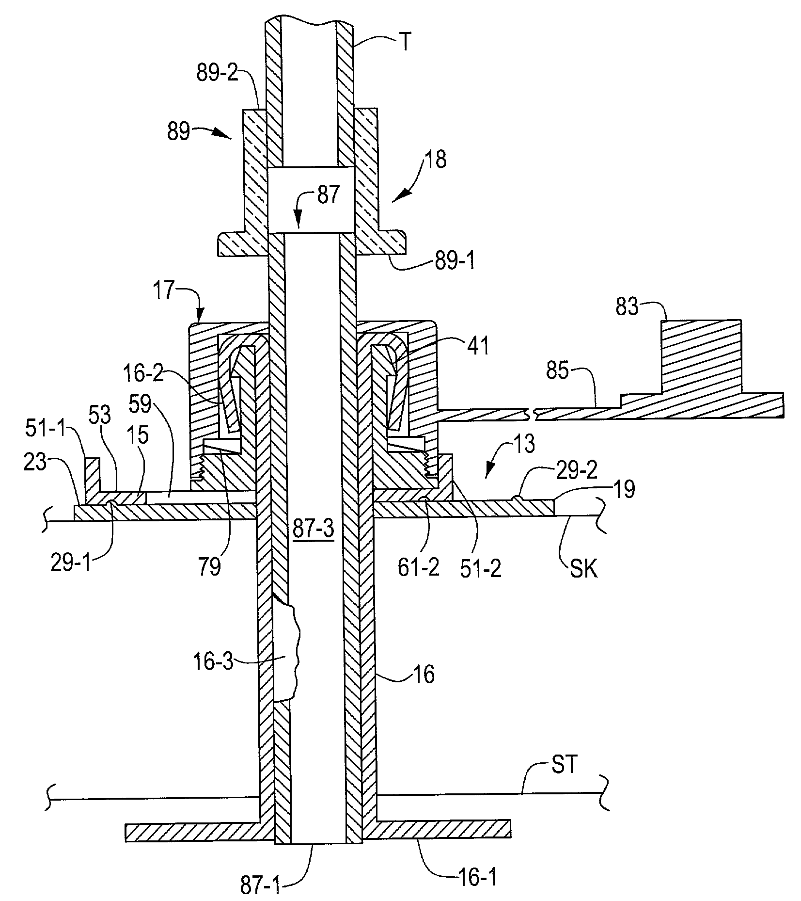
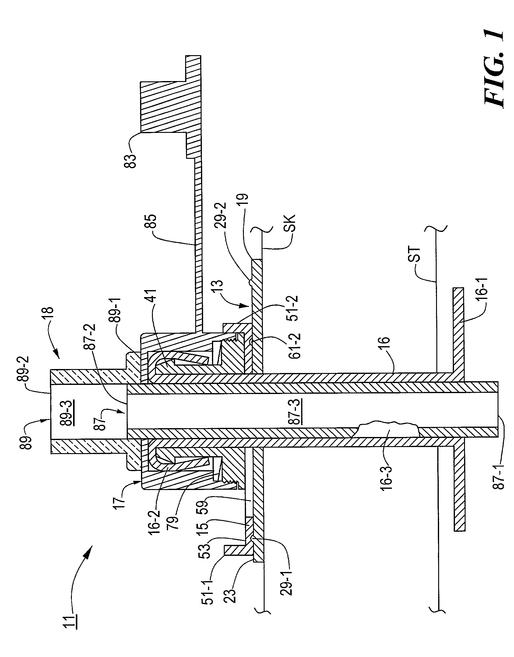
[0048]Referring now to FIG. 1, there is shown a section view, broken away in part, of a low profile medical catheter assembly constructed according to the teachings of the present invention, said low profile medical catheter assembly being represented generally by reference numeral 11.

[0049]Assembly 11, which is shown implanted in a patient as a low profile, convertible, percutaneous endoscopic gastrostomy (PEG) device, comprises a body 13, a clamp 15, a gastrostomy feeding tube 16, a cap 17, and an inner sleeve assembly 18.

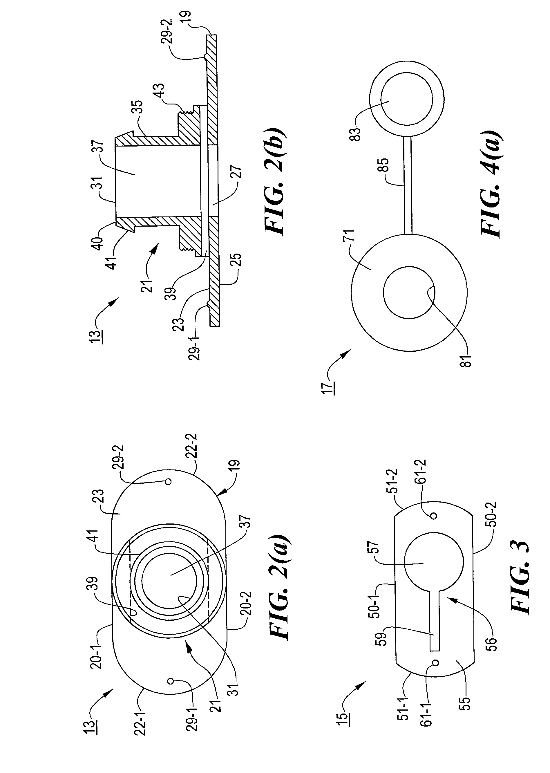
[0050]Referring now to FIGS. 1, 2(a) and 2(b), body 13, which is a unitary structure preferably made of molded medical grade plastic, is shaped to include a base 19 and a sleeve 21. Base 19, which is appropriately sized to engage the skin SK of the patient surrounding a stoma tract so as to serve as an external bolster, is a quasi-rectangular member having a pair of straight sides 20-1 and 20-2, a pair of rounded ends 22-1 and 22-2, a top surface 23, a bottom sur...

second embodiment

[0074]Referring now to FIGS. 18 and 19, there are shown section views of a low profile medical catheter assembly constructed according to the teachings of the present invention, said low profile medical catheter assembly being represented generally by reference numeral 201.

[0075]Assembly 201 is similar in many respects to assembly 11, the principal differences between the two assemblies being that (i) assembly 201 does not include clamp 15, (ii) cap 17 is replaced with a cap 203, and (iii) inner sleeve assembly 18 is replaced with an inner sleeve assembly 205.

[0076]Cap 203, which is similar in many respects to cap 17, differs notably from cap 17 in that cap 203 further includes a collar 207 extending upwardly from top surface 71 and surrounding opening 81. A helical thread 209, whose purpose will become apparent below, is formed on the inside surface of collar 207.

[0077]Inner sleeve assembly 205, which is similar in certain respects to inner sleeve assembly 18, differs notably from ...


PUM


Abstract

A medical catheter assembly including a removable inner sleeve. In one embodiment, the assembly is a low profile percutaneous endoscopic gastrostomy (PEG) device and comprises a body, a clamp, a feeding tube, a cap and an inner sleeve assembly. The body includes a base portion and a sleeve portion, the base portion being dimensioned to engage the skin of a patient and having a transverse bore, the sleeve portion extending upwardly from the base portion and having a longitudinal slot aligned with the transverse bore and a transverse slot intersecting the longitudinal bore. The clamp, which is slidably mounted on the base portion and across the transverse slot of the sleeve, comprises a plate having a transverse opening. The transverse opening has a wide region and a narrow region, the two regions being alternately alignable with the longitudinal bore to open and to close, respectively, the feeding tube. The feeding tube has a distal end adapted to be anchored to the inside of a patient and a proximal end inserted up through the base portion and the sleeve portion, including the transverse opening of the clamp situated within the sleeve, and then inverted over the top edge of the sleeve. The cap is then mounted on top of the sleeve so as to secure the inverted end of the catheter to the exterior of the sleeve. The cap is provided with an opening through which access to the catheter may be gained. An inner sleeve, sized to engage the inside surface of the feeding tube, is removably inserted through the cap and the feeding tube, the inner sleeve having a proximal end to which a tubular fitting is secured. Food and / or medications are dispensed to the patient through the fitting and the inner sleeve and, in this manner, prevent clogging of the feeding tube.

Description

BACKGROUND OF THE INVENTION[0001]The present invention relates generally to medical catheters, such as gastrostomy feeding tubes, and relates more particularly to medical catheter assemblies, such as percutaneous endoscopic gastrostomy (PEG) devices.[0002]Certain patients are unable to take food and / or medications transorally due to an inability to swallow. Such an inability to swallow may be due to a variety of reasons, such as esophageal cancer, neurological impairment and the like. Although the intravenous administration of food and / or medications to such patients may be a viable short-term approach, it is not well-suited for the longterm. Accordingly, the most common approach to the long-term feeding of such patients involves gastrostomy, i.e., the creation of a feeding tract or stoma between the stomach and the upper abdominal wall. Feeding is then typically performed by administering food through a catheter or feeding tube that has been inserted into the feeding tract, with th...

Claims


Application Information

Patent Timeline
Patent Type & Authority Patents(United States)
IPC IPC(8): A61M5/32A61M25/18A61M39/00A61M5/00A61J15/00A61M25/02
CPCA61J15/0015A61M25/02A61J15/0057A61J15/0026A61M2025/0233
Inventor CHU, MICHAEL S. H.BOUPHAVICHITH, LADDVANH
Owner BOSTON SCI SCIMED INC
Why Eureka
Try Eureka