Method and system for improving the spatial resolution for strain imaging

Inactive Publication Date: 2006-05-23
SIEMENS MEDICAL SOLUTIONS USA INC
View PDF11 Cites 14 Cited by
  • Summary
  • Abstract
  • Description
  • Claims
  • Application Information

AI Technical Summary

Benefits of technology

[0008]A system and method in accordance with the present invention provides for strain quantification based on conventional B-mode images. Using this technique, the strain of regions of interest (ROI) defined by users can be determined and quantitative comparisons can be effectively made in real time. The strain quantification can be used to determine tissue's properties and can potentially be applied in breast imaging as well as cardiac imaging.

Problems solved by technology

However, there are several problems with this type of method for quantifying the strain.
First, there are artifacts that can be generated based on the probe usage over several points.
Second, the system is time-consuming and tedious.
Thirdly, other areas can distort the measured strain due to shadows, cavities and other areas of anomalies within the area being imaged.
But there are technical difficulties for accurately determining the difference of displacements over these local blocks and generate the poor signal to noise ratio (SNR) for the strain images and therefore quantifying the strain measurements.
These differences will cause additional errors for our motion estimation.

Method used


Image

  • Method and system for improving the spatial resolution for strain imaging
  • Method and system for improving the spatial resolution for strain imaging
  • Method and system for improving the spatial resolution for strain imaging

Examples


Embodiment Construction

[0013]The present invention relates to medical imaging and more specifically to a technique for quantifying tissue strain images. The following description is presented to enable one of ordinary skill in the art to make and use the invention and is provided in the context of a patent application and its requirements. Various modifications to the preferred embodiment and the generic principles and features described herein will be readily apparent to those skilled in the art. Thus, the present invention is not intended to be limited to the embodiment shown but is to be accorded the widest scope consistent with the principles and features described herein.

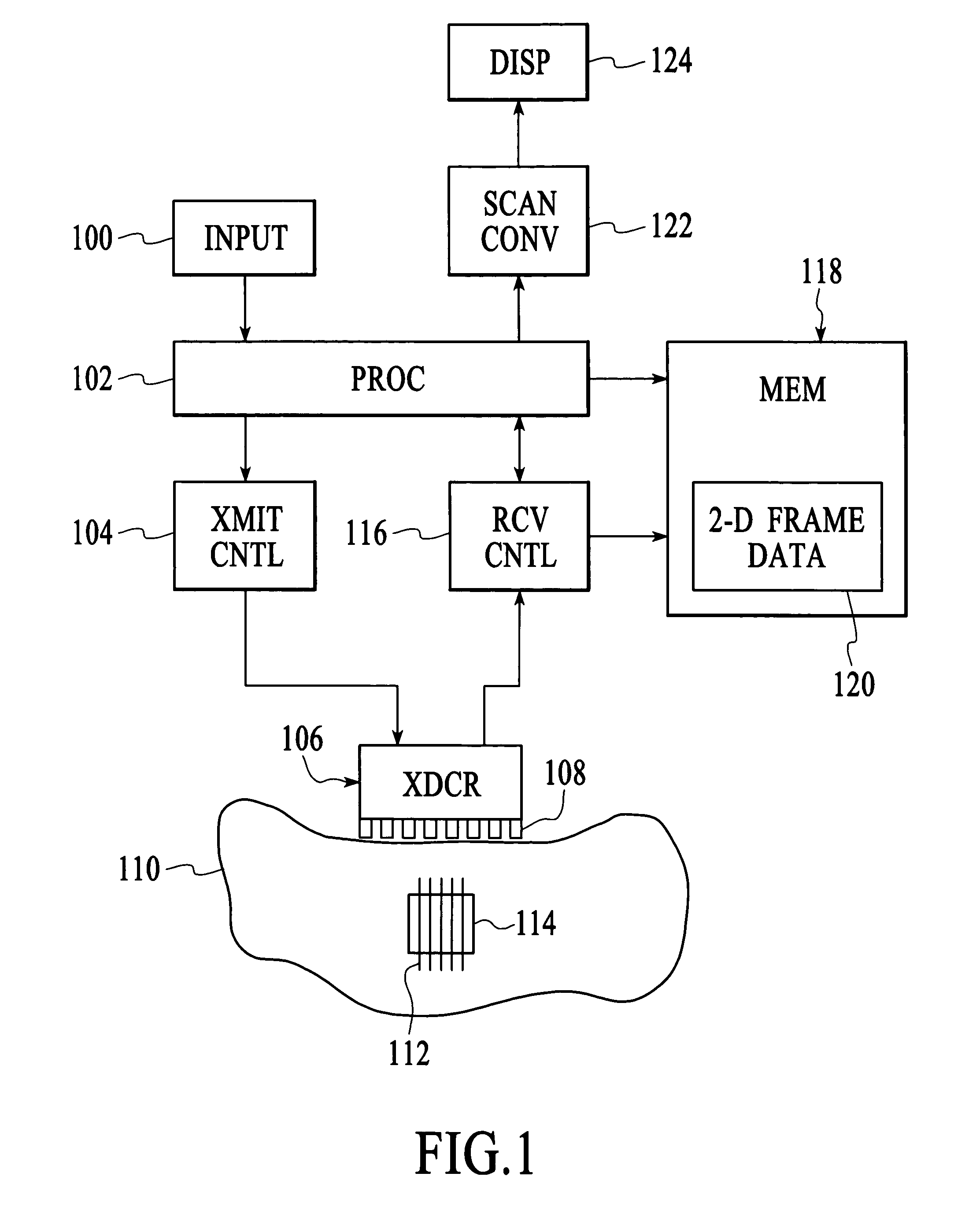
[0014]FIG. 1 is a simplified block diagram of an ultrasound system according to the present invention. The user enters the various conventional scan parameters into an input unit 100, which typically includes such devices as a keyboard, knobs and buttons. The input unit is connected to a processing system 102, which will typically be...


PUM


Abstract

A method and system for quantification of strain imaging is disclosed. The method and system comprises performing a motion analysis on the at least two regions of interest. The method and system further includes providing a strain estimate for each of the at least two regions of interest and comparing the strain estimates of each of the at least two regions to quantify the strain for the at least two regions of interest. A system and method in accordance with the present invention provides for strain quantification based on conventional B-mode images. Using this technique, the strain of regions of interest (ROI) defined by users can be determined and quantitative comparisons can be effectively made in real time. The strain quantification can be used to determine tissue's properties and can potentially be applied in breast imaging as well as cardiac imaging.

Description

FIELD OF THE INVENTION[0001]The present invention relates generally to medical imaging and more specifically to a technique for quantifying ultrasound strain imaging.BACKGROUND OF THE INVENTION[0002]Strain imaging has been used in ultrasound medical imaging to differentiate between hard tissue and soft tissue. For example, since tumors are generally harder than normal tissues, strain imaging may be used evaluate solid breast masses. Strain images are created by comparing echo data obtained before and after a slight compression of the tissue. The results of the comparison are displayed as an image on which the hard areas appear dark and soft areas appear bright. Other black-and-white and other color scale may be employed for display. Typically strain imaging is accomplished by comparing the hard and soft tissue in a region of interest on a pixel by pixel basis using conventional ultrasound equipment.[0003]Typically strain images are made from strain measure point by point, pixel by p...

Claims


Application Information

Patent Timeline
IPC IPC(8): G06K9/00A61B8/08G01S7/52G01S15/89
CPCA61B8/0825A61B8/0833A61B8/485G01S15/8981G01S7/52042G01S7/5206G01S7/52036
Inventor CHEN, JIAN-FENGVON BEHREN, PATRICK L.
Owner SIEMENS MEDICAL SOLUTIONS USA INC
Why Eureka
Try Eureka