Scan type digital X-ray imaging apparatus

a digital x-ray imaging and scan-type technology, applied in the direction of instruments, diaphragms, radiation diagnostics, etc., can solve the problems of inability to accurately separate the soft tissue area, inability to make clear images of the soft tissue, and complicated image processing, so as to improve the digital x-ray image, improve the dynamic range of the image, and improve the effect of the imag

Inactive Publication Date: 2006-09-05
MORITA MFG CO LTD
View PDF11 Cites 31 Cited by
  • Summary
  • Abstract
  • Description
  • Claims
  • Application Information

AI Technical Summary

Benefits of technology

[0009]The object of the present invention in order to solve the above-mentioned problems is to provide a scan type digital X-ray imaging apparatus wherein the dynamic range of an X-ray image

Problems solved by technology

However, according to such an apparatus, when trying to make an image of a hard tissue such as a skull and a dental jaw bone clear by emitting a X-ray slit beam uniformly onto the head to be scanned, the images of a soft tissue such as skin and lips haven't been made clear.
However, it has been difficult to adjust the position of the soft tissue filter on an actual soft tissue area of the patient's head so that accurate separation of the sof

Method used


Image

  • Scan type digital X-ray imaging apparatus
  • Scan type digital X-ray imaging apparatus
  • Scan type digital X-ray imaging apparatus

Examples


Embodiment Construction

[0085]Embodiments of the present invention will be explained with reference to the attached drawings.

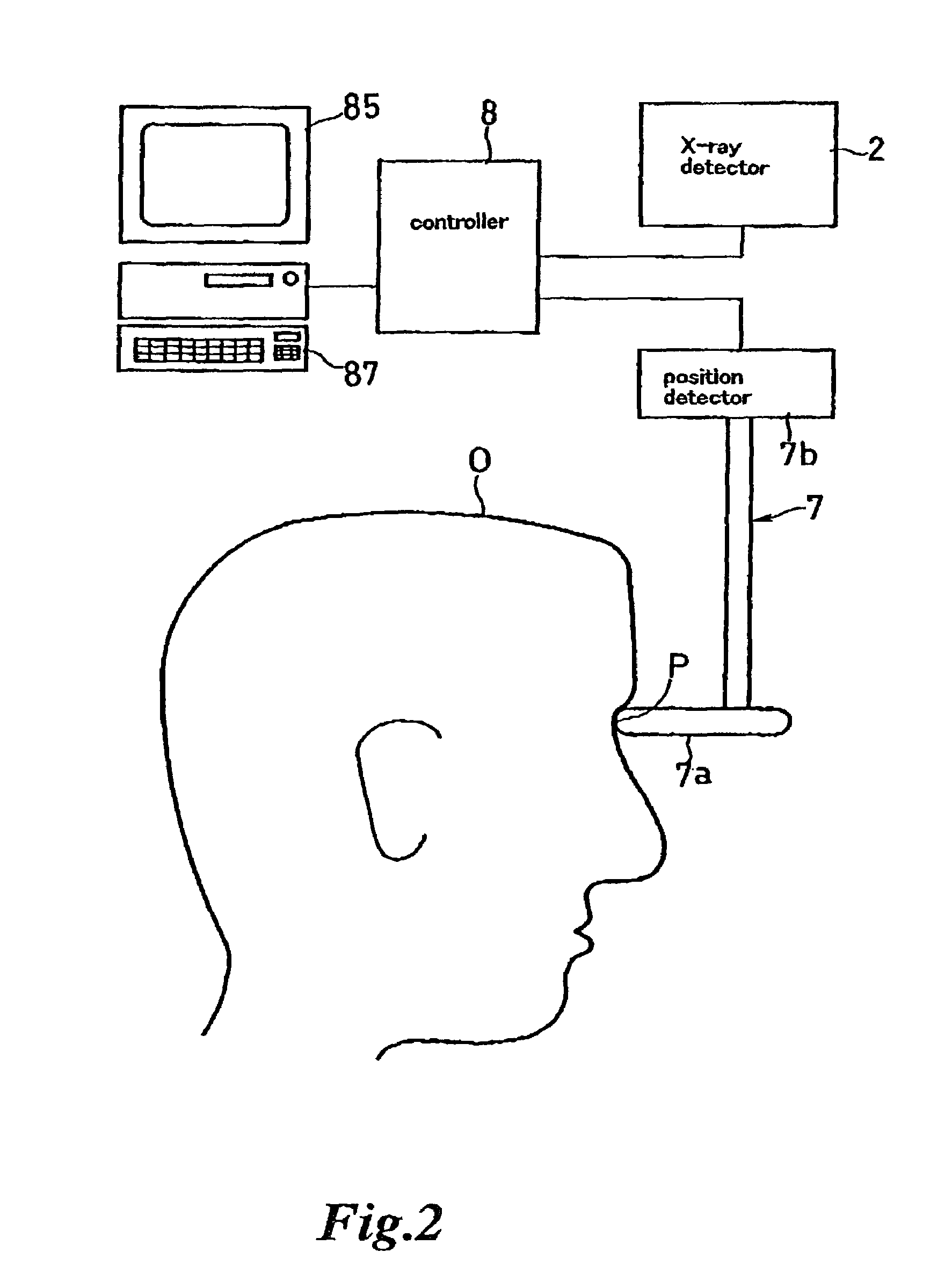
[0086]Referring to the FIGS. 1–14, embodiments wherein a scan type digital X-ray imaging apparatus for medical use of the present invention is applied to a dental cephalometric imaging apparatus will be explained hereinafter, however, the present invention is not limited to dental use alone but is widely applied to an X-ray imaging apparatus for general medical use, such as a mammography, and slit radiography, and so on.

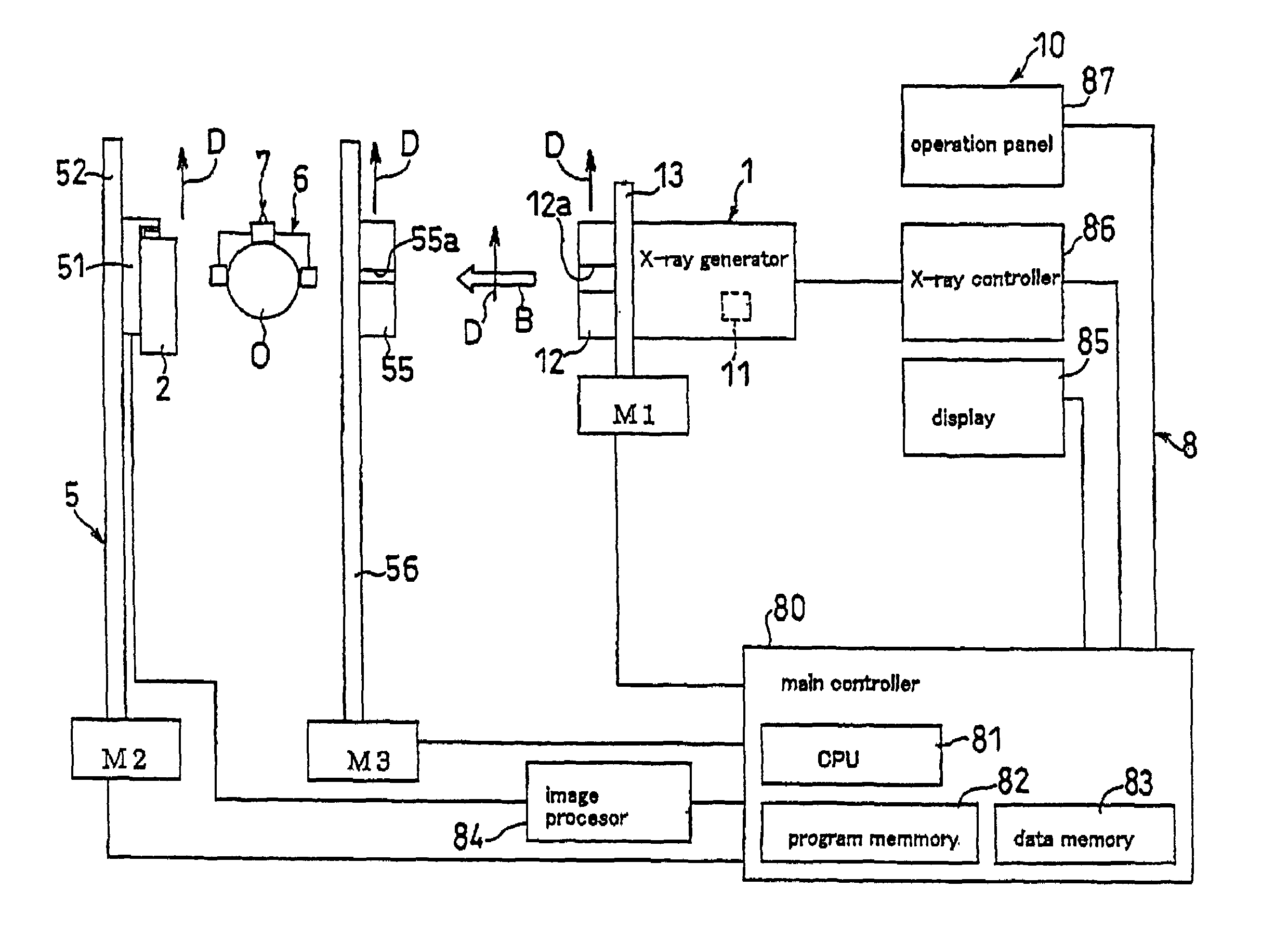
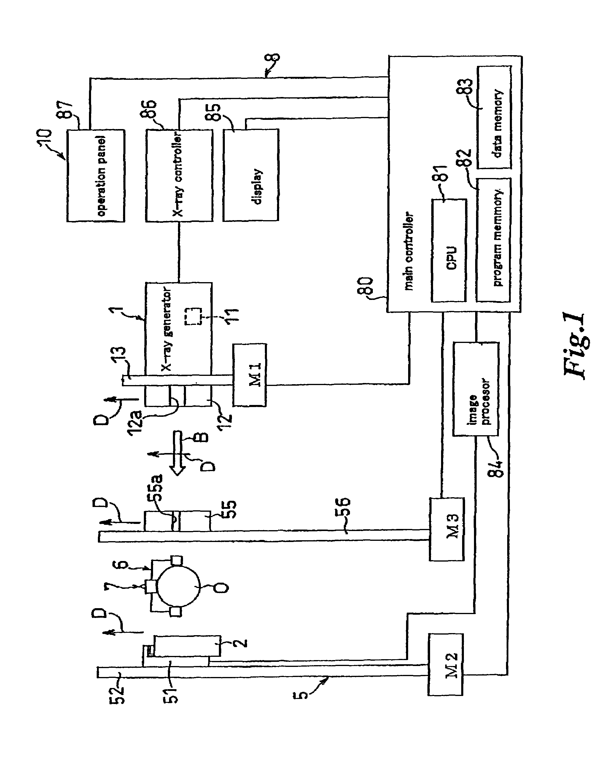
[0087]FIG. 1 is a block diagram showing an entire construction of one embodiment of a scan type digital X-ray imaging apparatus for medical use according to the present invention.

[0088]The scan type digital X-ray imaging apparatus for medial use 10 has an X-ray generator 1, an X-ray detector 2 for receiving X-ray slit beams B emitted from the X-ray generator 1 and transmitted through an object O and for outputting the received X-ray data in the form of digital or digi...


PUM


Abstract

A scan type digital X-ray imaging apparatus comprising an X-ray generator for emitting X-ray slit beams, an X-ray detector for detecting X-ray transmitted through an object to be examined, and an object fixing means disposed between the X-ray generator and the X-ray detector, for holding the object in an appropriate position for X-ray photography, in which X-ray beam scanning is performed in a manner that the X-ray slit beam is emitted from the X-ray generator for the object while the X-ray generator and the X-ray detector are synchronously moved in the same direction with the object interposed therebetween, thereby producing an X-ray image of the object. In the X-ray imaging apparatus, the scanning speed of the X-ray slit beam is controlled depending on the X-ray transmitted amount through the object to be examined detected by the X-ray detector during the X-ray beam scanning is performed, whereby soft and hard tissues appearing in the X-ray image are amended in each contrast and/or in each density.

Description

BACKGROUND OF THE INVENTION[0001]1. Field of the Invention[0002]The present invention relates to an improvement of a scan type digital X-ray imaging apparatus which has an X-ray detector capable of outputting digitalizable data or digital data upon receiving X-ray like a dental digital cephalometric X-ray imaging apparatus and wherein the X-ray slit beam scans an object to be examined while the X-ray slit beam emitted from the X-ray generator and the X-ray detector are synchronously moved in the same direction and an X-ray image of the object is produced.[0003]2. Prior Art[0004]Conventionally, an X-ray slit beam has scanned the head of the patient, which is an object to be examined, to obtain an X-ray image by a scan type digital X-ray imaging apparatus for medical use for example a dental cephalometric imaging apparatus disclosed in JP-A-7-143981. However, according to such an apparatus, when trying to make an image of a hard tissue such as a skull and a dental jaw bone clear by em...

Claims


Application Information

Patent Timeline
IPC IPC(8): A61B6/14A61B6/00A61B6/06H05G1/30
CPCA61B6/14H05G1/30A61B6/501A61B6/4429A61B6/06
Inventor SONOBE, KOUICHIHAYASHI, TAKESHIHONJYO, MAKOTO
Owner MORITA MFG CO LTD
Why Eureka
Try Eureka