Doppler ultrasound method and apparatus for monitoring blood flow and hemodynamics

a technology of hemodynamics and ultrasound method, which is applied in the direction of instruments, ultrasonic/sonic/infrasonic diagnostics, and using reradiation. it is not easy to accurately and the user of ultrasound equipment finds it difficult to properly orient and position the ultrasound transducer or probe on the patient. it is difficult to locate these particular windows or properly orient the ultrasound probe, etc. user does not know

Inactive Publication Date: 2006-10-31
TERUMO CARDIOVASCULAR SYST CORP
View PDF47 Cites 61 Cited by
  • Summary
  • Abstract
  • Description
  • Claims
  • Application Information

AI Technical Summary

Problems solved by technology

Typically, a user of ultrasound equipment finds it rather difficult to properly orient and position an ultrasound transducer or probe on the patient, as well as to select a depth along the ultrasound beam corresponding to the desired location where blood flow is to be monitored.
The user of the ultrasound equipment may find it difficult to locate these particular windows or to properly orient the ultrasound probe once the particular window is found.
Commonly, the user does not know if he is looking in the correct direction at the wrong depth, the wrong direction at the right depth, or whether the ultrasound window is too poor for appreciating blood flow at all.
Proper location and orientation of the Doppler ultrasound probe, and the proper setting of depth parameters, is typically by trial and error.
Not only does this make the use of Doppler ultrasound equipment quite inconvenient and difficult, it also creates a risk that the desired sample volume may not be properly located, with the corresponding diagnosis then being untenable or potentially improper.
However, in conventional Doppler ultrasound systems, regions having abnormal flow may be displayed ambiguously.
Both the aliased velocities and the clutter signals can severely compromise detection of peak blood flow velocity and other hemodynamic parameters.
The previously described issues with conventional Doppler ultrasound systems are often due to artifacts resulting from Doppler signal processing.
A possible cause of artifacts is the presence of a bruit signal that often accompanies the pathological condition of vasospasm, a condition that results in a constriction of the vessel lumen and results in high velocity blood flow.
This approach however is time consuming in that it requires a Fourier transform to be computed at every analyzed depth
Another potential cause of artifacts is high velocity aliasing due to the Doppler shift frequency of detected blood flow exceeding the Nyquist frequency of the Doppler ultrasound system, the result of which is to bias the detected mean velocity to zero.
However, in conventional Doppler ultrasound systems, increasing the Doppler PRF comes with a tradeoff of reducing the maximum interrogation depth, which is limited by the round trip distance an ultrasound pulse can travel before a subsequent ultrasound pulse is launched by the system.

Method used


Image

  • Doppler ultrasound method and apparatus for monitoring blood flow and hemodynamics
  • Doppler ultrasound method and apparatus for monitoring blood flow and hemodynamics
  • Doppler ultrasound method and apparatus for monitoring blood flow and hemodynamics

Examples


Embodiment Construction

[0023]The following describes a method and apparatus for providing Doppler ultrasound information to a user, such as in connection with measuring blood velocities to quickly detect hemodynamically significant deviations from normal values across a range of depth. Certain details are set forth to provide a sufficient understanding of the invention. However, it will be clear to one skilled in the art that the invention may be practiced without these particular details. In other instances, well-known circuits, control signals, timing protocols, and software operations have not been shown in detail in order to avoid unnecessarily obscuring the invention.

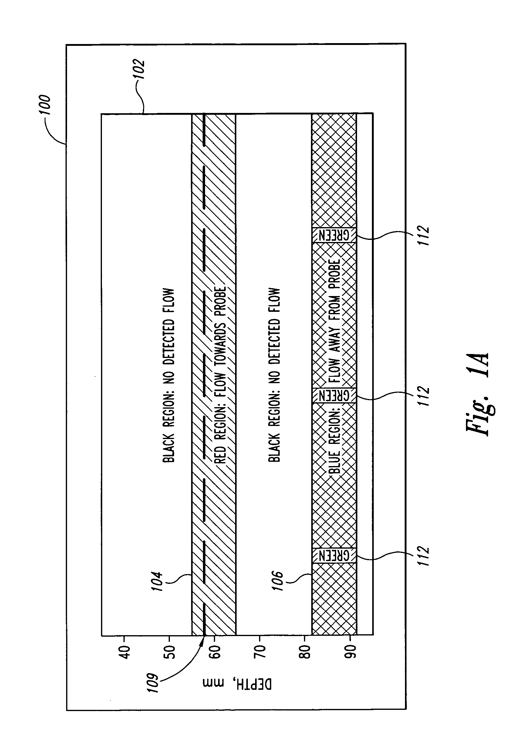
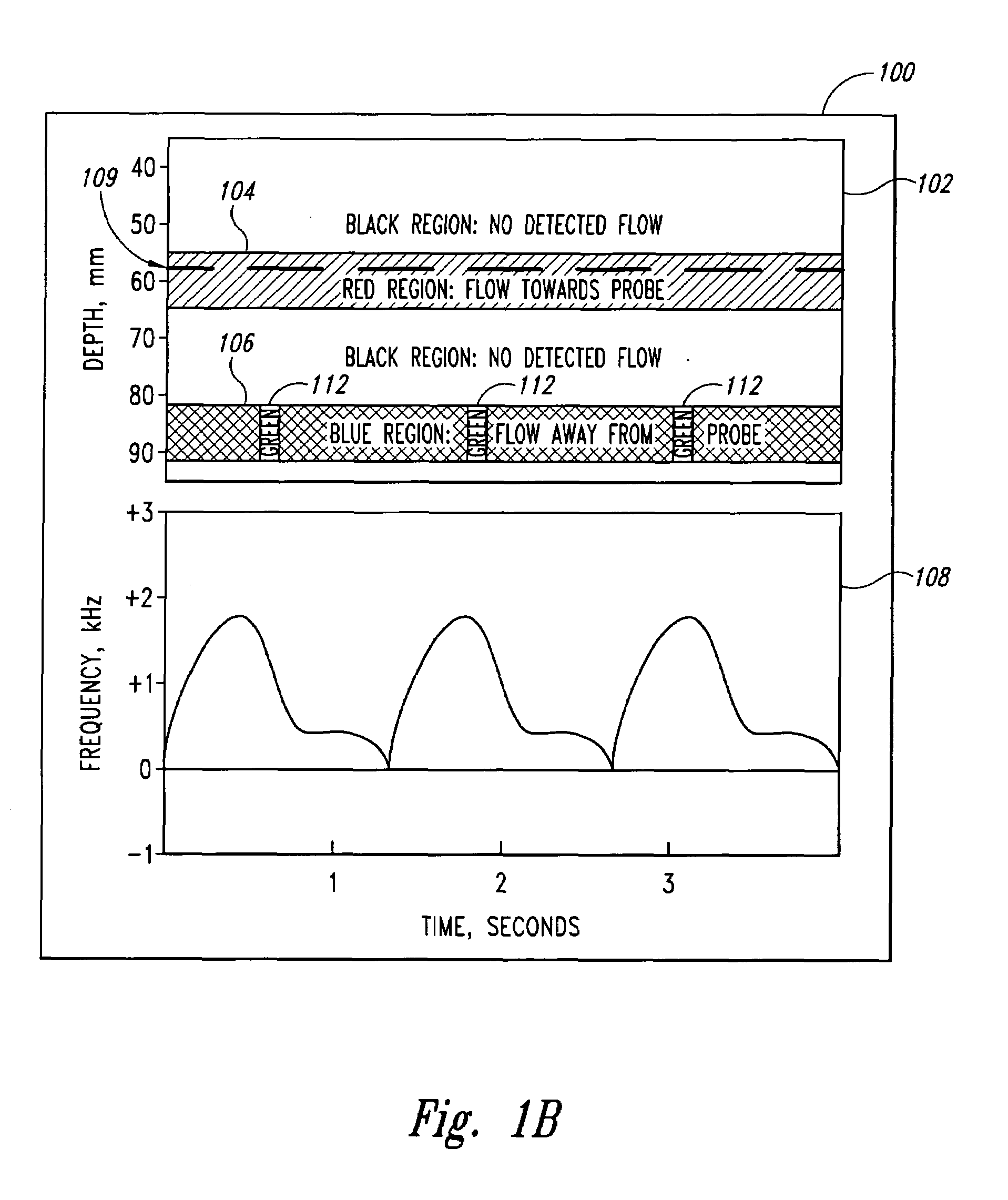
[0024]FIG. 1A is an Aiming Mode Display 100 depicting a display mode of Doppler ultrasound information in accordance with an embodiment of the invention. In this display mode, as shown on the Aiming Mode Display 100, a depth-mode display 102 depicts, with color, blood flow away from and towards the ultrasound probe at various depths alon...


PUM


Abstract

A pulse Doppler ultrasound system and associated methods are described for monitoring blood flow and hemodynamics. The Doppler ultrasound system includes an ultrasound probe to emit ultrasound signals and detect reflected signals therefrom and further includes a processor coupled to the ultrasound probe and operable to process the detected reflected signals and calculate therefrom blood flow data for a plurality of locations at time intervals, the processor further operable to identify locations at which blood flow having a hemodynamic characteristic is present based on the blood flow data calculated for a plurality of the time intervals. A user interface coupled to the processor provides blood flow information based on the blood flow data, the blood flow information representative of detected blood flow and the presence of the hemodynamic characteristic.

Description

STATEMENT AS TO GOVERNMENT RIGHTS[0001]The disclosed invention was made with support from the United States Government, which has certain fights in the invention pursuant to Grant No. 5R 44HL057108-03 awarded by the National Institutes of Health.TECHNICAL FIELD[0002]The invention relates generally to medical monitoring and diagnostic procedures and devices, and more particularly to a Doppler ultrasound method and apparatus for monitoring blood flow and hemodynamics.BACKGROUND OF THE INVENTION[0003]Doppler ultrasound has been used to measure blood flow velocity for many years. The well-known Doppler shift phenomenon provides that ultrasonic signals reflected from moving targets will have a shift in frequency directly proportional to the target velocity component parallel to the direction of the ultrasound beam. The frequency shift is the same for any object moving at a given velocity, whereas the amplitude of the detected signal is a function of the acoustic reflectivity of the movin...

Claims


Application Information

Patent Timeline
Patent Type & Authority Patents(United States)
IPC IPC(8): A61B8/06A61BA61B8/02
CPCA61B8/06G01S7/52071G01S15/8981A61B8/0816
Inventor MOEHRING, MARK A.FARNSWORTH, JOSEPH H.ZACHARIAH, AJAY PHILIPBARON, JR., HENRY JAMES
Owner TERUMO CARDIOVASCULAR SYST CORP
Why Eureka
Try Eureka