

Intradermal delivery of active agents by needle-free injection and electroporation
- Summary
- Abstract
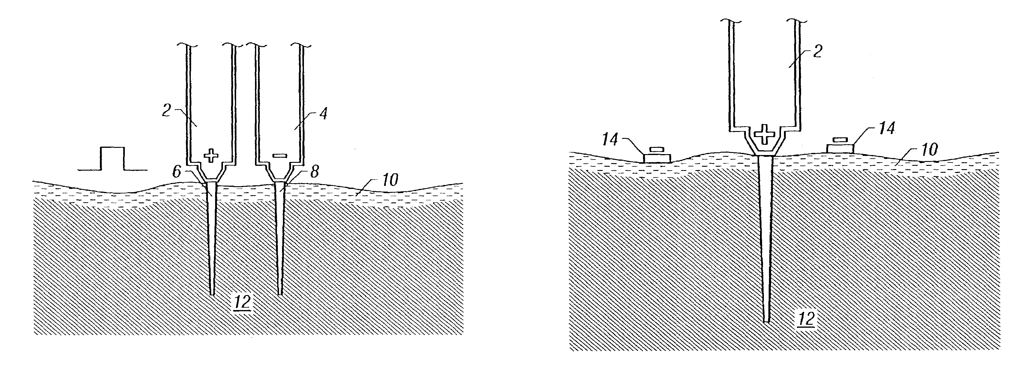
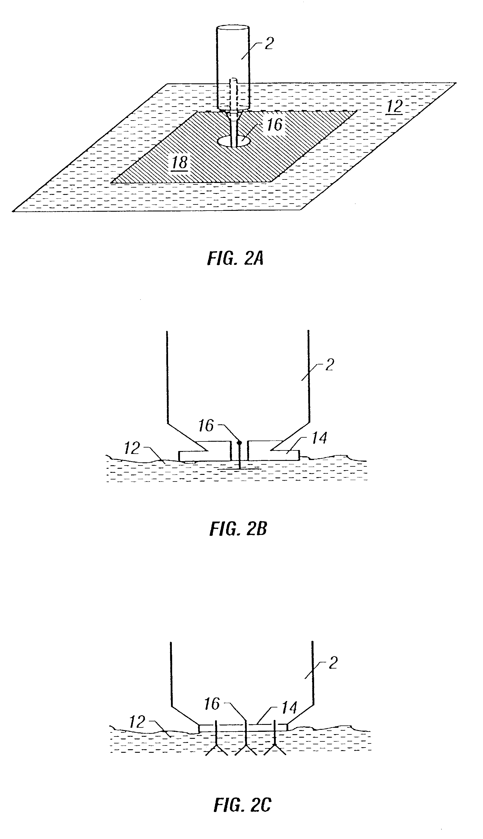
- Description
- Claims
- Application Information
AI Technical Summary
Benefits of technology
Problems solved by technology
Method used
Image

Examples
example 1
[0093]The following study was conducted to compare quantatively the level of gene expression when a DNA-containing plasmid is administered by intradermal injection and by needle-free injector. Electroporation of the injection site at various voltage levels was used to determine and compare electroporation enhancement of gene expression when DNA is injected by the two methods tested.
I. Luciferase reporter gene. To quantify the level of gene expression, the luciferase reporter gene was used in the first study.
[0094]Animals. Four to six week old male and female outbred pigs weighing 20 to 40 pounds were purchased from the Prairie Swine Center (University of Saskatchewan, Saskatoon, Saskatchewan). The animals were housed and treated in compliance with the Canadian Council for Animal Care. Animals were randomly assigned to six groups of five animals each.
[0095]Electroporation. To determine the effect of electroporation on expression of plasmid DNA, electroporation was performed using the...
example 2
[0104]To study the effects of electroporation in the context of a DNA vaccine, a DNA-prime / DNA-boost / protein-boost strategy was evaluated for enhancement of immune responses in large animals, and compared the responses achieved following standard protein vaccination. The strategy used resembled DNA-prime / protein-boost strategies previously used in non-human primates, which yielded outstanding results for both malaria (12) and HIV vaccinations (1).
[0105]Immunization protocols. For immunization studies, animals were selected and care for as above. Pigs were randomly assigned to six groups of five animals each. The pigs were anesthetized with halothane prior to DNA injection and electroporation and treated as follows: The animals in Group 1 each received 250 μg pHBsAg in 100 μl 0.1 M phosphate buffered saline (PBS) by a dermal BIOJECT B 2000° needle-free injection device (Bioject, Inc. Portland, Oreg.) at each of two abdominal sites for a total of 500 μg pHBsAg. The animals in Group 2 ...
PUM
 Abstract
Description
Claims
Application Information