
Devices and methods for maintaining collateral channels in tissue

a tissue and collateral channel technology, applied in the field of tissue collateral channel maintenance devices and methods, can solve the problems of copd-afflicted individuals, loss of muscle strength, and inability to perform common daily activities, and achieve the effect of optimizing the effect of collateral channels

Inactive Publication Date: 2007-02-13
BRONCUS MEDICAL
View PDF240 Cites 240 Cited by
  • Summary
  • Abstract
  • Description
  • Claims
  • Application Information

AI Technical Summary

Benefits of technology

This patent describes a method and device for improving gas flow in a diseased lung by creating collateral channels within the respiratory system. The invention allows for the creation of channels that can help to decompress a hyperinflated lung. The method involves selecting a site for the creation of a channel and creating the channel by removing a portion of the airway wall or forming a hole, slit, skive, or cut flap. The invention also includes methods for detecting blood vessels and marking the site of the channel. The technical effects of the invention include improved gas flow, reduced pressure in the airways, and the ability to create channels that can help to decompress a hyperinflated lung.

Problems solved by technology

Those inflicted with COPD face disabilities due to the limited pulmonary functions.
Usually, individuals afflicted by COPD also face loss in muscle strength and an inability to perform common daily activities.
Since the damage to the lungs is irreversible, there is little hope of recovery.
Most times, the physician cannot reverse the effects of the disease but can only offer treatment and advice to halt the progression of the disease.
However, these conducting airways do not take part in gas exchange because they do not contain alveoli.
If the lungs' ability to recoil is damaged, the lungs cannot contract and reduce in size from their inflated state.
As a result, the lungs cannot evacuate all of the inspired air.
Also, the bio-chemical breakdown of the walls of the alveolar walls causes a loss of radial support for airways which results in a narrowing of the airways on expiration.
As a result of the COPD, the airways close prematurely at an abnormally high lung volume.
Consequently, this increased residual gas volume interferes with the ability of the lung to draw in additional fresh gas during inspiration.
Yet, while the resistance to collateral ventilation may be decreased in an emphysematous lung the decreased resistance does not assist the patient in breathing due to the inability of the gasses to enter and exit the lungs as a whole.
Yet, bronchodilator drugs are only effective for a short period of time and require repeated application.
Moreover, the bronchodilator drugs are only effective in a certain percentage of the population of those diagnosed with COPD.
Unfortunately, aside from the impracticalities of needing to maintain and transport a source of oxygen for everyday activities, the oxygen is only partially functional and does not eliminate the effects of the COPD.
Moreover, patients requiring a supplemental source of oxygen are usually never able to return to functioning without the oxygen.
However, lung reduction surgery is an extremely traumatic procedure which involves opening the chest and thoracic cavity to remove a portion of the lung.
Both bronchodilator drugs and lung reduction surgery fail to capitalize on the increased collateral ventilation taking place in the diseased lung.

Method used


Image

  • Devices and methods for maintaining collateral channels in tissue
  • Devices and methods for maintaining collateral channels in tissue
  • Devices and methods for maintaining collateral channels in tissue

Examples


Embodiment Construction

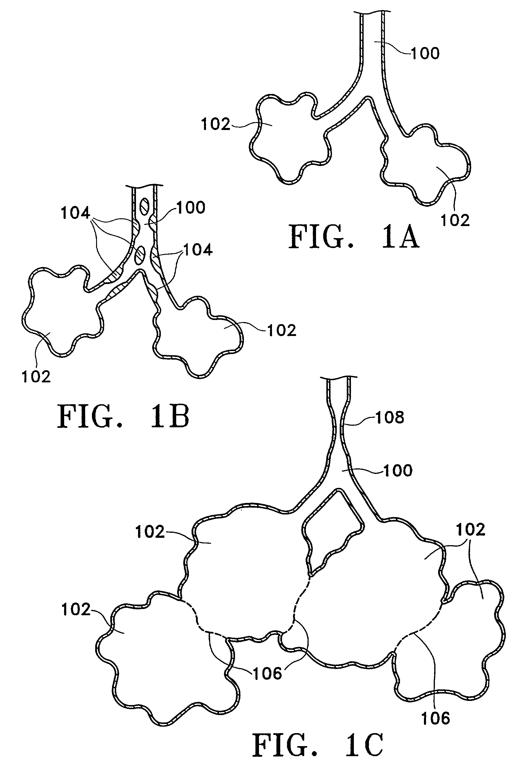
[0054]Prior to considering the invention, simplified illustrations of various states of a natural airway and a blood gas interface found at a distal end of those airways are provided in FIGS. 1A–1C. FIG. 1A shows a natural airway 100 which eventually branches to a blood gas interface 102. FIG. 1B illustrates an airway 100 and blood gas interface 102 in an individual having COPD. The obstructions 104 impair the passage of gas between the airways 100 and the interface 102. FIG. 1C illustrates a portion of an emphysematous lung where the blood gas interface 102 expands due to the loss of the interface walls 106 which have deteriorated due to a bio-chemical breakdown of the walls 106. Also depicted is a constriction 108 of the airway 100. It is generally understood that there is usually a combination of the phenomena depicted in FIGS. 1A–1C. More usually, the states of the lung depicted in FIGS. 1B and 1C are often found in the same lung.

[0055]The following illustrations are examples of...


PUM


Abstract

The devices and methods of placement of such devices disclosed herein are directed to altering gaseous flow within a lung to improve the expiration cycle of, for instance, an individual having Chronic Obstructive Pulmonary Disease. More particularly, these devices produce and maintain collateral openings or channels through the airway wall so that oxygen depleted / carbon dioxide rich air is able to pass directly out of the lung tissue to facilitate both the exchange of oxygen ultimately into the blood and / or to decompress hyper-inflated lungs. The medical kits disclosed herein are also directed to produce and maintain collateral openings through airway walls.

Description

CROSS-REFERENCE TO RELATED APPLICATIONS[0001]This application claims the benefit of U.S. Provisional Patent Application Ser. No. 60 / 269,130, filed on Feb. 14, 2001, which is incorporated herein by reference in its entirety.FIELD OF THE INVENTION[0002]The invention is directed to devices and methods of placing such devices to altering gaseous flow within a lung to improve the expiration cycle of an individual, particularly individuals having Chronic Obstructive Pulmonary Disease (COPD). More particularly, methods and devices are disclosed to produce and to maintain collateral openings or channels through the airway wall so that oxygen depleted / carbon dioxide rich air is able to pass directly out of the lung tissue to facilitate both the exchange of oxygen ultimately into the blood and / or to decompress hyper-inflated lungs. The invention is also directed to medical kits disclosed which produce and maintain collateral openings through airway walls.BACKGROUND OF THE INVENTION[0003]The t...

Claims


Application Information

Patent Timeline
Patent Type & Authority Patents(United States)
IPC IPC(8): A61M29/00A61F2/06A61B8/06A61B8/12A61B17/00A61B17/064A61B17/11A61B17/32A61B18/00A61B18/14A61B19/00A61F2/04A61F2/24A61F2/90A61F2/92
CPCA61B8/445A61B8/12A61B17/064A61B18/1485A61B18/1492A61F2/07A61F2/90A61F2/92A61B5/489A61B17/11A61B17/320068A61B18/1477A61B19/52A61B2017/00106A61B2017/00252A61B2017/00575A61B2017/00809A61B2017/22067A61B2018/00214A61B2018/00541A61B2018/00601A61B2018/00738A61B2018/00982A61B2018/1417A61B2018/1425A61B2018/1472A61B2019/545A61B2218/002A61F2/2412A61F2/91A61F2002/043A61F2002/061A61F2002/8483A61F2002/072A61F2220/0008A61F2220/0016A61F2230/005A61F2230/0058A61F2230/0067A61F2230/0076A61F2230/0078A61B8/06A61B90/36A61B2090/395
Inventor COOPER, JOEL D.DAVENPORT, JAMES M.LOOMAS, BRYANTANAKA, DONKAPLAN, GARY
Owner BRONCUS MEDICAL
Learn More