Intubation tube placement assessment device

a technology for assessing devices and intubation tubes, which is applied in the direction of medical devices, respiratory apparatus, other medical devices, etc., can solve the problems of inadvertent esophageal intubation, inability to accurately detect the position of the ett in the patient, and inability to perform inadvertent intubation, etc., to achieve the effect of improving specificity, sensitivity and quick discernmen

Inactive Publication Date: 2007-02-20
UNIV OF FLORIDA RES FOUNDATION INC
View PDF18 Cites 30 Cited by
  • Summary
  • Abstract
  • Description
  • Claims
  • Application Information

AI Technical Summary

Benefits of technology

[0009]The present invention comprises a connection device useful in connecting a CO2 detection device and a volume displacement device in line with an endotracheal tube (ETT). The device further comprises at least one positive pressure response valve and at least one negative pressure response valve to allow a user to control the movement of gas. By combining two or more existing ETT placement detectors in one unit, the present device allows a user to rapidly obtain alternate quantitative measurements of ETT placement in a patient in wide variety of clinical settings, both in and out of a hospital. The primary benefit of the present device is the ability to quickly discern with almost complete certainty the location of an ETT. Since incorrect placement of an ETT is not uncommon in situations of low blood flow, particularly cardiac arrest, this device offers several advantages over existing device, including improved specificity, sensitivity and full operation without the need for an electrical hookup. The benefit to the patient in any environment is obvious.

Problems solved by technology

Because of the close proximity of the esophagous to the trachea, an intubationist may accidentally place the ETT into an esophagous.
However, if the incorrect placement is not recognized and the ETT repositioned, within a few minutes irreversible brain damage and / or death may result, due to the lack of oxygen.
However, even under such controlled conditions inadvertant esophageal intubations have been reported.
Under such stressful and often chaotic conditions, misplacement of an ETT in a patient is not uncommon.
Each of these methods have utility in a limited range of clinical conditions.
However, these devices are less reliable when there is a pulmonary embolis or a patient is in cardiac arrest.
(Garnett, A R et al., JAMA 1987; 257: 512–515) Further, traditional capnometers are expensive, sophisticated, and fragile instruments requiring careful calibration and a source of power, making their use in out-of hospital procedures impractical.
Thus, in emergency-type settings, a capnometer may be inadequate.
However, even if the ETT is properly placed, the EasyCap, like the more expensive capnometer, is inadequate in those instances where a patient lacks a pulse or has very poor pulmonary perfusion because without CO2 exchange from pulmonary arterial blood to alveoli, insufficient CO2 will be exhaled to produce a color change.
However, when the ETT is placed in the trachea this negative pressure is incapable of causing the more rigid trachea to collapse, thereby allowing free exchange of air.
Further, these devices are useful in patients experiencing cardiac arrest because the test results do not depend upon the presence of CO2 in exhaled gas (Haridas, R P Update in Anaesthesia 1997;7:6(1)) However, regurgitation of gastric air, distension of the esophagus with air, or an EDD that is not airtight may cause the bulb to re-inflate, giving a false impression of tracheal intubation when the tube is in fact in the esophagus.
(Haridas, R P Update in Anaesthesia 1997; 7:6(3)) Further, although these devices are highly accurate when used in a hospital setting, studies indicate that they are only 50% accurate when used in the field by paramedics (Pelicio, M.
The National Association of EMS Physicians has recognized that no single technique for assessing ETT tube placement currently used is completely reliable in all circumstances.

Method used


Image

  • Intubation tube placement assessment device
  • Intubation tube placement assessment device
  • Intubation tube placement assessment device

Examples


Embodiment Construction

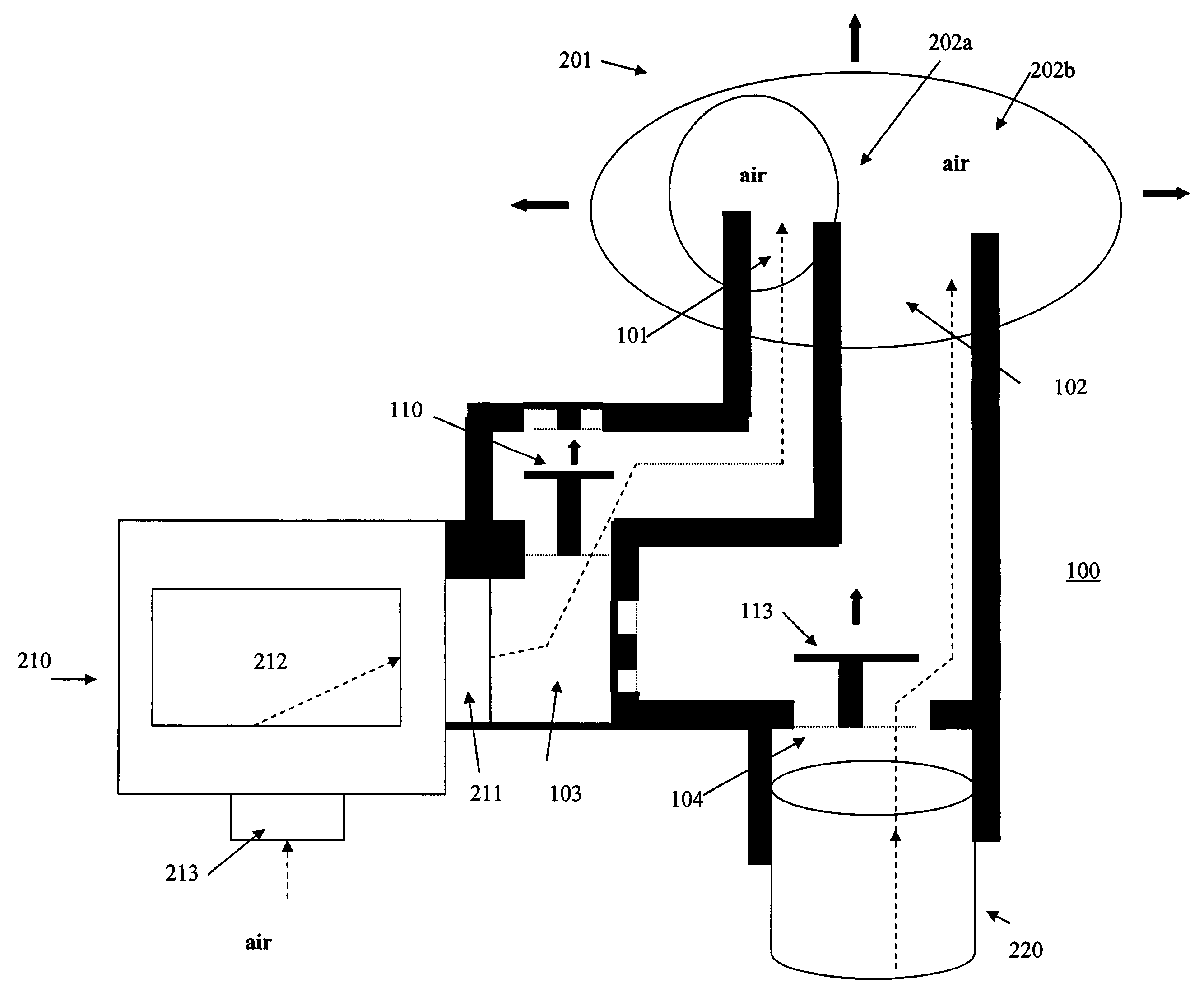
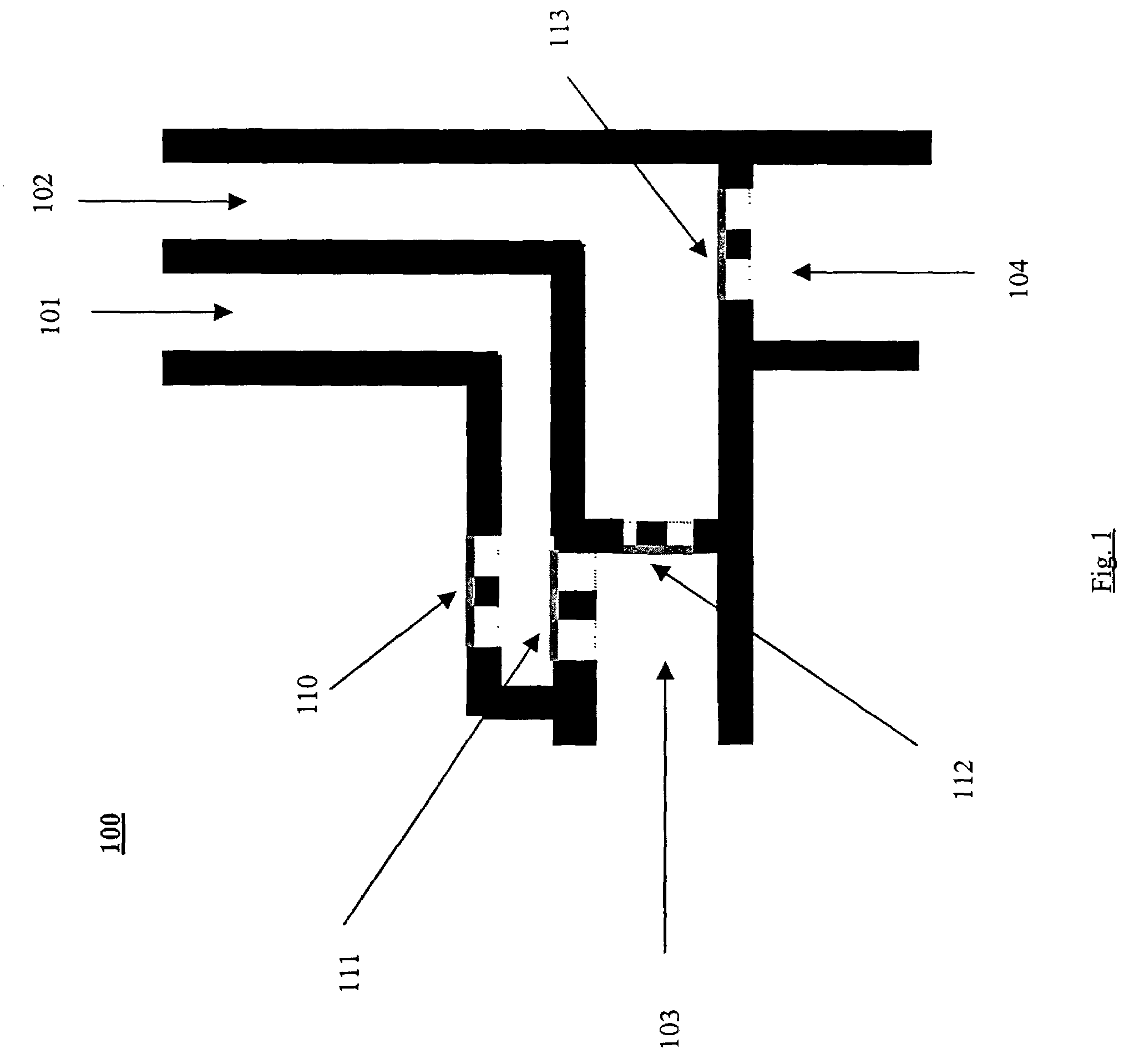
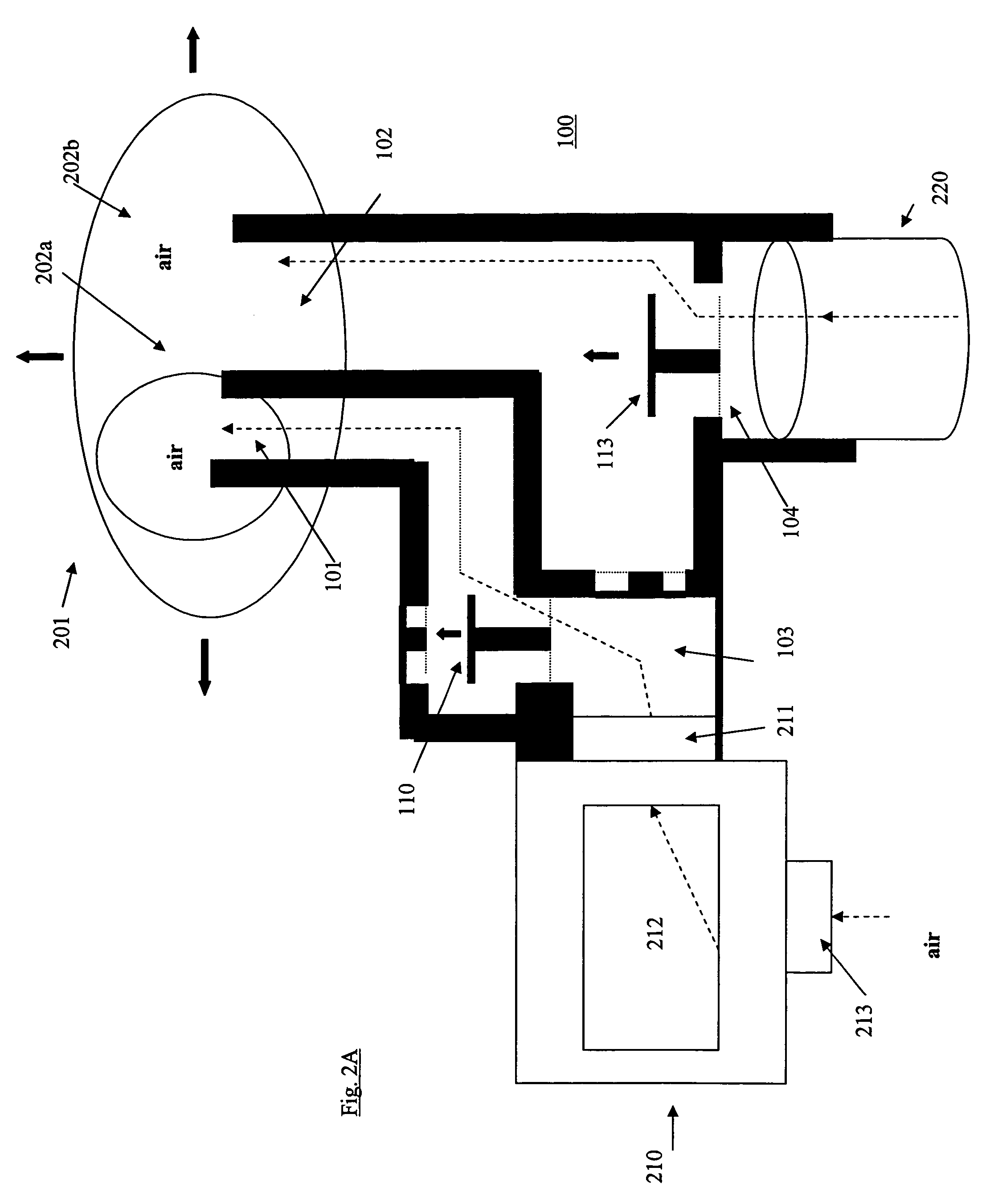
[0015]FIG. 1 shows a novel device, generally represented at 100, used to connect both a CO2 detection device and a volume displacement detector in line with an ETT. In a preferred embodiment the device comprises first 101 and second 102 volume displacement detector portals, a CO2 detector portal 103 and an endotracheal tube portal 104. The portals 101, 102, 103 and 104 are configured to connect with other appropriate devices via slip fittings, friction fittings, threaded fittings, or similar detachable fittings. The device comprises first 111 and second 113 positive pressure response valves which open when positive pressure is applied to the device 100 (see FIGS. 2B and 3B). First 110 and second 112 negative response valves are also incorporated into the device 100 and configured such that they open when negative pressure is applied to the device 100. (see FIGS. 3A and 3B) Preferably, the device is manufactured from inexpensive material such that it may be disposed of following use....


PUM


Abstract

A novel connection device, and method for its use, for connecting a volume displacement detection device and a gas composition detection device in line with an intubation tube is disclosed. The connection device allows a user to combine two systems commonly employed by medical personnel to assess the location of an intubation tube placed into a patient. The two systems have alternate means of assessing the location of an intubation tube in a patient and therefore, when combined into a single unit via the connection device, improve the accuracy of placing a tube in a desired location while reducing or eliminating complications associated with misplacement. A kit comprising components that may be assembled to provide a device for assessing the proper placement of an intubation tube in a patient is also disclosed. The kit may be used in any environment, but is particularly well suited for emergency, pre-hospital settings requiring intubation where quick assembly of the components is essential.

Description

FIELD OF THE INVENTION[0001]The present invention is directed to a device used to connect two or more intubation placement detectors in line with an endotracheal tube (ETT) to provide rapid, alternative methods of assessing whether an ETT has been properly placed in the trachea of a patient.BACKGROUND OF THE INVENTION[0002]Ventilation of the lungs is essential to life. Patients in need of medical attention often require assistance with breathing as a result of injury, trauma, or airway obstruction. In an apneic patient, the lungs must be artificially ventilated to ensure proper oxygenation and exchange of gasses within the body. Endotracheal intubation is the technique of placing an endotracheal tube (ETT) into the trachea of a patient, for the purposes of establishing an airway. It is estimated that over 18 million intubations are preformed each year in the US, the majority in the operating room. Because of the close proximity of the esophagous to the trachea, an intubationist may ...

Claims


Application Information

Patent Timeline
Patent Type & Authority Patents(United States)
IPC IPC(8): A61M16/00A61M16/04
CPCA61M16/0488A61M2016/0413A61M2230/432
Inventor MELKER, RICHARDGRAVENSTEIN, DIETRICHGABRIELLI, ANDREA
Owner UNIV OF FLORIDA RES FOUNDATION INC
Why Eureka
Try Eureka