Method and apparatus for processing a computed tomography image of a lung obtained using contrast agent

a computed tomography and lung technology, applied in tomography, instruments, diagnostics, etc., can solve the problems of inability to dependably determine the degree of stenosis, the inability to identify the thrombosis, and the inability to diagnose the disease, so as to improve the diagnostic reliability

Active Publication Date: 2007-04-10
SIEMENS HEALTHCARE GMBH
View PDF8 Cites 45 Cited by
  • Summary
  • Abstract
  • Description
  • Claims
  • Application Information

AI Technical Summary

Benefits of technology

[0013]An object of the invention is to provide an additional evaluation technique for CT images in the diagnosis of PE on the basis of computer tomog

Problems solved by technology

This standard technique has the following problems:Blockages can be overlooked;The hemodynamic effect of the thrombosis cannot be identified;Blockages at small vessels (sub-segmental) cannot be recognized in the CT image due to the

Method used


Image

  • Method and apparatus for processing a computed tomography image of a lung obtained using contrast agent
  • Method and apparatus for processing a computed tomography image of a lung obtained using contrast agent
  • Method and apparatus for processing a computed tomography image of a lung obtained using contrast agent

Examples


Embodiment Construction

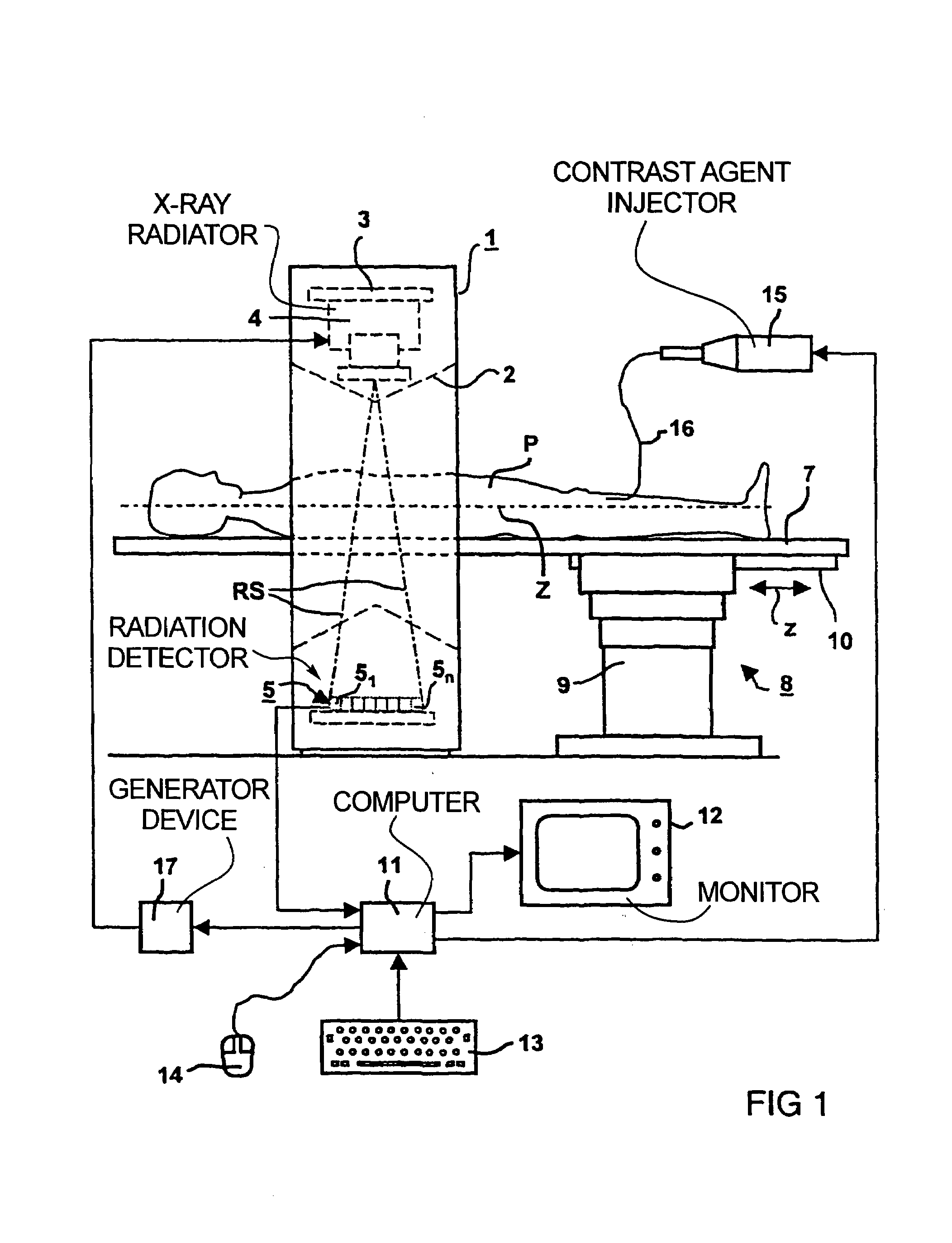
[0046]FIG. 1 shows an x-ray CT apparatus having a gantry 1 with a measurement opening 2 that is surrounded by a live ring 3 on which an x-ray radiator 4 and a detector system are attached. The detector system has a radiation detector fashioned in a known way and curved around an axis that preferably proceeds parallel to a system axis Z and through the focus of the x-ray radiator 4. The radiation detector 5 has a number of lines 51 through 5n of detector elements, each forming a row of detector elements. A pyramidal x-ray beam RS that is indicated dot-dashed and that strikes the detector 5 emanates from the x-ray radiator 4. The gantry 1 having the x-ray radiator 4 and the radiation detector 5, and at least the support plate 7 of a support mechanism provided for the acceptance of an examination subject, for example of a patient P, are adjustable relative to one another in the direction of the longitudinal axis of the support plate 7 proceeding parallel to the system axis Z. This adju...


PUM


Abstract

In a method and apparatus for image processing proceeding from a computed tomography (CT) image of a lung as an original image that is registered using a contrast agent, pulmonary parenchyma pixels are determined, the pulmonary parenchyma pixels are presented in false colors, and the remaining image regions are presented in the gray scale values of the original image.

Description

BACKGROUND OF THE INVENTION[0001]1. Field of the Invention[0002]The present invention is directed to a method for image processing proceeding from a computer tomography (CT) image of a lung registered with a contrast agent, we well as a CT apparatus for the implementation of such a method.[0003]2. Description of the Prior Art[0004]Computed tomography is increasingly acquiring significance for the diagnosis of pulmonary embolisms. CT angiographies of the vessels in the thorax are analyzed.[0005]For diagnosis of pulmonary embolisms (PE), a CT angiography of the lung is implemented. Contrast agent is injected into the patient with a contrast agent pump. After pausing a few seconds, a spiral CT of the lung is implemented, a stack (series) of axial images, i.e. transverse tomograms, preferably representing body slices adjacent to one another and following one another in the direction of the longitudinal patient axis are acquired. The vessels filled with contrast agent can be clearly seen...

Claims


Application Information

Patent Timeline
IPC IPC(8): G06K9/00A61B6/03G06T1/00G06T7/00G06T7/60
CPCG06T7/0012A61B6/027Y10S128/922
Inventor KLOTZ, ERNSTNIETHAMMER, MATTHIASSCHALLER, STEFAN
Owner SIEMENS HEALTHCARE GMBH
Why Eureka
Try Eureka