Method and magnetic resonance tomography apparatus for graphic planning of angiographic exposures using a contrast agent

a magnetic resonance tomography and contrast agent technology, applied in the direction of instruments, measurements using nmr, magnetic variable regulation, etc., can solve the problems of poor image quality, measurement not ensuing, and limit the contrast that can be obtained by non-augmented mr imaging, so as to facilitate or optimize the time planning of a contrast agen

Active Publication Date: 2007-11-06
SIEMENS HEALTHCARE GMBH
View PDF2 Cites 31 Cited by
  • Summary
  • Abstract
  • Description
  • Claims
  • Application Information

AI Technical Summary

Benefits of technology

[0017]It is an object of the present invention to facilitate or optimize the time planning

Problems solved by technology

Particularly in the case of angiographic exposures (i.e. exposures of the blood vessels in the human body, specifically in organs with blood circulation), limits exist as to the contrast that can be obtained by non-augmented MR imaging.
This can result in the measurement not ensuing at the optimum point in time.
The consequence thereof is a poor image quality due

Method used


Image

  • Method and magnetic resonance tomography apparatus for graphic planning of angiographic exposures using a contrast agent
  • Method and magnetic resonance tomography apparatus for graphic planning of angiographic exposures using a contrast agent
  • Method and magnetic resonance tomography apparatus for graphic planning of angiographic exposures using a contrast agent

Examples


Embodiment Construction

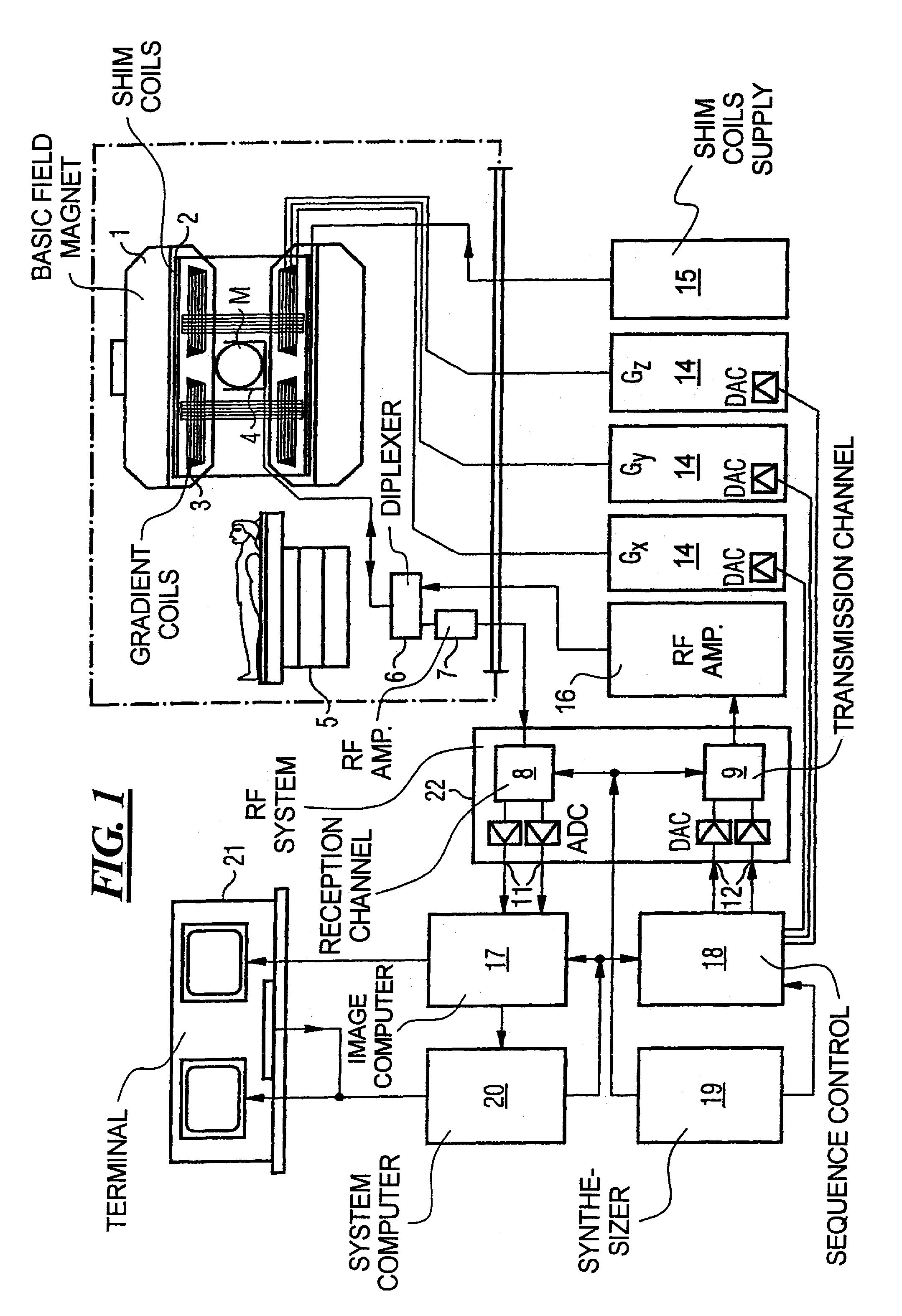
[0033]FIG. 1 is a schematic illustration of a magnetic resonance tomography apparatus with improved contrast behavior for MR time-of-flight angiography exposures according to the present invention. The basic components of the magnetic resonance tomography apparatus corresponds to those of a conventional tomography apparatus, with the differences described below. A basic field magnet 1 generates a temporally constant, strong magnetic field for the polarization or alignment of the nuclear spins in the examination region of a subject such as, for example, a part of a human body to be examined. The high homogeneity of the basic magnetic field required for a magnetic resonance measurement is defined in a spherical measuring volume M into which the parts of the human body to be examined are introduced. Shim plates of ferromagnetic material are attached at a suitable location for achieving the homogeneity requirements and, in particular, for eliminating time-invariable influences. Time-var...


PUM


Abstract

In a method and processing device for a magnetic resonance tomography apparatus, with which a test bolus measurement is conducted for a subsequent angiography measurement using contrast agent, a test bolus pop-up is displayed in an interactive graphic user interface, and a time curve of at least the arterial contrast agent concentration is automatically determined from the test bolus measurement and presented in the test bolus pop-up, for time planning of the subsequent angiography measurement.

Description

BACKGROUND OF THE INVENTION[0001]1. Field of the Invention[0002]The present invention is directed in general to nuclear magnetic resonance tomography (MRT) as employed in medicine for examining patients. The present invention is more specifically directed to a magnetic resonance tomography apparatus with a device for graphic planning of angiographic MRT measurements that are made using a contrast agent.[0003]2. Description of the Prior Art[0004]MRT is based on the physical phenomenon of nuclear magnetic resonance and has been utilized for more than 15 years as an imaging method in medicine and biophysics. In this examination method, the subject is exposed to a strong, constant magnetic field. As a result, the nuclear spins of the atoms in the subject align, these having been previously irregularly oriented. Radiofrequency energy can then excite these “ordered” spins to a specific resonance. This resonance generates the actual measured signal in MRT that is picked up by suitable rece...

Claims


Application Information

Patent Timeline
IPC IPC(8): G06K9/00G01R33/28A61B5/055G01R33/54G01R33/563
CPCA61B5/055G01R33/5635G01R33/546
Inventor HORGER, WILHELMLAUB, GERHARD
Owner SIEMENS HEALTHCARE GMBH
Why Eureka
Try Eureka