Steerable ultrasound catheter

a catheter and ultrasound technology, applied in the field of medical devices and methods, can solve the problems of continuing to be difficult to develop a catheter and the difficulty of manipulating the currently available ultrasound catheter devices, and achieve the effects of enhancing the manipulation of the catheter body, enhancing flexibility, and enhancing the disruption of blood vessel obstruction

Active Publication Date: 2008-02-26
FLOWCARDIA
View PDF43 Cites 204 Cited by
  • Summary
  • Abstract
  • Description
  • Claims
  • Application Information

AI Technical Summary

Benefits of technology

[0008]Ultrasound catheter devices and methods provide enhanced disruption of blood vessel obstructions. Generally, ultrasound catheters include an elongate flexible catheter body with one or more lumens, an ultrasound transmission member extending longitudinally through the catheter body lumen, and a distal head coupled with the transmission member and positioned adjacent the distal end of the catheter body for disrupting occlusions. Improved features include but are not limited to catheter bodies and ultrasound transmission members with increasing flexibility toward their distal ends, devices that bend concomitantly with one or more bends in a guidewire and / or a blood vessel, a proximal housing coupled with an ultrasound catheter body for enhancing manipulation of the catheter body, a guidewire lumen configured to allow contact between a guidewire and an ultrasound transmission member, distal heads with improved guidewire lumens and the like.
[0010]The increasing flexibility of the catheter body and the ultrasound transmission wire toward the distal end of the catheter device may be achieved by any suitable fabrication method. In some embodiments, for example, the ultrasound transmission wire, catheter body, or both may be tapered, such that their cross-sectional diameters decrease distally. In one embodiment, for example, the cross-sectional diameter of a catheter body may range from between about 0.102 cm and about 0.178 cm along its proximal end to between about 0.076 cm and about 0.127 cm along its distal end, and the cross-sectional diameter of an ultrasound transmission member may range from between about 0.051 cm and about 0.102 cm near its proximal end to between about 0.013 cm and about 0.038 cm near its distal end. Additionally or alternatively, the wall thickness of the catheter body may decrease from proximal to distal. For example, in one embodiment, the wall thickness of a catheter body may measure about 0.007 cm to about 0.020 cm along its proximal portion and about 0.005 cm to about 0.013 cm along its distal portion. In these or other embodiments, various combinations of materials may be used to give the catheter body, transmission wire or both a desired flexibility profile. Providing an ultrasound catheter with a catheter body and ultrasound transmission wire that both increase in flexibility toward the distal end of the device will enhance navigation of the distal end of the device through tortuous blood vessels. In one embodiment, for example, the distal portion of the device will be sufficiently flexible to pass, without kinking, through at least 5 cm length of a blood vessel having at least one bend and an inner diameter of between about 2 mm and about 5 mm. A “tortuous vessel,” for the purposes of this application, means having at least one bend or curve, and the bend or curve may have any angle or radius of curvature. In some embodiments, for example, at least one bend in a vessel may have a radius of about 1.0 cm or less. In some instances, of course, a tortuous blood vessel will have multiple bends or curves.
[0018]Having a guidewire lumen that exits from the center of the distal head is advantageous for advancing the guidewire as well as the catheter device. Angling the guidewire lumen away from the longitudinal axis of the catheter body allows a guidewire and the ultrasound transmission member to pass through a narrower catheter body, thus decreasing the diameter of the catheter device and facilitating passage through tortuous vessels. In some embodiments, the guidewire lumen in the distal head includes a cavity in which a distal end of a guidewire tube of the catheter body is disposed. In some embodiments, the cavity extends through the distal end of the distal head, such that the distal end of the guidewire tube is flush with the distal end of the distal head. In other embodiments, the cavity extends partially through the distal head, such that the distal end of the guidewire tube is disposed proximal to the distal end of the distal head.

Problems solved by technology

For example, it continues to be challenging to develop a catheter that is stiff enough to be pushable or “steerable” through vasculature yet flexible enough, at least along part of its length, to navigate small, tortuous blood vessels, such as the coronary arteries or tortuous peripheral vasculature.
It is also sometimes difficult to manipulate currently available ultrasound catheter devices, for example to twist or torque the proximal end of the catheter to move the distal end in a way that helps with navigation through the vasculature.

Method used


Image

  • Steerable ultrasound catheter
  • Steerable ultrasound catheter
  • Steerable ultrasound catheter

Examples


Embodiment Construction

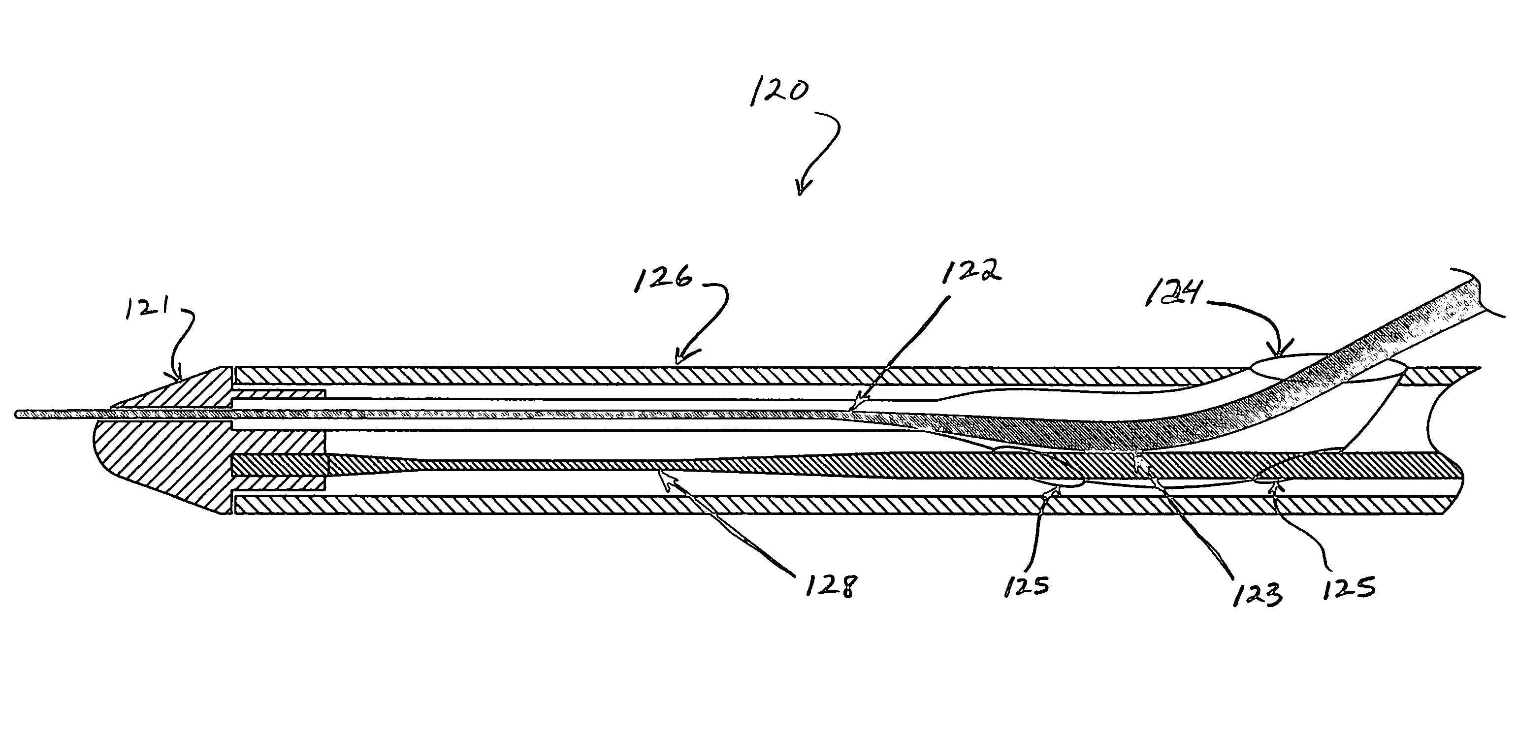
[0031]Ultrasound catheter devices and methods of the present invention generally provide for enhanced treatment of occlusive intravascular lesions. Catheter devices generally include a catheter body, an ultrasound energy transmission member disposed within the catheter body and a distal head coupled with the energy transmission member and disposed adjacent the distal end of the catheter body. The ultrasound transmission member transmits ultrasound energy from an ultrasound transducer to the distal head, causing the head to vibrate and, thus, disrupt vascular occlusions. A number of improved features of such ultrasound catheter devices are described more fully below.

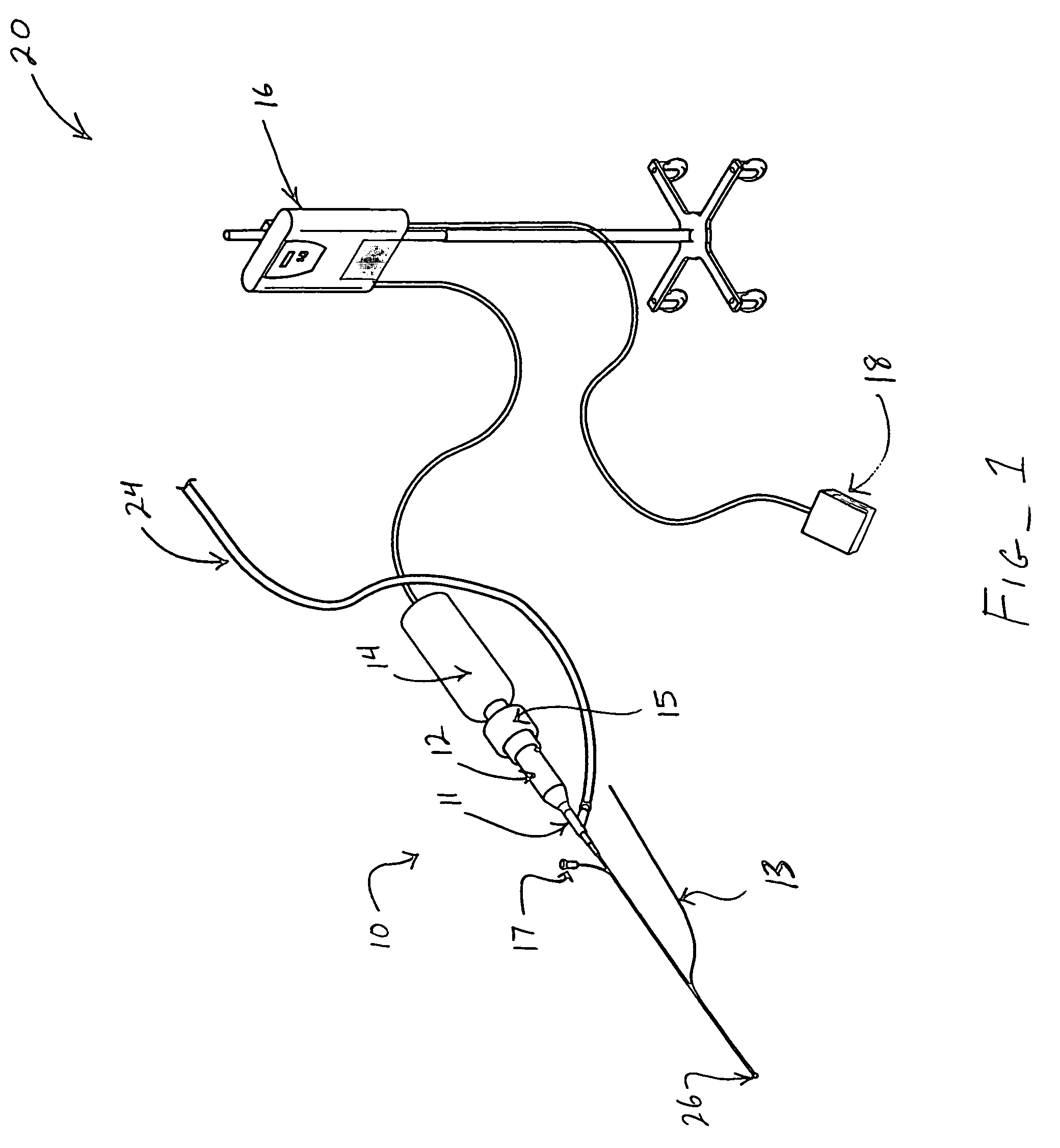
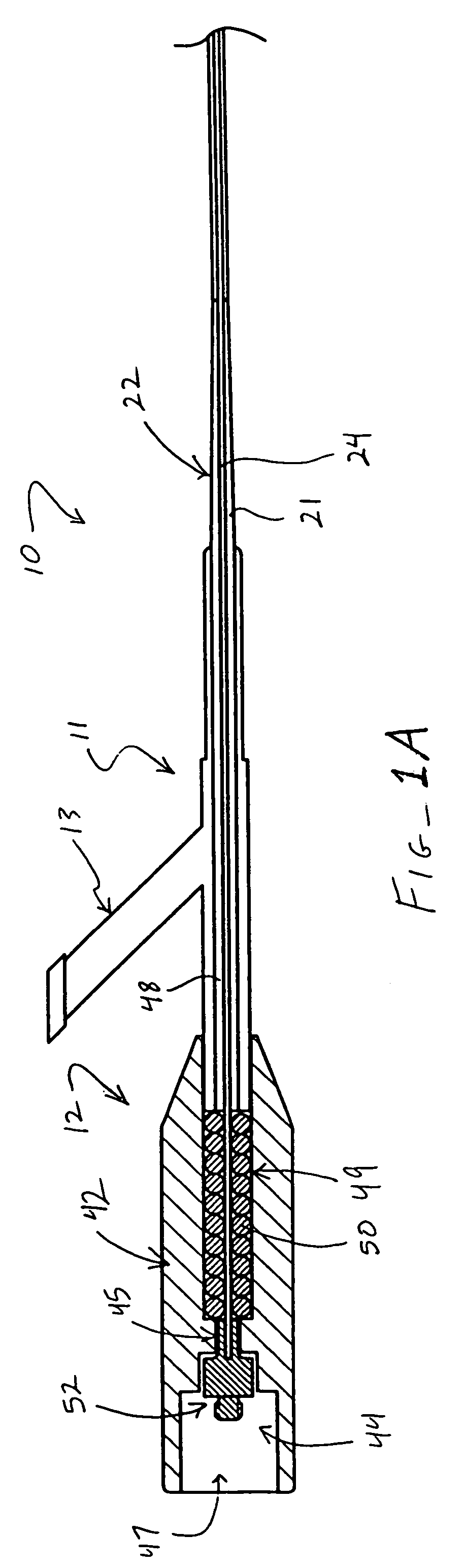
[0032]Referring now to FIG. 1, one embodiment of an ultrasound catheter system 20 suitably includes an ultrasound catheter device 10, including a proximal end connector 12 for coupling device 10 with an ultrasound transducer 14, and an ultrasound generator 16 coupled with transducer 14 and a foot-actuated on / off switch 18...


PUM


Abstract

Ultrasound catheter devices and methods provide enhanced disruption of blood vessel obstructions. Ultrasound catheter devices generally include an elongate flexible catheter body with one or more lumens, an ultrasound transmission member extending longitudinally through the catheter body lumen and a distal head coupled with the transmission member and positioned adjacent the distal end of the catheter body for disrupting occlusions. Improved features of ultrasound catheters include catheter bodies and ultrasound transmission members with increasing distal flexibility, guidewire tubes allowing contact between a guidewire and an ultrasound transmission member, distal heads with improved guidewire lumens, and torquable proximal housings for enhancing disruption of blood vessel occlusions.

Description

CROSS REFERENCE TO RELATED APPLICATIONS[0001]This application is related to the following pending U.S. patent applications, the full disclosures of which are all hereby incorporated by reference: application Ser. No. 10 / 229,371, filed Aug. 26, 2002, entitled “Ultrasound Catheter for Disrupting Blood Vessel Obstructions”; application Ser. No. 10 / 345,078, filed Jan. 14, 2003, entitled “Ultrasound Catheter and Methods for Making and Using Same”; application Ser. No. 10 / 375,903, filed Feb. 26, 2003, entitled “Ultrasound Catheter Apparatus”; and application Ser. No. 10 / 410,617, filed Apr. 8, 2003, entitled “Improved Ultrasound Catheter Devices and Methods.”BACKGROUND OF THE INVENTION[0002]The present invention relates generally to medical devices and methods. More specifically, the present invention relates to ultrasound catheter devices and methods for treating occlusive intravascular lesions.[0003]Catheters employing various types of ultrasound transmitting members have been successful...

Claims


Application Information

Patent Timeline
Patent Type & Authority Patents(United States)
IPC IPC(8): A61B17/32A61B17/20A61B8/00A61B8/14A61M25/00A61B17/00A61B17/22A61B17/30A61M
CPCA61B17/22004A61B17/320068A61B17/2202A61B2017/00243A61B2017/22014A61M25/0054A61B2017/308A61B2017/32007A61B2017/320071A61B17/3207
Inventor NITA, HENRYSARGE, JEFF
Owner FLOWCARDIA
Why Eureka
Try Eureka