Connective tissue substitutes, method of preparation and uses thereof

a technology of connective tissue and substitutes, applied in the field of tissue engineering, can solve the problems of affecting the patient's recovery, affecting the patient's quality of life, and affecting the quality of life of the patient, and achieving the effects of improving the quality of li

Inactive Publication Date: 2008-02-26
UNIV DE MONTREAL +1
View PDF25 Cites 13 Cited by
  • Summary
  • Abstract
  • Description
  • Claims
  • Application Information

AI Technical Summary

Problems solved by technology

Yet despite their wide use, surgically implanted prostheses present many attendant risks to the patient.
The major problem with these techniques is that another part of the body, or the joint of the donor is often significantly weakened after biopsied to get grafts.
Long term drawbacks of this approach are that chronic pain, patellar fractures, knee instability and cartilage degeneration.
It has been found that it is difficult to provide long-term solution using these materials to permanently replace connective tissues.
However, persistent inflammatory reactions occur following wear off of particles upon time post-implantation.
Additionally, they do not readily breakdown and are not readily integrated with the body via remodeling by tissue cells.
However, since this implant is constructed without anchoring extremities, the anchorage capability is limited.

Method used


Image

  • Connective tissue substitutes, method of preparation and uses thereof
  • Connective tissue substitutes, method of preparation and uses thereof
  • Connective tissue substitutes, method of preparation and uses thereof

Examples


example i

Preparation of Anterior Crutiate Ligament

Material and Methods

LF Isolation and Culture

[0072]Torn ACL biopsies are collected from the host. The biopsies are kept at 4° C. for no longer than 24-48 hrs before cell isolation. The ACL biopsy is weighted and cut into small pieces after removal of the periligamentous tissues. The fragments are digested with 0.125% collagenase containing 2 mM CaCl2 (1 ml of enzymatic solution / mg of tissue) for 20 hrs, under gentle agitation, at 37° C. A 0.1% trypsin solution (1 ml / mg of hydrated tissue) is then added to the cellular suspension for 1 hr. The enzymes are dissolved in Dulbecco's Modification of Eagle's™ medium (Gibco), pH 7.4, containing antibiotics.

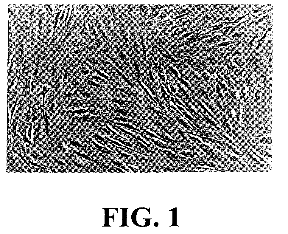
[0073]The ligament fibroblasts (LF) isolated from ACL biopsies are cultured in DME supplemented with 10% fetal calf serum (FCS), 100 IU / ml penicillin G and 25 μg / ml gentamicin (FIG. 1).

[0074]When LF primary cultures reach about 85% confluence, the cells are detached from their culture flasks using 0...

example ii

Preparation of Connective Tissues

Material and Methods

Dermal Fibroblasts Isolation and Culture

[0098]The dermal fibroblasts (DF) isolated from the dermis of skin biopsies, enzymatically (same procedure described in Example I) or by explants, are cultured in DME supplemented with 10% fetal calf serum (FCS), 100 IU / ml penicillin G and 25 μg / ml gentamicin.

[0099]When DF primary cultures reach about 85% confluence, the cells are detached from their culture flasks using 0.05% trypsin-0.01% EDTA solution (pH 7.8), for about 10 min at 37° C. The DF suspensions are centrifuged twice at 200×g for 10 min. The cell pellets are resuspended in complete culture medium and the cells are counted. The cellular viability is determined using the trypan blue exclusion method.

[0100]Up until now, the DF were isolated and cultured from skin biopsies of more than hundred patients and 10 animals (goats, dogs, and rabbits) with 100% success. The cells maintained their morphology for more than 7 passages in cult...

example iii

Preparation of Periodontal Ligament Substitute

Material and Methods

Fibroblasts Isolation and Culture

[0108]Dermal fibroblasts (DF), ligament fibroblasts (LF), or fibroblasts from other sources (e.g. mucosa of the mouth) can be isolated and cultured in DME supplemented with 10% fetal calf serum (FCS), 100 IU / ml penicillin G and 25 μg / ml gentamicin.

[0109]When the cells primary cultures reach about 85% confluence, they are detached from their culture flasks using 0.05% trypsin-0.01% EDTA solution (pH 7.8), for about 10 min at 37° C. The cell suspensions are centrifuged twice at 200×g for 10 min. The cell pellets are resuspended in complete culture medium and the cells are counted. The cellular viability is determined using the trypan blue exclusion method.

Preparation of the Peridontal Ligament Substitutes' Tooth Anchors

[0110]Teeth pieces are washed and sterilized according to the procedure described in Example I.

[0111]Holes are made in each tooth, as previously described. A sterile tooth...


PUM

PropertyMeasurementUnit
frequencyaaaaaaaaaa
pHaaaaaaaaaa
pHaaaaaaaaaa

Abstract

The present invention relates to connective tissue substitute implant and method of preparation thereof. The implant is essentially composed of two bone anchors joined at the proximal ends by matrix layers and / or filaments coated by supplementary biocompatible matrix coating layer which can contain living stem cells isolated from injured connective tissue.

Description

CROSS REFERENCE TO RELATED APPLICATION[0001]This application claims the benefit, under 35 U.S.C. § 119(e), of U.S. provisional application No. 60 / 252,588 filed Nov. 24, 2000.BACKGROUND OF THE INVENTION[0002](a) Field of the Invention[0003]The invention relates to the field of tissue engineering, production of connective tissue linked to natural bones or synthetic bone substitutes (tendons, ligaments, cartilage, etc.) can benefit from the invented procedure. The procedure of the present invention is carried out to produce bioengineered connective tissue substitutes. Connective tissue substitutes (CTS) of the invention may be constructed for replacement of ligaments, and most particularly cruciate ligaments or tendons.[0004](b) Description of Prior Art[0005]Researchers in the surgical arts have been working for many years to develop new techniques and materials for use as grafts to replace or repair damaged or torn tissue structures, particularly bones and connective tissues, such as ...

Claims


Application Information

Patent Timeline
Patent Type & Authority Patents(United States)
IPC IPC(8): A61F2/08A61L27/24A61L27/38C12N5/077
CPCA61F2/08A61L27/24A61L27/3608A61L27/3641A61L27/3687A61L27/3691A61L27/3804A61L27/3839A61L27/3843A61L27/386A61L27/3886C12N5/066A61F2/0811C12N2533/54
Inventor GOULET, FRANCINERANCOURT, DENISCLOUTIER, REJEANTREMBLAY, JULIEAUGER, FRANCOIS A.NORMAND, ALBERTGERMAN, LUCIELAMONTAGNE, JEANBOUCHARD, MARCLANGELIER, EVEDUPUIS, DANIELBOUCHARD, STEPHANEISLAM, NAZRULSTEVENS, LOUIS-MATHIEULAVERTY, SHEILALUSSIER, BERTRANDBELZIL, ANNE-MARIETREMBLAY, PIERROT
Owner UNIV DE MONTREAL
Why Eureka
Try Eureka