Neural proteins as biomarkers for traumatic brain injury

a neuron and brain injury technology, applied in the field of reliable detection and identification of biomarkers, can solve the problems of inability to quickly employ in an emergency room environment, high cost of ct and mri, and most tbi survivors' long-term impairmen

Active Publication Date: 2008-07-08
UNIV OF FLORIDA RES FOUNDATION INC +1
View PDF10 Cites 82 Cited by
  • Summary
  • Abstract
  • Description
  • Claims
  • Application Information

AI Technical Summary

Benefits of technology

[0018]In another preferred embodiment an expanded panel of biomarkers are used to provide highly enriched information of mechanism of injury, modes of cell death (necrosis versus apoptosis), sites of injury, sites and status of different cell types in the nervous system and enhanced diagnosis (better selectivity and specificity). This invention is an important and significant improvement over existing technologies focused on small panel (e.g. a four-marker panel:-MBP-Thrombomodulin-S100B-NSE from Syn X Pharma (Mississauga, Canada)- or single markers (e.g. S100B from DiaSorin (Sweden)).

Problems solved by technology

However, despite dramatically improved emergency triage systems based on these outcome measures, most TBI suffer long term impairment and a large number of TBI survivors are severely affected despite predictions of “good recovery” on the GOS.
In addition, CT and MRI are expensive and cannot be rapidly employed in an emergency room environment.
Accordingly, the neural pathways of a mammal are particularly at risk if neurons are subjected to mechanical or chemical trauma or to neuropathic degeneration sufficient to put the neurons that define the pathway at risk of dying.
Stroke is a leading cause of death in the developed world.
It may be caused by reduced blood flow or ischemia that results in deficient blood supply and death of tissues in one area of the brain (infarction).
The ability to detect such injury is lacking in the prior art.
Mammalian neural pathways also are at risk due to damage caused by neoplastic lesions.
Transformed cells of neural origin generally lose their ability to behave as normal differentiated cells and can destroy neural pathways by loss of function.
Metastatic tumors, which are a significant cause of neoplastic lesions in the brain and spinal cord, also similarly may damage neural pathways and induce neuronal cell death.

Method used


Image

  • Neural proteins as biomarkers for traumatic brain injury
  • Neural proteins as biomarkers for traumatic brain injury
  • Neural proteins as biomarkers for traumatic brain injury

Examples


example 1

Detection of Neural Proteins UCH-L1, p24, and Alpha-Synuclein in CSF of Rodents Following TBI

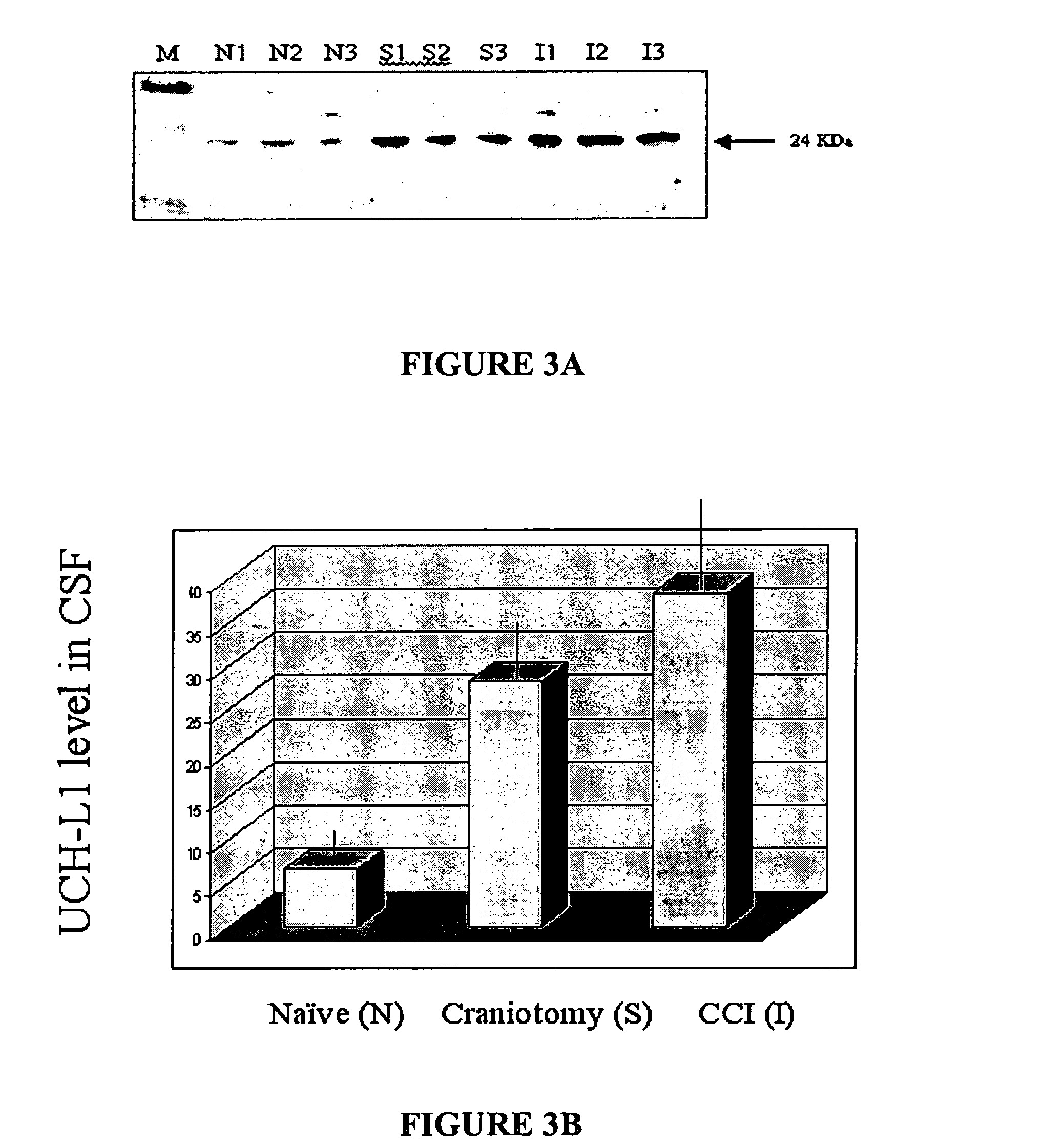
[0269]TBI was induced in rodents as described above. Following TBI or sham operation or naïve rats, samples of CSF were collected and analyzed for presence of three novel neural protein biomarkers (e.g. UCH-L1 (FIG. 3), p24 (FIG. 4) and alpha-synuclein (FIG. 5). Results, shown in FIGS. 3-5, demonstrated independent or concurrent accumulation of UCH-L1 (see FIG. 3), p24 (see FIG. 4) and alpha-synuclein (see FIG. 5), in the CSF of rodents after TBI. Significantly less of these neural proteins were observed in sham-injured and naïve controls. Each lane in the blots represents a different animal. The sensitivity of this assay permits detection of inter-animal differences, which is valuable for prediction of outcome. The results of this study demonstrated that after TBI, neural proteins accumulated in the CSF in sufficient levels to be easily detectable on Western blots or by other immunoassays s...

example 2

Detection of Neural Proteins UCH-L1 and p24 in CSF of Human TBI

[0270]Accumulation of novel neural markers (UCH-L1 and p24) was analyzed in samples of human CSF taken at 24 hr after TBI. From five patients who experienced severe TBI and five neurological controls (normal pressure hydrocephalus. As in the rodent models of TBI, the neural proteins examined (UCH-L1 and p24) were prominent in CSF samples TBI. Levels of these neural proteins were much higher in the TBI patients than in the control patients (e.g. UCH-L1 (FIG. 6), p24 (FIG. 7). These data demonstrated that after TBI, neural proteins accumulated in human CSF in sufficient levels to be easily detectable on Western blots or by other immunoassays such as ELISA.


PUM

PropertyMeasurementUnit
timeaaaaaaaaaa
pHaaaaaaaaaa
pHaaaaaaaaaa

Abstract

The present invention identifies biomarkers that are diagnostic of nerve cell injury and / or neuronal disorders. Detection of different biomarkers of the invention are also diagnostic of the degree of severity of nerve injury, the cell(s) involved in the injury, and the subcellular localization of the injury.

Description

CROSS-REFERENCE TO RELATED APPLICATIONS[0001]The present application claims the priority of U.S. Provisional Patent application No. 60 / 562,944 filed Apr. 15, 2004; and entitled “NEURAL PROTEINS AS BIOMARKERS FOR NERVOUS SYSTEM INJURY AND OTHER NEURAL DISORDERS” which is incorporated herein by reference in its entirety.[0002]The invention was made with government support under National Institutes of Health Grant #R01 NS39091, National Institutes of Health Grant #R01 NS40182, U.S. Army Grant #DAMD 17-99-1-9565, and U.S. Army Grant #DAMD 17-01-1-0765. The government has certain rights in the invention.FIELD OF THE INVENTION[0003]The invention provides for the reliable detection and identification of biomarkers, important for the diagnosis and prognosis of damage to the nervous system (central nervous system (CNS) and peripheral nervous system (PNS)), brain injury and neural disorders. The protein / peptide profile in patients with damage to nerves and brain cells are distinguished from n...

Claims


Application Information

Patent Timeline
Patent Type & Authority Patents(United States)
IPC IPC(8): G01N33/53C12Q1/00G01N33/537G01N33/542G01N33/566G01N33/567C12Q1/68G01N33/543G01N33/68
CPCC12Q1/6883G01N33/6896G01N2800/52G01N2800/28
Inventor HAYES, RONALDWANG, KEVIN KA-WANGLIU, MING CHENOLI, MONIKA
Owner UNIV OF FLORIDA RES FOUNDATION INC
Why Eureka
Try Eureka