Fusion partner for production of monoclonal rabbit antibodies
a monoclonal antibody and fusion partner technology, applied in the field of fusion partners for the production of rabbit monoclonal antibodies, can solve the problems of hampered approaches, relative few monoclonal antibodies, and inability to use this approach for producing rabbit monoclonal antibodies
- Summary
- Abstract
- Description
- Claims
- Application Information
AI Technical Summary
Benefits of technology
Problems solved by technology
Method used
Image
Examples
example 1
[0060]In initial experiments, 240E-1 cells were fused with spleen cells obtained from an immunized rabbit. Following HAT medium selection, hybridoma colonies were isolated and tested for antibody production. Several antibody-secreting hybridoma clones were obtained. After expansion and repeated passaging of the hybridoma cell lines, it was found that antibody secretion was greatly diminished in a majority of the clones. Similar results were reported by others (Liguori et al., 2001, supra). Furthermore, after subculturing the 240E-1 cells, their efficiency as a fusion partner decreased. Similarly, Rief et al. (1998), supra, reported a low yield of hybridomas, and only one out of six positive hybridomas persisted after subcloning. The 240E-1 cell line is therefore somewhat unstable, and impractical for routine use.
[0061]240E-1 cells were subjected to repeated rounds of subcloning. Subclones were selected for high cloning efficiency and robust growth, as well as for morphological chara...
example 2
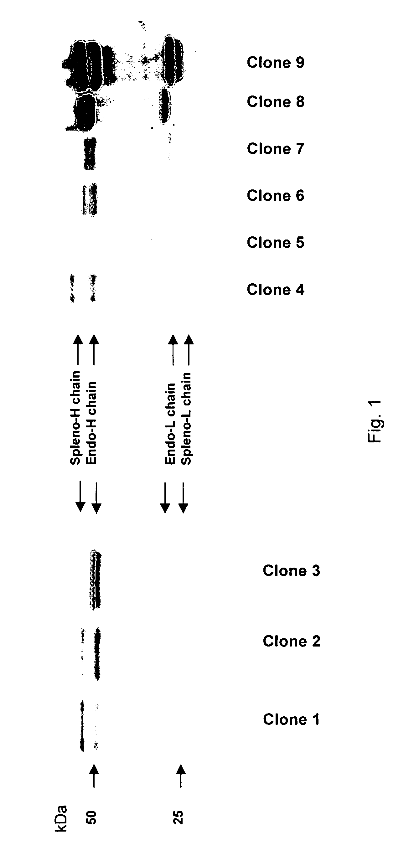
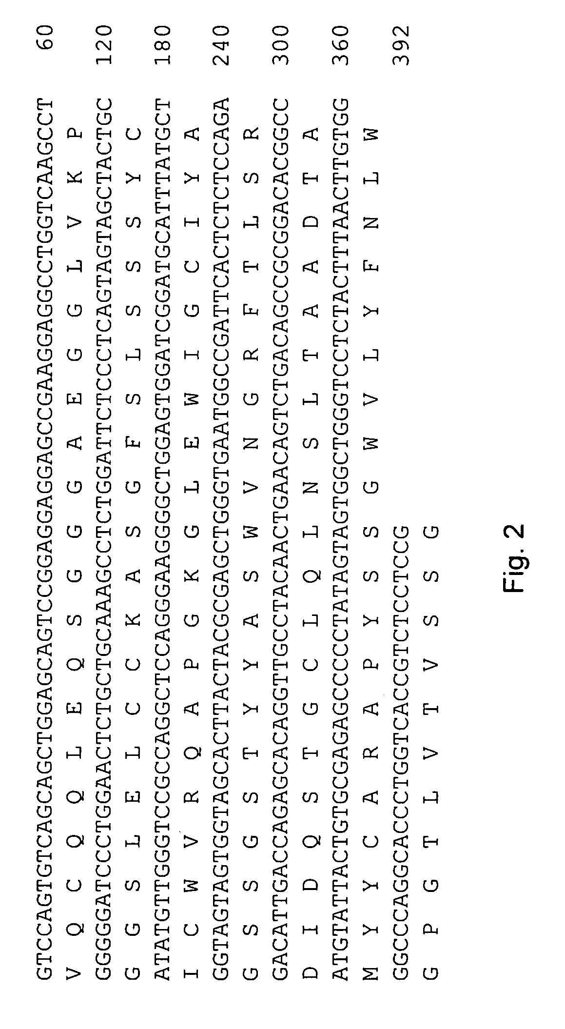
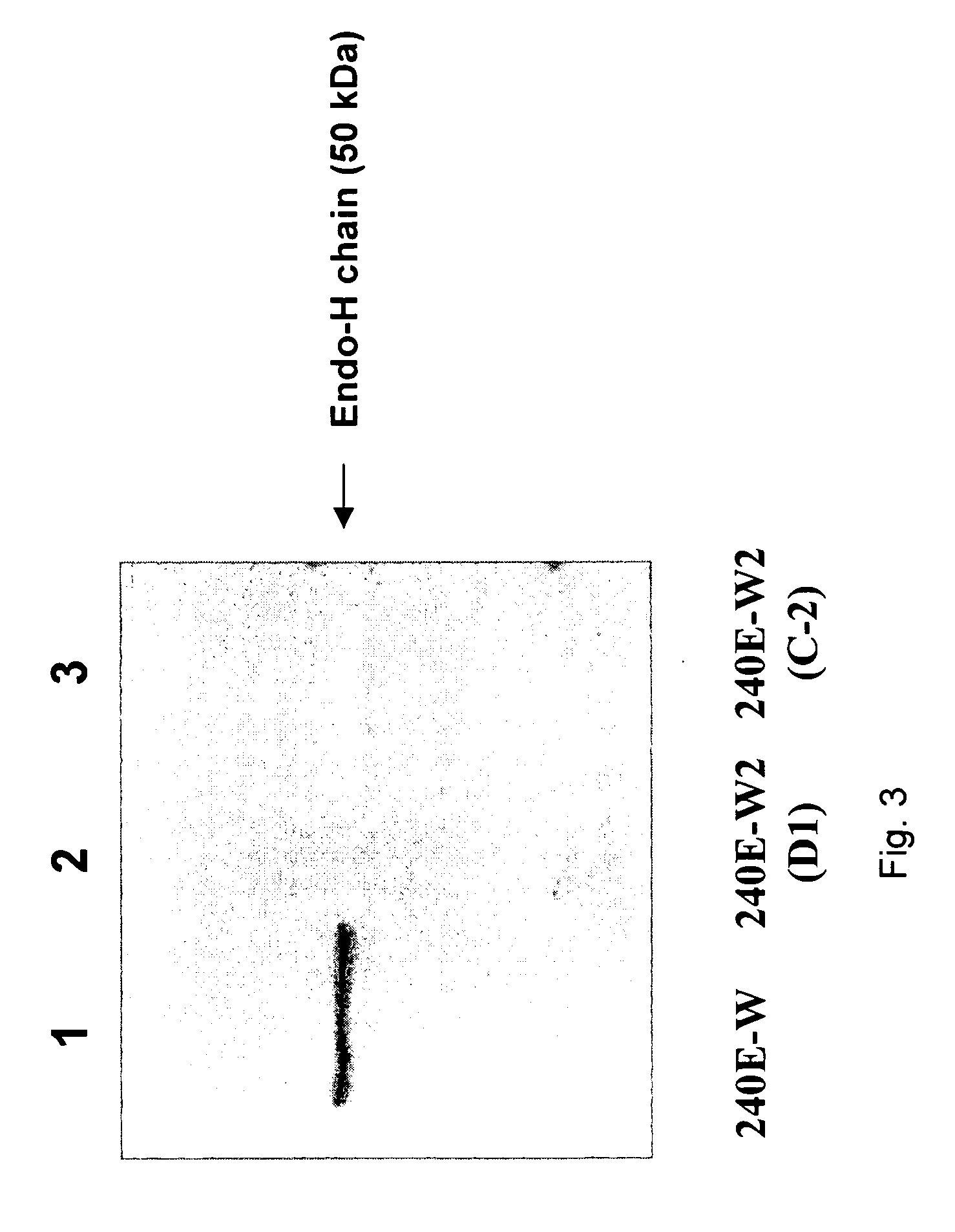
[0062]The 240E-W cell line and the original 240E-1 cell line, express a significant amount of an endogenous IgG heavy chain (endo-IgH), which is readily detectable at the protein and mRNA level (FIG. 3). The expression of endogenous IgH persists in hybridomas derived from 240E-W, and is often equal to or higher than the expression of the splenocyte-derived IgH (spleno-IgH) comprising the heavy chain of the desired antibody secreted by the hybridoma (FIG. 1). As shown in FIG. 1, endo-IgH and spleno-IgH are, in most cases, readily distinguishable by SDS polyacrylamide gel electrophoresis (SDS-PAGE). The nucleotide sequence and deduced amino acid sequence of endo-IgH was determined (FIG. 2), and the identity of the lower band in FIG. 1 as endo-IgH was confirmed by protein sequencing. Endo-IgH can associate with the splenocyte-derived Ig light chain (spleno-IgL), as demonstrated by the presence of the intact 150 kDa IgG complex on non-reducing SDS-PAGE (data not shown). Therefore, in ma...
example 3
[0064]Fusion of 240E-W cells with rabbit splenocytes typically yields a subset of hybridoma clones that do not secrete any detectable IgG. Since even in the absence of secreted IgG the cells may contain intracellular IgG heavy chains, several of these clones were tested by immunocytochemistry for the presence of intracellular IgG. Briefly, this was performed by culturing subclones in duplicate 96-well plates, and processing one of the plates for immunocytochemistry by drying, fixation with 0.4% paraformaldehyde, permeabilization with 0.5% Triton X-100, and immunoperoxidase staining with polyclonal goat-anti-rabbit IgG antibodies. Several clones were identified that contained only low levels of IgG, and only in a subset of the cells. One of these hybridoma clones was selected for subcloning and further testing. The cells were grown in standard growth medium ((Spieker-Polet et al., Proc. Natl. Acad. Sci. 1995 92:9348-52), and subcloned by the limiting-dilution method. Single-cell subc...
PUM
| Property | Measurement | Unit |
|---|---|---|
| dissociation constant | aaaaa | aaaaa |
| dissociation constant | aaaaa | aaaaa |
| dissociation constant | aaaaa | aaaaa |
Abstract
Description
Claims
Application Information
 - R&D
- Intellectual Property
- Life Sciences
- Materials
- Tech Scout
- Unparalleled Data Quality
- Higher Quality Content
- 60% Fewer Hallucinations
Browse by: Latest US Patents, China's latest patents, Technical Efficacy Thesaurus, Application Domain, Technology Topic, Popular Technical Reports.
© 2025 PatSnap. All rights reserved.Legal|Privacy policy|Modern Slavery Act Transparency Statement|Sitemap|About US| Contact US: help@patsnap.com


