Antibodies directed against programmed death-1 (PD-1)
a technology of anti-programmed death and anti-lag-3, which is applied in the direction of antibacterial agents, drug compositions, antibody medical ingredients, etc., can solve the problems of limited human efficacy of these potential therapies, and achieve the effects of increasing the activity of anti-tim-3 antagonists, low levels, and increasing the activity of anti-lag-3 antagonists
- Summary
- Abstract
- Description
- Claims
- Application Information
AI Technical Summary
Benefits of technology
Problems solved by technology
Method used
Image
Examples
example 1
[0093]This example demonstrates a method of generating monoclonal antibodies directed against human PD-1.
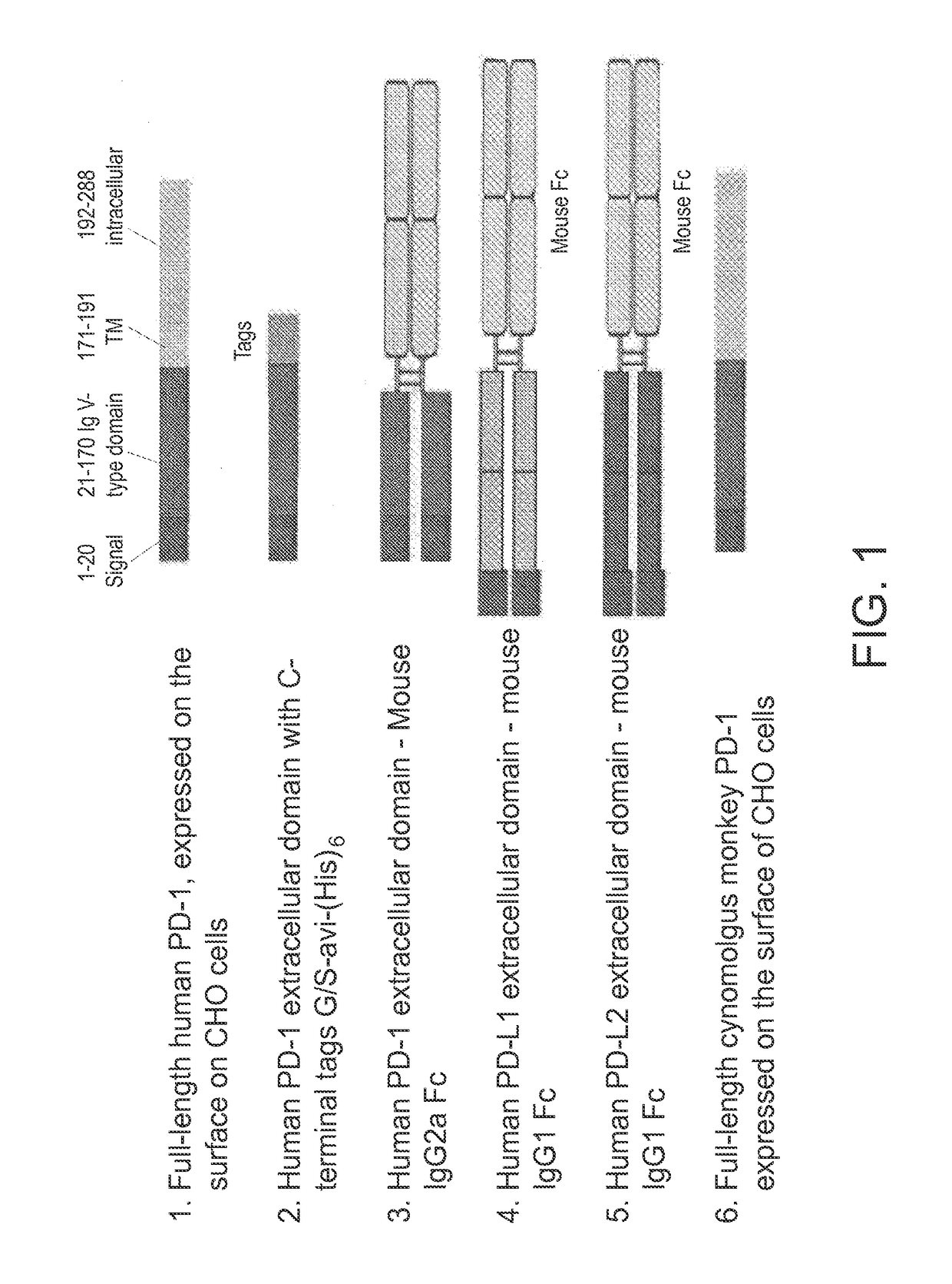
[0094]Several forms of genes encoding human PD-1 and its ligands PD-L1 and PD-L2 were generated as antigens for use in mouse immunization, hybridoma screening, and affinity maturation of CDR-grafted antibodies, and are schematically depicted in FIG. 1. Full-length human and cynomolgus monkey PD-1 genes were expressed with their native leader sequence and no added tags using a ubiquitous chromatin opening element (UCOE) single expression vector with hygromycin selection (Millipore, Billerica, Mass.). CHO-K1 cells were stably transfected with Lipofectamine LTX (Life Technologies, Carlsbad, Calif.) according to the manufacturer's instructions. Following selection with hygromycin, cells expressing PD-1 on the cell surface were identified by flow cytometry using a PE-conjugated mouse antibody to human PD-1 (BD Bioscience, Franklin Lakes, N.J.) and subcloned. Subclones were then select...
example 2
[0101]This example describes the design and generation of CDR-grafted and chimeric anti-PD-1 monoclonal antibodies.
[0102]Subclones of the hybridomas which produced PD-1-binding antibodies with PD-L1 blocking activity as described in Example 1 were isotyped, subjected to RT-PCR for cloning the antibody heavy chain variable region (VH) and light chain variable region (VL), and sequenced. Specifically, RNA was isolated from cell pellets of hybridoma clones (5×10⁵ cells / pellet) using the RNEASY™ kit (Qiagen, Venlo, Netherlands), and cDNA was prepared using oligo-dT-primed SUPERSCRIPT™ III First-Strand Synthesis System (Life Technologies, Carlsbad, Calif.). PCR amplification of the VL utilized a pool of 9 or 11 degenerate mouse VL forward primers (see Kontermann and Dubel, eds., Antibody Engineering, Springer-Verlag, Berlin (2001)) and a mouse κ constant region reverse primer. PCR amplification of the VH utilized a pool of 12 degenerate mouse VH forward primers (Kontermann and Dubel, sup...
example 3
[0106]This example demonstrates affinity maturation of monoclonal antibodies directed against PD-1.
[0107]CDR-grafted antibodies derived from the original murine monoclonal antibodies, (9A2, 10B11, and 6E9) were subjected to affinity maturation via in vitro somatic hypermutation. Each antibody was displayed on the surface of HEK 293c18 cells using the SHM-XEL deciduous system (see Bowers et al., Proc. Natl. Acad. Sci. USA, 108: 20455-20460 (2011); and U.S. Patent Application Publication No. 2013 / 0035472). After establishment of stable episomal lines, a vector for expression of activation-induced cytosine deaminase (AID) was transfected into the cells to initiate somatic hypermutation as described in Bowers et al., supra. After multiple rounds of FACS sorting under conditions of increasing antigen binding stringency, a number of mutations in the variable region of each antibody were identified and recombined to produce mature humanized antibodies with improved properties.
[0108]A panel...
PUM
| Property | Measurement | Unit |
|---|---|---|
| in vivo half life | aaaaa | aaaaa |
| in vivo half life | aaaaa | aaaaa |
| in vivo half life | aaaaa | aaaaa |
Abstract
Description
Claims
Application Information
 - R&D
- Intellectual Property
- Life Sciences
- Materials
- Tech Scout
- Unparalleled Data Quality
- Higher Quality Content
- 60% Fewer Hallucinations
Browse by: Latest US Patents, China's latest patents, Technical Efficacy Thesaurus, Application Domain, Technology Topic, Popular Technical Reports.
© 2025 PatSnap. All rights reserved.Legal|Privacy policy|Modern Slavery Act Transparency Statement|Sitemap|About US| Contact US: help@patsnap.com


