System and method for the non-destructive assessment of the quantitative spatial distribution of components of a medical device

a medical device and quantitative spatial distribution technology, applied in chemical machine learning, specific gravity measurement, instruments, etc., can solve the problems of ischemic injury, stroke or myocardial infarction, angioplasty is the abrupt closure of the vessel, and chronic problems such as restnosis, and achieve high throughput raman

Active Publication Date: 2009-01-13
CARDINAL HEALTH SWITZERLAND 515 GMBH
View PDF8 Cites 10 Cited by
  • Summary
  • Abstract
  • Description
  • Claims
  • Application Information

AI Technical Summary

Problems solved by technology

More severe blockage of blood vessels in such individuals often leads to hypertension, ischemic injury, stroke, or myocardial infarction.
A limitation associated with percutaneous transluminal coronary angioplasty is the abrupt closure of the vessel, which may occur immediately after the procedure and restenosis, which occurs gradually following the procedure.
Additionally, restenosis is a chronic problem in patients who have undergone saphenous vein bypass grafting.
However, this assumption proved invalid because the influence of mixing and drying that occurs during manufacture was not considered.
Both techniques are coating surface limited and do not adequately represent the three-dimensional system.
The techniques also lack the chemical specificity to positively differentiate drug from polymer matrix components.
Although NIR and FTIR imaging provide chemical selectivity, both suffer from poor spatial resolution and cannot depth profile through coatings.

Method used


Image

  • System and method for the non-destructive assessment of the quantitative spatial distribution of components of a medical device
  • System and method for the non-destructive assessment of the quantitative spatial distribution of components of a medical device
  • System and method for the non-destructive assessment of the quantitative spatial distribution of components of a medical device

Examples


Embodiment Construction

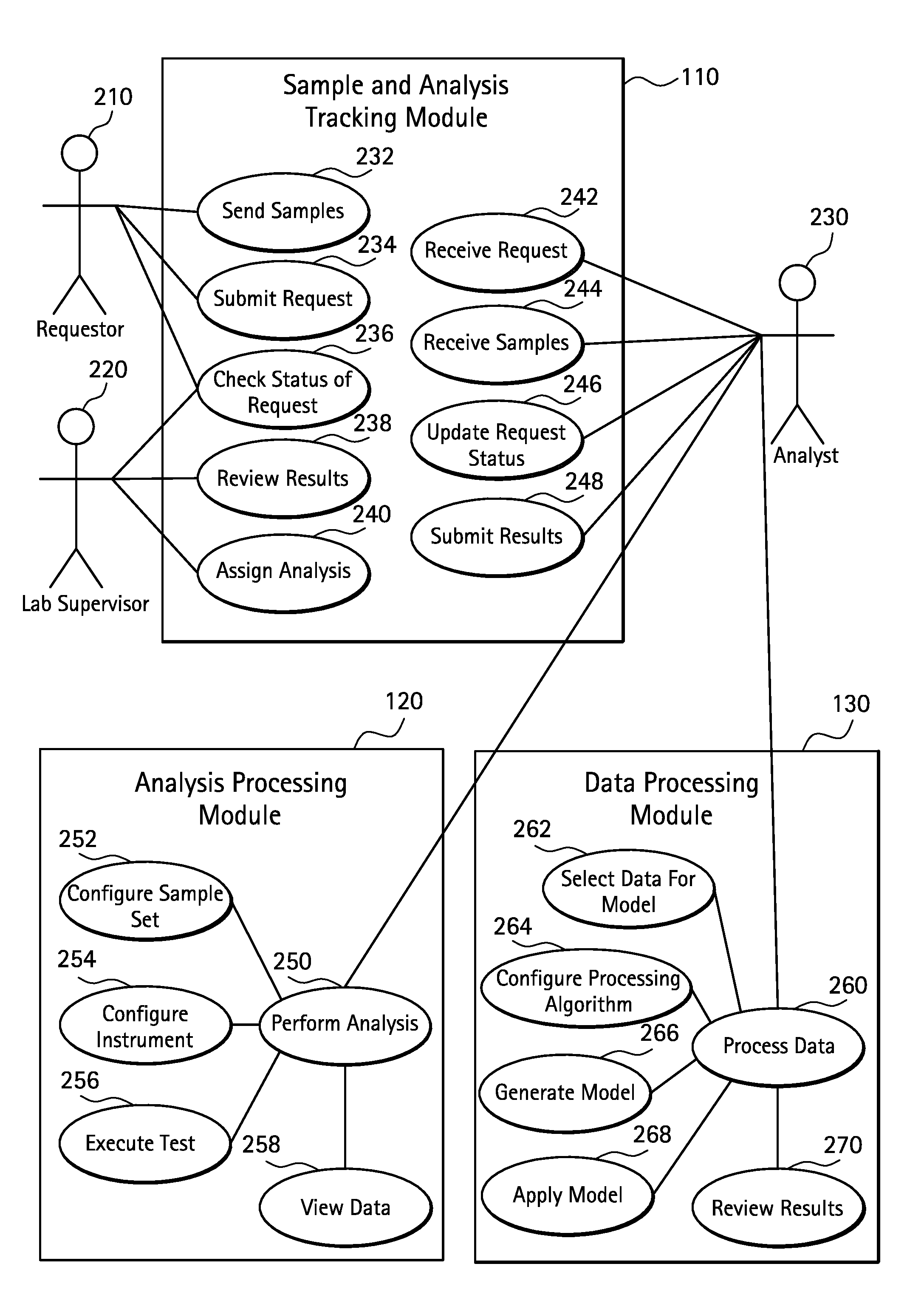
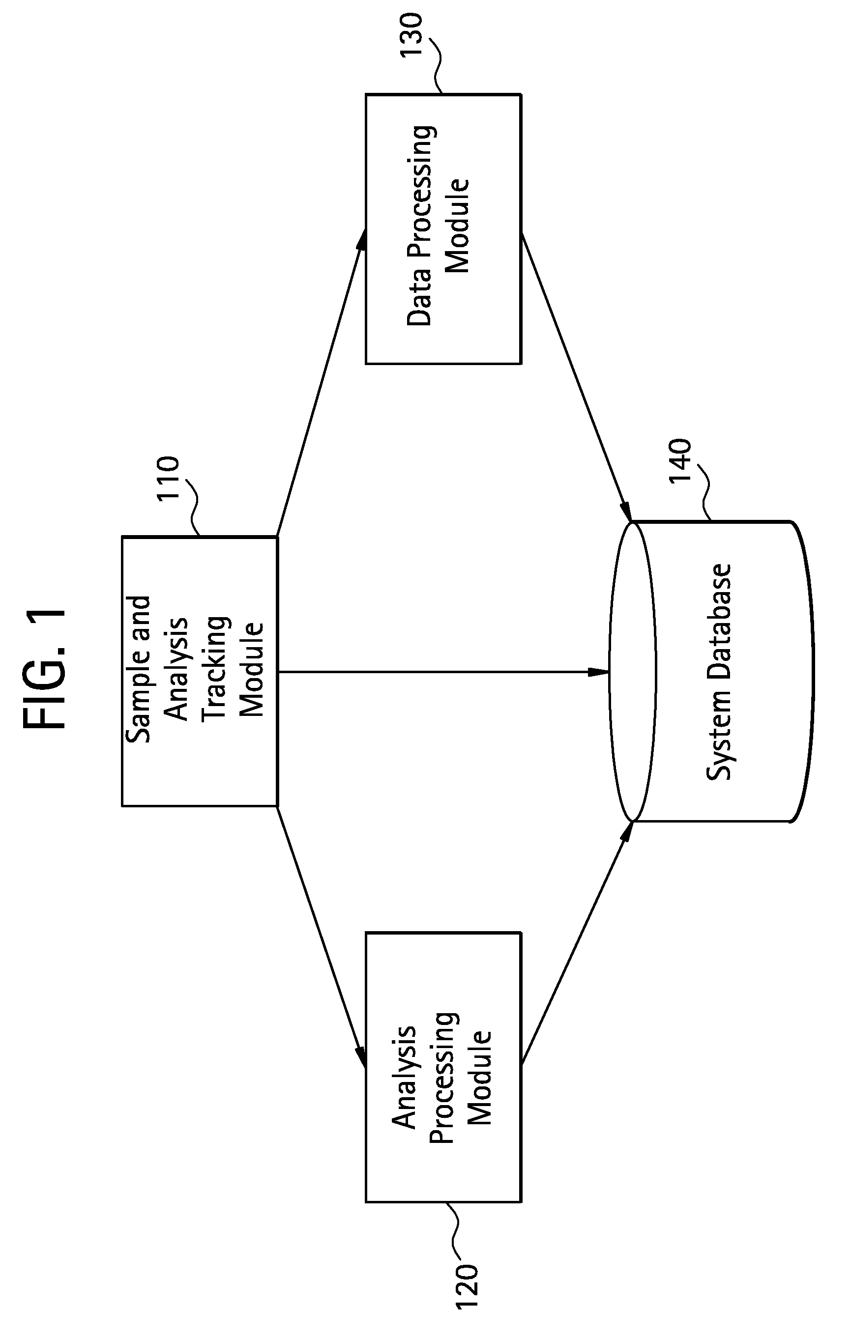
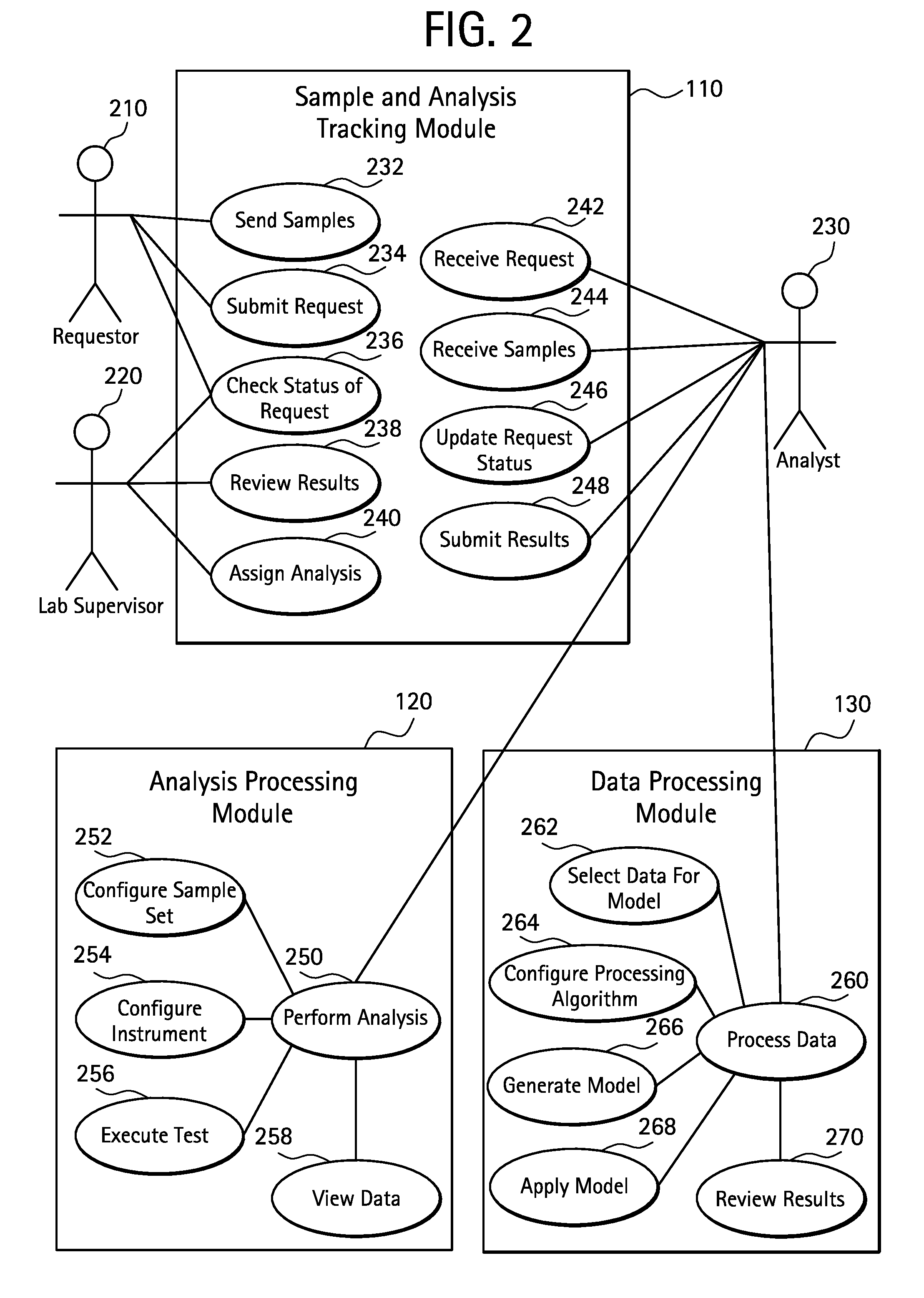
[0052]The schematic diagram of the system of the present invention is set forth FIG. 1. There are three separate modules and a system database. Sample and analysis tracking module 110 enables the user of the system to input data regarding the sample to be analyzed and tracks the progress of the sample through the analysis process. The sample and analysis tracking module 110 will store information about the sample such as a description of the sample (formulation of API and matrix components and process type of sample device), fabrication date, and the analysis date in the system database 140. The analysis processing module 120 comprises an instrument to collect the data and a also retains a description of how the data is collected including the type of instrument, sample positioning, number of observations, and type of data collected. The data processing module 130 processes the data collected from the analysis processing module 120 analytically. The output from the data processing m...


PUM


Abstract

A method and system for the non-destructive analysis of medical devices uses a confocal Raman microscope and other non-destructive analytical tools to assess the spatial distribution of components of an object such as the distribution of an active pharmaceutical ingredient (API) within a polymer matrix. In a preferred embodiment, confocal Raman spectroscopy was used to differentiate each component found in the sirolimus-eluting coronary stent. The unique spectral features identified for each component were then used to develop three separate calibration curves to describe the solid phase distribution found on drug-polymer coated stents. The calibration curves were obtained by analyzing confocal Raman spectral depth profiles from a set of 16 unique formulations of drug-polymer coatings sprayed onto stents and planar substrates.

Description

CROSS REFERENCE TO RELATED APPLICATION[0001]This application claims the benefit of U.S. Provisional Application No. 60 / 895,242 filed Mar. 16, 2007.FIELD OF THE INVENTION[0002]The present invention relates to a system and method for the non-destructive, chemically specific, profiling and assessment of the spatial distribution of the therapeutic agents and other components of a substance applied to an object such as a medical device. More particularly, the invention relates to a system and method to determine that therapeutic agent or agents have been accurately applied to a medical device such as a drug-eluting stent so as to confirm the elution profile of the stent over time thereby providing an empirical link between the macroscopic properties of the device with the microscopic distribution of components within the device.BACKGROUND OF THE INVENTION[0003]Many individuals suffer from circulatory disease caused by a progressive blockage of the blood vessels that profuse the heart and...

Claims


Application Information

Patent Timeline
Patent Type & Authority Patents(United States)
IPC IPC(8): G06F15/00
CPCG01N21/65G06F19/327G01N21/95G06F19/703G06F19/707G16H40/20G16C20/20G16C20/70
Inventor BALSS, KARIN MARIALLANOS, GERARDMARYANOFF, CYNTHIA ANNEPAPANDREOU, GEORGEVESELOV, VLADIMIR
Owner CARDINAL HEALTH SWITZERLAND 515 GMBH
Why Eureka
Try Eureka