
An imaging method of tumor targeting based on enhancement effect of zinc ion signals

A tumor-targeted imaging and signal enhancement technology, applied in the field of medical imaging, can solve problems such as harm to the human body, and achieve the effects of low biological toxicity, various sources, and easy operation.

Active Publication Date: 2015-05-06
SOUTHEAST UNIV
View PDF7 Cites 0 Cited by
  • Summary
  • Abstract
  • Description
  • Claims
  • Application Information

AI Technical Summary

Problems solved by technology

However, there are still many problems in the fluorescence imaging of tumors in vivo. For example, toxic fluorescent chemical dyes or nano-quantum dots are used as fluorescent markers of tumor tissue, which is harmful to the human body; or some fluorescent markers are non-toxic but cannot pass through the veins. Injection reaches the targeted tumor site

Method used


Image

  • An imaging method of tumor targeting based on enhancement effect of zinc ion signals
  • An imaging method of tumor targeting based on enhancement effect of zinc ion signals
  • An imaging method of tumor targeting based on enhancement effect of zinc ion signals

Examples


Embodiment 1

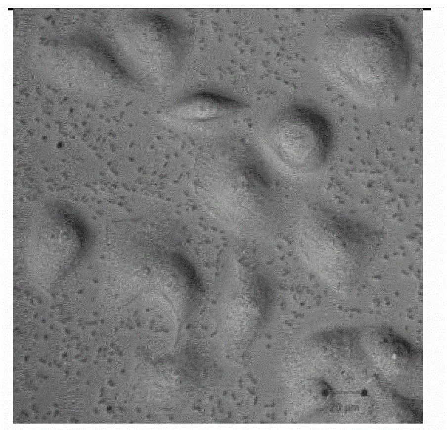
[0028] Dissolve zinc gluconate in PBS solution with pH=7.4 to make 1mmol / L zinc gluconate solution, take hepatoma cell (HepG2) as the research object, put hepatoma cell (HepG2) and zinc gluconate solution in cell culture After incubating at 37°C in the box for 12 hours, the intensity and distribution of cell autofluorescence were detected with a confocal fluorescence microscope, and Raman imaging was performed on liver cancer cells (HepG2) through a Raman microscope, and the signal data of the fluorescence spectrum and the Raman spectrum were used to determine Qualitative and quantitative analysis of the structure or chemical composition of hepatocellular carcinoma cells (HepG2).

Embodiment 2

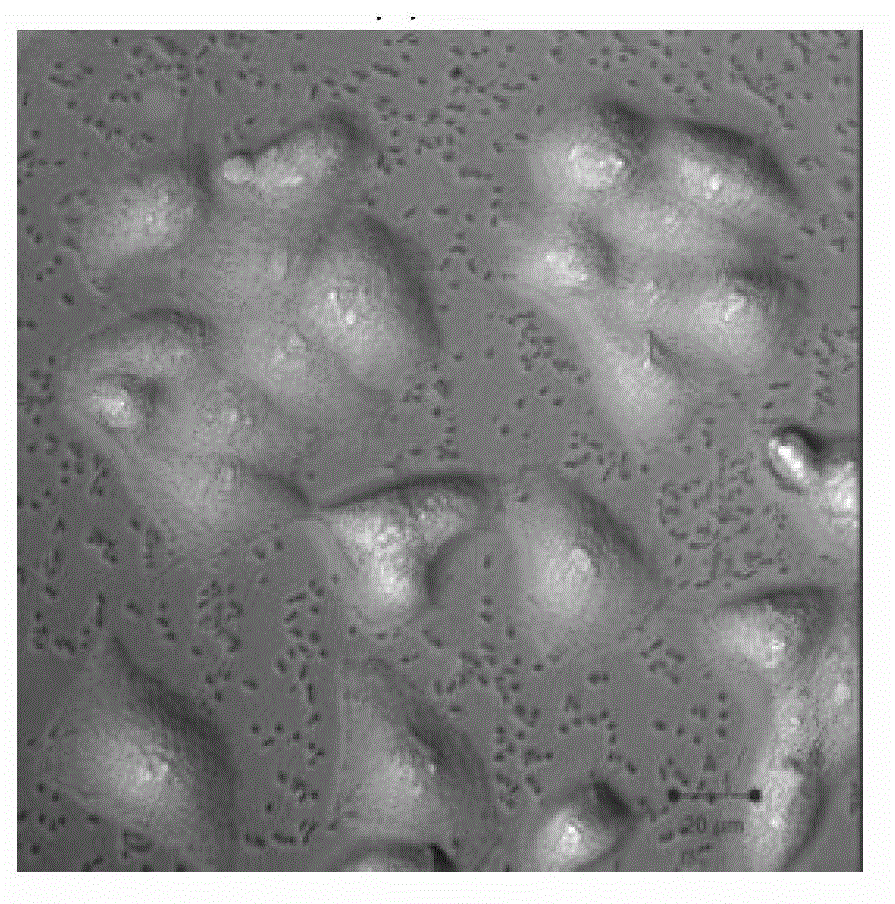
[0030] Dissolve zinc phosphate in PBS solution with pH = 7.4 to make 50mmol / L zinc phosphate solution, take cervical cancer cells (HeLa) as the research object, put cervical cancer cells (HeLa) and zinc phosphate solution in the cell culture incubator After incubating at 37°C for 24 hours, the intensity and distribution of cell autofluorescence were detected with an ultrasonic imager, Raman imaging was performed on cervical cancer cells (HeLa) through a Raman microscope, and the signal data of the fluorescence spectrum and the Raman spectrum were compared. Qualitative and quantitative analysis of cervical cancer cell (HeLa) structure or chemical composition.

Embodiment 3

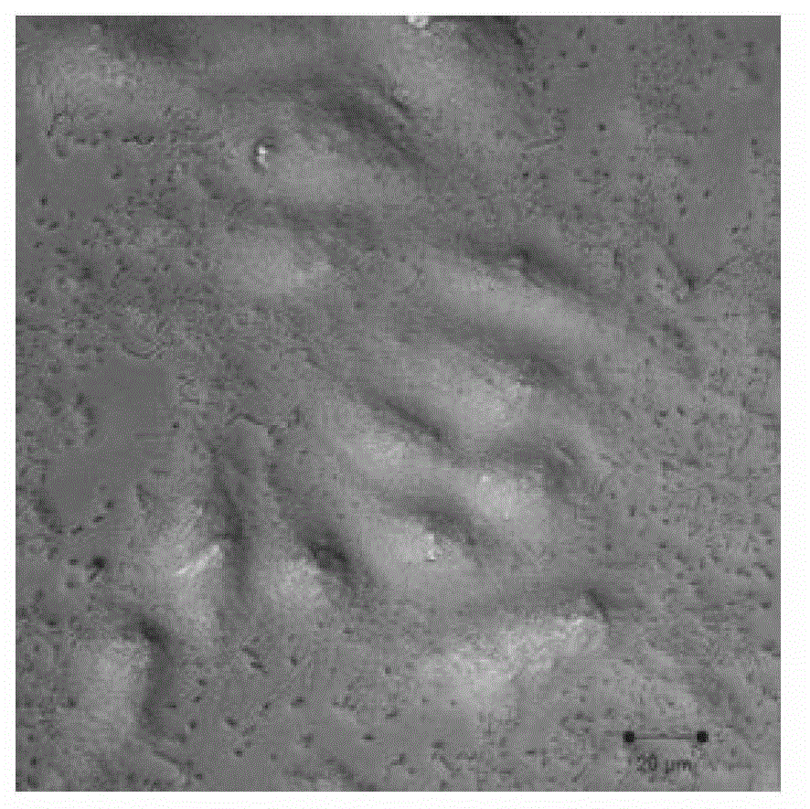
[0032] Dissolve zinc chloride in PBS solution with pH=7.4 to make 100mmol / L zinc chloride solution, take leukemia cells (K562) as the research object, put leukemia cells (K562) and zinc chloride solution in cell culture After incubating at 37°C in the box for 48 hours, use a fluorescence imager to detect the intensity and distribution of cell autofluorescence, use a Raman spectrometer to perform Raman imaging on leukemia cells (K562), and use the signal data of the fluorescence spectrum and Raman spectrum to compare Qualitative and quantitative analysis of the structure or chemical components of leukemia cells (K562).


PUM


Abstract

The invention discloses an imaging method of tumor targeting based on enhancement effect of zinc ion effects. When the imaging method of the tumor targeting based on the enhancement effect of the zinc ion signals is applied at the cellular level, a zinc saline solution is manufactured firstly. Then the saline solution is arranged into a cell incubator together with cancer cells or cancer tissues for incubation. After high resolution fluorescence microscopic imaging and Raman imaging are carried out, qualitative and quantitative analysis is performed on the structure or chemical constituents of the cancer cells or the cancer tissues through the signal data of a fluorescence spectrum and a Raman spectrum. When the imaging method of the tumor targeting based on the enhancement effect of the zinc ion signals is applied at the animal model level, a tumor animal model is constructed and the zinc saline solution is manufactured, after part of the tumor location of an experimental animal is injected with the zinc saline solution, a confocal fluorescence microscope, an ultrasonic imaging device, a fluorescence imaging device, a Raman spectrometer or a Raman microscope are used for monitoring the signal information of the tumor cells or the cancer tissues and processing and analyzing the signal information. Due to the fact that the zinc ions are adopted as the imaging signal enhancer, the imaging method of the tumor targeting based on the enhancement effect of the zinc ion signals has small toxicity on a living body, and has tumor targeting capability.

Description

technical field [0001] The invention relates to a method in the field of medical imaging, in particular to a tumor targeting imaging method based on the signal enhancement effect of zinc ions. Background technique [0002] Cancer is a serious threat to human health and life safety. Early detection, early diagnosis and early treatment are important preventive means to reduce the mortality of cancer patients. As an extremely important auxiliary means for the diagnosis and treatment of tumors, medical imaging has been widely used in the medical field. In vivo tumor fluorescence imaging can be used for direct biological imaging of tumors. This imaging method is simple, fast, low-cost and has the characteristics of visualization, so it has attracted more and more attention from the scientific community. However, there are still many problems in the fluorescence imaging of tumors in vivo. For example, toxic fluorescent chemical dyes or nano-quantum dots are used as fluorescent ma...

Claims


Application Information

Patent Timeline
Patent Type & Authority Patents(China)
IPC IPC(8): A61B5/00A61B8/08A61D7/00G01N21/64G01N21/65A61K49/00A61K49/22
Inventor 王雪梅李水红吴长宇高生平
Owner SOUTHEAST UNIV
Learn More