Anti-extravasation sheath and method

a technology of anti-extravasation and sheath, which is applied in the field of arthroscopic surgery, can solve the problems of premature collapse of the surgical field, long recovery time, pain and discomfort of patients, and fluid escaping into the soft tissues of the shoulder and the periscapular region can have adverse effects on patients, so as to reduce the amount of fluid extravasation occurring in the surrounding tissue, the surgical field is clear and the effect of reducing the amount of fluid extravasation

Active Publication Date: 2009-03-17
CANNUFLOW INC
View PDF22 Cites 345 Cited by
  • Summary
  • Abstract
  • Description
  • Claims
  • Application Information

AI Technical Summary

Benefits of technology

[0006]The devices and methods shown below provide for the minimization of fluid extravasation during arthroscopic surgery. The anti-extravasation inflow / outflow sheath allows a surgeon to drain fluids from the soft tissue surrounding the surgical field while also providing fluid inflow and outflow to a surgical site.
[0007]The anti-extravasation inflow / outflow sheath is a multi-lumen tube into which an arthroscopic surgical instrument such as an arthroscope is inserted. The proximal portion of the sheath is provided with fluid ports, a manifold and other means of controlling the flow of fluid inside the sheath. The distal portion of the inflow / outflow sheath is provided with a plurality of inflow / outflow holes. The body or central portion of the sheath is provided with a plurality of drainage apertures. Each inflow / outflow hole communicates with one or more of the lumens inside the tube, thereby allowing fluid to flow between the surgical field and sources or sinks located outside the patient. Each drainage aperture communicates with one or more of the drainage lumens inside the tube, thereby allowing fluid to drain from the tissue surrounding the surgical site and sources or sinks located outside the patient. The anti-extravasation inflow / outflow sheath allows the surgeon to maintain a clear surgical field and eliminate the need for a third irrigation instrument while reducing the amount of fluid extravasation occurring in surrounding tissue.

Problems solved by technology

Fluid escaping into the soft tissues of the shoulder and the periscapular region can have adverse effects on the patient.
These effects cause longer recovery time as well as pain and discomfort in patients.
Extravasation occurring during surgery can also cause premature collapse of the surgical field forcing surgeons to rush procedures.

Method used


Image

  • Anti-extravasation sheath and method
  • Anti-extravasation sheath and method
  • Anti-extravasation sheath and method

Examples


Embodiment Construction

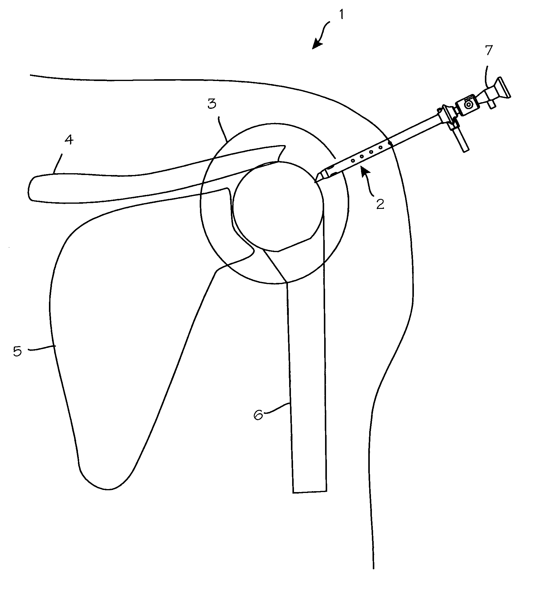
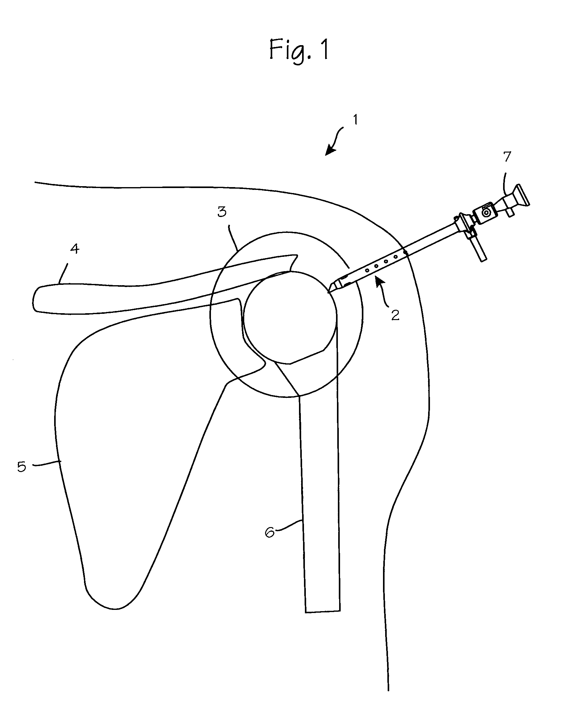
[0021]FIG. 1 illustrates a method of performing arthroscopic surgery on a patient's shoulder 1 using the anti-extravasation sheath 2. The anti-extravasation sheath is shown inserted into the joint capsule 3 of a shoulder of a patient. Various anatomical landmarks are depicted including the patient's clavicle 4, scapula 5 and humerus 6. An arthroscopic instrument 7 such as an arthroscope is disposed within the anti-extravasation sheath.

[0022]During arthroscopic shoulder surgery, the surgeon introduces the arthroscope into the shoulder via a first portal in order to visualize the surgical field. A trimming instrument is introduced through a second portal to remove or trim tissue that the surgeon determines should be removed or trimmed. Optionally, an irrigating instrument may be introduced through a third portal in order to distend the joint, and / or irrigate the surgical field to maintain a clear view. Other arthroscopic instruments used in arthroscopic surgery include endoscopes, awl...


PUM


Abstract

The methods shown provide for the minimization of extravasation during arthroscopic surgery. Use of an anti-extravasation sheath having at least one drainage aperture allows a surgeon to drain excess fluids from the tissue surrounding the surgical field during an arthroscopic surgical procedures when the drainage aperture is disposed within the tissue surrounding an arthroscopic surgical field outside of the joint capsule. The method of performing arthroscopic surgery may further include providing fluid inflow and outflow to the joint capsule through inflow / outflow holes in the anti-extravasation sheath.

Description

FIELD OF THE INVENTIONS[0001]The inventions described below relate to the field arthroscopic surgery and more specifically, to fluid management during arthroscopic shoulder surgery.BACKGROUND OF THE INVENTIONS[0002]During minimally invasive surgeries, surgical instruments such as trocars, cannulas, and optical medical devices, including endoscopes, cystoscopes, arthroscopes, laparoscopes, etc., are inserted through small incisions or portals in a patient's body or body cavity and manipulated to perform surgical procedures within the patient.[0003]Minimally invasive surgical procedures are safer than open surgery and result in quicker patient recovery, shorter hospital stays, and lower health care costs. Accordingly, minimizing invasiveness continues to be of importance, and there is a continuing need for devices and methods that achieve this objective.[0004]One area that has benefited from minimally invasive surgical techniques is shoulder surgery. Shoulder surgery has evolved over ...

Claims


Application Information

Patent Timeline
Patent Type & Authority Patents(United States)
IPC IPC(8): A61B1/015
CPCA61B1/00135A61B1/317A61B17/3421A61B1/015A61B17/3474A61B2017/00849A61B2017/00853A61B2017/00858A61B2017/3441A61B2217/005A61B2217/007A61M1/0084A61M1/85A61B17/0218A61B17/3423
Inventor KUCKLICK, THEODORE R.
Owner CANNUFLOW INC
Why Eureka
Try Eureka