Non human animals with human-glial chimeric brains

a technology of glial chimeric brain and human glial chimeric, which is applied in the field of nonhuman animals with human glial chimeric brain, can solve the problems of unclear remyelination strategy, achieve widespread central myelination, influence the phenotype and survival of the recipient animal, and increase the number of glial chimeric cells

Active Publication Date: 2009-04-28
UNIVERSITY OF ROCHESTER
View PDF2 Cites 21 Cited by
  • Summary
  • Abstract
  • Description
  • Claims
  • Application Information

AI Technical Summary

Benefits of technology

[0009]In the work corresponding to the present invention, using a newly developed set of approaches to both cell acquisition and transplantation, a far more extensive and higher density cell engraftment was attempted than any previously noted. The aim in doing so was to achieve sufficiently widespread central myelination to influence the phenotype and survival of the recipient animals. To avoid rejection as a complicating variable in these experiments, shiverer mice were crossed with rag2 null immunodeficient mice (Shinkai et al., “RAG2-Deficient Mice Lack Mature Lymphocytes Owing to Inability to Initiate V(D)J Rearrangement,”Cell 68:855-867 (1992)), thereby generating an immunodeficient line of congenitally hypomyelinated mice in which to assess graft efficacy and effect. Using these double homozygous rag2− / −×shiverershi / shi mice, a multi-site injection protocol, with concurrent bilateral hemispheric and cerebellar cell injections delivered at birth was established. This procedure resulted in widespread donor cell engraftment throughout the neuraxis, with infiltration of the forebrain, brainstem and cerebellum, and ultimately the spinal cord and roots. The engrafted human glial progenitor cells exhibited robust, efficient, and functional myelination, with progressive ensheathment of host axons and restoration of normal nodes of Ranvier and attendant conduction velocities. This ultimately led to the high efficiency myelination of the major intracerebral, ascending and descending tracts, the cranial nerves and intracranial ganglia, and the spinal cord to the thoracolumbar level. Most notably, the implanted animals exhibited a substantial recovery of normal neurological phenotype, such that a fraction were frankly rescued by perinatal transplantation, surviving well over a year until sacrificed for histology, which revealed both a remyelinated—and essentially humanized—central white matter. The neurological recovery and sustained survival of these transplanted mice was in sharp contrast to the fate of their untreated controls, which uniformly died by 5 months of age. Together, these data represent the first outright rescue of a congenital leukodystrophy, or indeed of any hereditary-metabolic disorder, by means of a stem or progenitor cell transplantation. Such results indicate that neonatal glial progenitor cell transplantation may prove an effective means of treating disorders of both hereditary and perinatal hypomyelination.

Problems solved by technology

However, the potential utility of this approach to the development of clinical remyelination strategies has been unclear, since previous studies have failed to note significant brainstem, cerebellar or spinal engraftment from intracerebral grafts, and no effect on disease phenotype or survival has yet been reported in hypomyelinated mice as a consequence of progenitor cell transplantation (reviewed in Keyoung et al., “Glial Progenitor-Based Repair of Demyelinating Neurological Diseases,”Neurosurg. Clin. NA 18:93-104 (2007) and Goldman, S. A., “Stem and Progenitor Cell-Based Therapy of the Human Central Nervous System,”Nature Biotech.

Method used


Image

  • Non human animals with human-glial chimeric brains
  • Non human animals with human-glial chimeric brains
  • Non human animals with human-glial chimeric brains

Examples


example 1

Cell Extraction

[0063]Fetal oligodendrocyte progenitor cells (“OPCs”) were extracted from second trimester human fetuses (19 to 22 weeks gestational age, g.a.), obtained at abortion as described (Windrem et al., “Fetal and Adult Human Oligodendrocyte Progenitor Cell Isolates Myelinate the Congenitally Dysmyelinated Brain,”Nature Medicine 10:93-97 (2004), which is hereby incorporated by reference in its entirety). The forebrain ventricular / subventricular zones were rapidly dissected from the remaining brain parenchyma, the samples chilled on ice, and the minced samples then dissociated using papain / DNAse as described (Keyoung et al., “Specific Identification, Selection and Extraction of Neural Stem Cells from the Fetal Human Brain,”Nature Biotechnology 19:843-850 (2001), which is hereby incorporated by reference in its entirety), always within 3 hours of extraction. The dissociates were then maintained overnight in minimal media of DMEM / F12 / N1 with 20 ng / ml FGF. A total of 5 tissue s...

example 2

Cell Sorting

[0064]Sorted glial progenitor cells were isolated from dissociated tissue using a dual immunomagnetic sorting strategy. On the day after dissociation the cells were incubated with mouse anti-PSA-NCAM (Chemicon) at 1:100. then washed and labeled with microbead-tagged rat anti-mouse IgM (Miltenyi Biotech), and removed by MACS depletion. The remaining PSA-NCAM− cells were next incubated 1:1 with MAb A2B5 supernatant (clone 105; ATCC, Manassas, Va.), for 20 minutes, then washed and labeled with microbead-tagged rat anti-mouse IgM (Miltenyi Biotech). All incubations were done on ice (Keyoung et al., “Specific Identification, Selection and Extraction of Neural Stem Cells from the Fetal Human Brain,”Nature Biotechnology 19:843-850 (2001) and Roy et al., “In Vitro Neurogenesis by Progenitor Cells Isolated from the Adult Human Hippocampus,”Nat Med 6:271-7. (2000), which are hereby incorporated by reference in their entirety). Magnetic separation of A2B5+ cells (MACS; Miltenyi) wa...

example 3

Transplantation and Husbandry

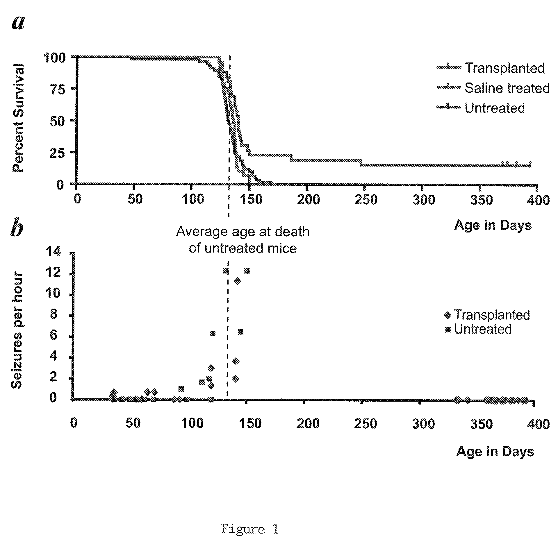
[0065]Homozygous shiverers were crossed to homozygous rag2 null immunodeficient mice (Shinkai et al., “RAG2-Deficient Mice Lack Mature Lymphocytes Owing to Inability to Initiate V(D)J Rearrangement,”Cell 68:855-867 (1992), which is hereby incorporated by reference in its entirety), to generate a line of shi / shi×rag2− / − myelin-deficient, immunodeficient mice. Newborn pups of this line were transplanted within a day of birth, using a total of 300,000 donor cells dispersed over 5 injection sites. The pups were first cryoanesthetized for cell delivery. 5×10⁴ donor cells in 0.5 μl HBSS were then injected at each of 4 locations in the forebrain subcortex, specifically into the anterior and posterior anlagen of the corpus callosum bilaterally. These injections were delivered to a depth of 1.0 to 1.2 mm ventrally, depending on the weight / size of the pup (which varied from 1-1.5 g). A fifth injection of 105 cells in 1 μl was delivered into the cerebellar peduncle...


PUM

PropertyMeasurementUnit
depthaaaaaaaaaa
body temperatureaaaaaaaaaa
diameteraaaaaaaaaa

Abstract

The present invention is directed to a non-human mammal with at least 30% of all of its glial cells in its corpus callosum being human glial cells and / or at least 5% of all of its glial cells in the white matter of its brain and / or brain stem being human glial cells. Methods of producing and using the non-human mammal are also disclosed.

Description

[0001]This application claims the benefit of U.S. Provisional Patent Application Ser. No. 60 / 885,094, filed Jan. 16, 2007.FIELD OF THE INVENTION[0002]The present application relates to non-human animals with human glial chimeric brains.BACKGROUND OF THE INVENTION[0003]Glial progenitor cells and the neural stem cells from which they derive may be isolated and transplanted into myelin-deficient hosts, as a means of introducing new oligodendrocytes able to myelinate host axons (Yandava et al., “Global Cell Replacement is Feasible Via Neural Stem Cell Transplantation: Evidence From the Dysmyelinated Shiverer Mouse Brain,”Proc Natl Acad Sci USA 96:7029-34 (1999); Learish et al., “Intraventricular Transplantation of Oligodendrocyte Progenitors Into a Fetal Myelin Mutant Results in Widespread Formation of Myelin,”Ann Neurol 46:716-22 (1999); Archer et al., “Myelination of the Canine Central Nervous System by Glial Cell Transplantation: A Model for Repair of Human Myelin Disease,”Nature Med...

Claims


Application Information

Patent Timeline
Patent Type & Authority Patents(United States)
IPC IPC(8): G01N33/00A01K67/00A01K67/033C12N15/00
CPCA01K67/0271C12N5/0623G01N33/5058G01N33/5088A01K2207/12A01K2207/15A01K2217/15A01K2227/105A01K2267/0356
Inventor GOLDMAN, STEVEN A.WINDREM, MARTHA
Owner UNIVERSITY OF ROCHESTER
Why Eureka
Try Eureka