Apparatus for endoscopic cardiac mapping and lead placement

a technology of endoscopic and endocardial surgery, applied in the direction of catheters, applications, therapy, etc., can solve the problems of further complicating the placement of electrodes by the beating heart movement, and achieve the effect of facilitating the placement of vacuum channels

Inactive Publication Date: 2009-04-28
MAQUET CARDIOVASCULAR LLC
View PDF206 Cites 179 Cited by
  • Summary
  • Abstract
  • Description
  • Claims
  • Application Information

AI Technical Summary

Benefits of technology

[0005]In accordance with the illustrated embodiments of the present invention, a substantially rigid cannula includes separate elongated lumens extending between distal and proximal ends of the cannula to provide an instrument channel and one or more separate vacuum channels at the distal end of the cannula. The instrument channel is sized to accommodate various surgical instruments including a device to anchor cardiac leads utilizing a hollow needle for penetrating the myocardium. The needle is configured for shallow penetration to avoid puncturing into a chamber of the heart with associated complications. The needle is sized to accommodate a guide channel housing epicardial pacing or defibrillating leads. Additionally, the cannula with separate lumens or channels therethrough may be incorporated into or disposed within an instrument channel of an endoscopic cannula that houses an endoscope aligned with a distal transparent tip. This assemblage of surgical instruments may be conveniently positioned through tissue disposed between a subxiphoid incision and a surgical site on the epicardium of a beating heart, or positioned through tissue disposed between a thoracotomy incision and a surgical site on the epicardium of a beating heart. In some cases, a laterally expandable sheath may be employed to form a working cavity in tissue to facilitate the placement of the vacuum channel and instrument channel at the surgical site on the epicardium, as described in the aforecited related applications.

Problems solved by technology

Movement of the beating heart further complicates electrode placement because of erratic movement of the heart as sites for electrode placements are analyzed and placement of pacing electrodes on the surface of a beating heart must be carefully performed to avoid puncture of a cardiac vein or coronary artery with concomitant complications.

Method used


Image

  • Apparatus for endoscopic cardiac mapping and lead placement
  • Apparatus for endoscopic cardiac mapping and lead placement
  • Apparatus for endoscopic cardiac mapping and lead placement

Examples


Embodiment Construction

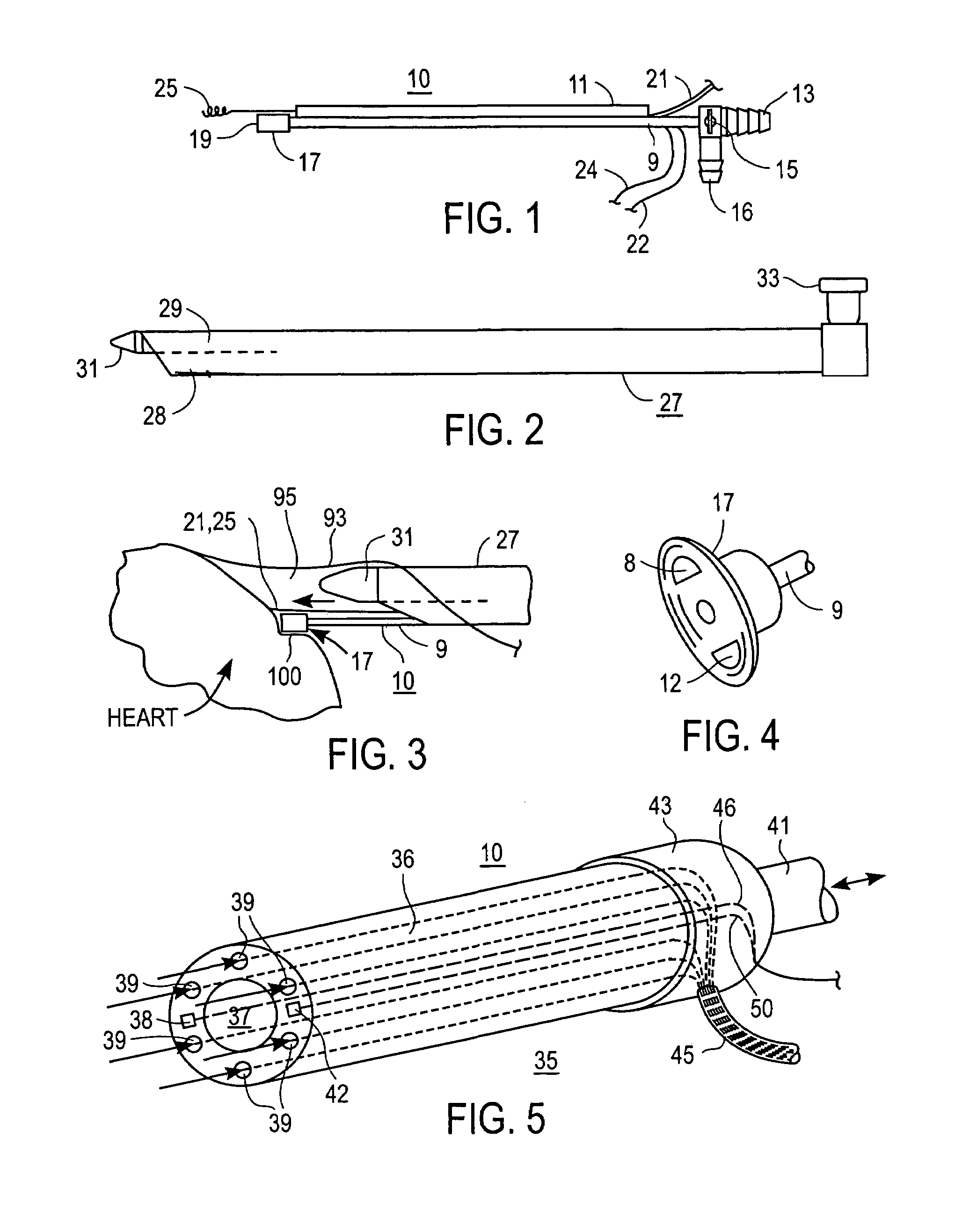
[0044]Referring now to FIG. 1, there is shown one embodiment of a suction assisted insertion cannula 10 according to the present invention including a closed channel 9 and a superior channel 11 attached to the closed channel. The closed channel 9 includes a suitable hose connection 13 and a three-way vacuum control valve 15 including an irrigation port 16 at the proximal end. A three-way valve 15 on the cannula 9 allows suction in the pod 17 to be turned on or off, and allows irrigation fluid such as saline to be injected through the suction pod 17 at the distal end while suction is turned off. The suction pod 17 includes a flexible, resilient suction cup with a porous distal face 19 or suction ports that serves as a vacuum port. The distal surface of the suction cup includes one or more surface electrodes 8, 12 as shown in FIG. 4, for contacting a surface of the heart. The surface electrodes 8, 12 carried by the suction cup 17 can be positioned against the epicardium to facilitate ...


PUM


Abstract

Apparatus and surgical methods establish temporary suction attachment to a target site on the surface of a beating heart for analyzing electrical signals or hemodynamic responses to applied signals at the target sites for enhancing the accuracy of placement of cardiac electrodes at selected sites and for enhancing accurate placement of a surgical instrument maintained in alignment with the suction attachment. A suction port on the distal end of a supporting cannula carries surface-contacting electrodes and provides suction attachment to facilitate temporary positioning of the electrodes in contact with tissue at the target site, and a clamping and release mechanism to facilitate anchoring a cardiac electrode on the moving surface of a beating heart at a selected site. Analyses of sensed signals or responses to applied signals at target sites promote epicardial mapping of a patient's heart for determining optimum sites at which to attach cardiac electrodes.

Description

RELATED APPLICATIONS[0001]This application is a continuation-in-part of application Ser. No. 10 / 174,454, entitled “Releasable Guide and Method for Endoscopic Cardiac Lead Placement” filed on Jun. 17, 2002 by A. Chin, which is a continuation-in-part of application Ser. No. 10 / 140,309, entitled “Methods And Apparatus For Endoscopic Cardiac Surgery”, filed on May 6, 2002 by A. Chin. et al, which is a continuation-in-part of application Ser. No. 09 / 635,721, entitled “Apparatus for Endoscopic Access”, filed on Aug. 9, 2000 by A. Chin, which claims the benefit of the filing of provisional application Nos. 60 / 150,737, on Aug. 25, 1999, and 60 / 148,130 on Aug. 10, 1999, each of which applications is incorporated herein in its entirety by this reference.TECHNICAL FIELD[0002]This invention relates to endoscopic cardiovascular surgical procedures and instruments, and more particularly to apparatus including a vacuum-assisted cannula and surgical instruments operable therewith, and to surgical p...

Claims


Application Information

Patent Timeline
Patent Type & Authority Patents(United States)
IPC IPC(8): A61N1/02A61B17/32A61BA61B1/01A61B17/00A61B17/06A61B17/22A61B17/30A61B17/34A61B18/00A61B18/14A61B19/00
CPCA61B17/00008A61B17/3403A61B17/3417A61B17/3421A61B19/201A61B1/00094A61B17/320068A61B17/3468A61B17/3478A61B18/1482A61B19/54A61B2017/00243A61B2017/00247A61B2017/061A61B2017/22077A61B2017/306A61B2017/308A61B2017/320044A61B2017/3445A61B2017/3488A61B2018/00291A61B2018/00392A61B2018/00982A61B2019/306A61B2019/462A61B17/0487A61B90/11A61B90/39A61B2090/036A61B2090/062
Inventor CHIN, ALBERT K.DAVIS, JOHN W.WESTLUND, RANDY W.
Owner MAQUET CARDIOVASCULAR LLC
Why Eureka
Try Eureka