

Method and apparatus for X-ray image correction
a technology of x-ray and image correction, applied in the direction of radiation beam directing means, instruments, applications, etc., can solve the problems of many problems to be solved, and achieve the effect of correcting the image of x-ray
- Summary
- Abstract
- Description
- Claims
- Application Information
AI Technical Summary
Benefits of technology
Problems solved by technology
Method used
Image

Examples
Embodiment Construction
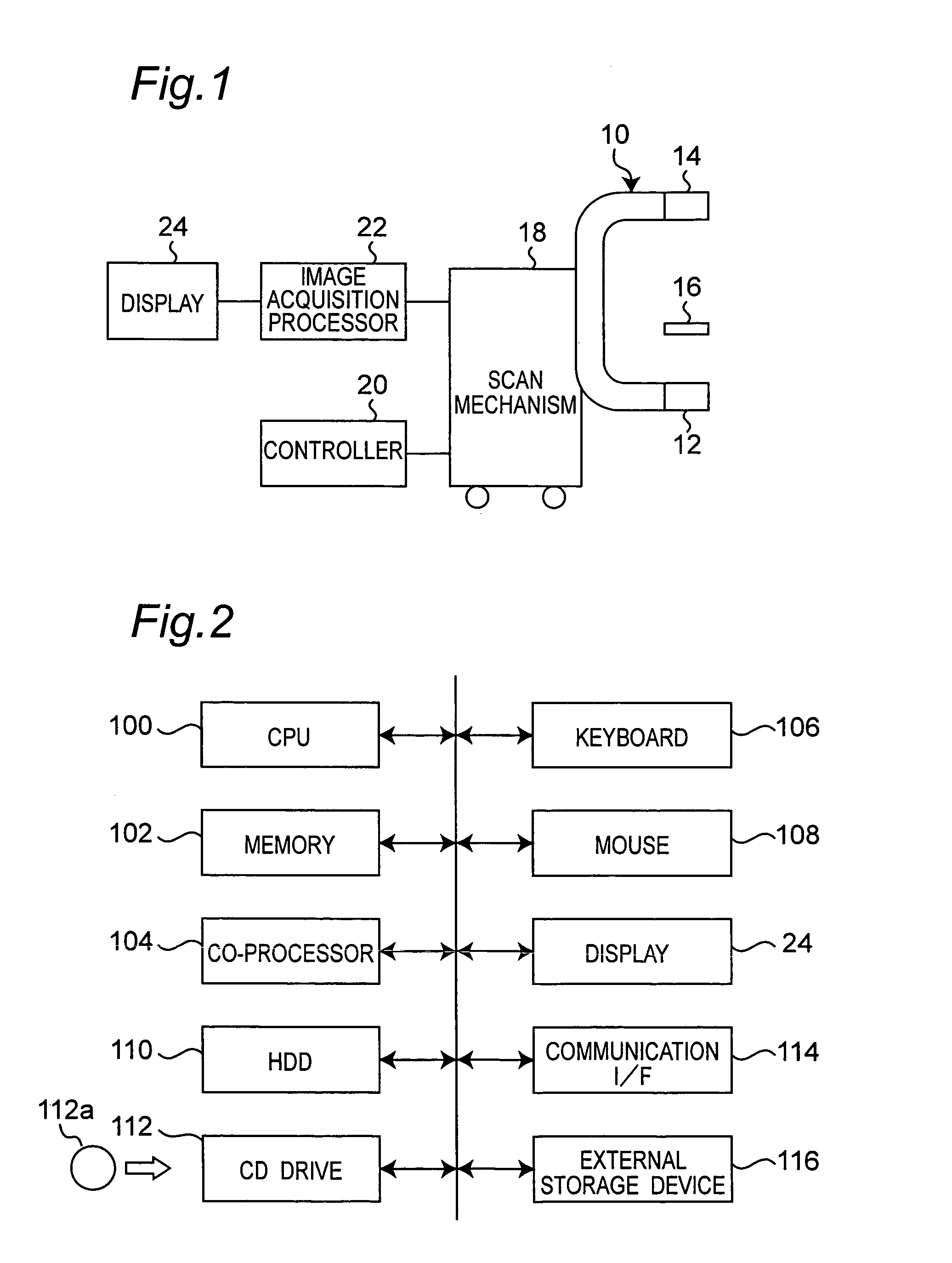
[0027]Referring now to the drawings, wherein like reference characters designate like or corresponding parts throughout the several views, FIG. 1 shows a portable X-ray CT scanner using a rotary arm 10 having a horizontal rotary axis. The rotary arm 10 has a shape similar to transversely tilted character U including two right-angle portions. In such a scanner, an X-ray source 12 is mounted at an end of the rotary arm 10, and a two-dimensional X-ray detector 14 is mounted at the other end of the rotary arm 10. The X-ray source 12 generates a cone beam of X-rays. The two-dimensional X-ray detector 14 is for example an X-ray photomultiplier. It receives incident X-rays to generate visible light which is imaged with a charge-coupled device (CCD) camera and outputs electric signals. A head rest 16 to support a person's head to be examined is positioned around a rotation center between the X-ray source 12 and the X-ray detector 14. This CT scanner is used for dentistry, and the person's h...
PUM
 Abstract
Description
Claims
Application Information