High affinity antibodies against HMGB1 and methods of use thereof

a technology of high affinity antibodies and antibodies, applied in the field of high affinity antibodies against hmgb1, can solve the problems of elevated serum hmg1 levels that are toxic, and achieve the effects of reducing bone loss and/or cartilage damag

Inactive Publication Date: 2009-09-08
MEDIMMUNE LLC
View PDF39 Cites 19 Cited by
  • Summary
  • Abstract
  • Description
  • Claims
  • Application Information

AI Technical Summary

Benefits of technology

[0044]In another embodiment of the present invention, the compositions described herein can inhibit a condition mediated or characterized by activation of an inflammatory cytokine cascade including both acute and chronic inflammatory conditions.
[0047]In still another embodiment of the present invention, the compositions described herein reduce bone loss and / or cartilage damage (by at least 10% or at least 15%, or at least 20%, or at least 30%, or at least 40%, or at least 50%, or at least 60%, or at least 70%, or at least 80%, or at least 90% ) more than a control composition in an animal arthritis model (e.g., rat AIA, mouse passive or active CIA models).
[0048]In still another embodiment of the present invention, the compositions described herein reduce bone loss and / or cartilage damage (by at least 10% or at least 15%, or at least 20%, or at least 30%, or at least 40%, or at least 50%, or at least 60%, or at least 70%, or at least 80%, or at least 90% ) more than a control composition in humans.
[0051]In yet another embodiment of the present invention, the compositions described herein are more protective (by at least 10% or at least 15%, or at least 20%, or at least 30%, or at least 40%, or at least 50%, or at least 60%, or at least 70%, or at least 80%, or at least 90% ) than a control composition in a mouse peritonitis model.

Problems solved by technology

More specifically, it is been demonstrated that bacterial endotoxin (lipopolysaccharide (LPS)) activates monocytes / macrophages to release HMG1 as a late response to activation, resulting in elevated serum HMG1 levels that are toxic.

Method used


Image

  • High affinity antibodies against HMGB1 and methods of use thereof
  • High affinity antibodies against HMGB1 and methods of use thereof
  • High affinity antibodies against HMGB1 and methods of use thereof

Examples


example 1

7.1 Example 1

Development and Physical Characterization of Human anti-HMG1 Antibodies

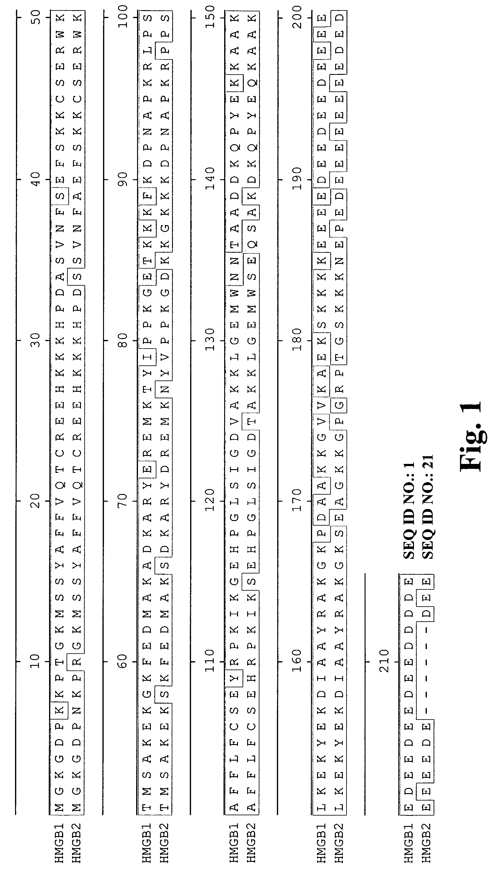
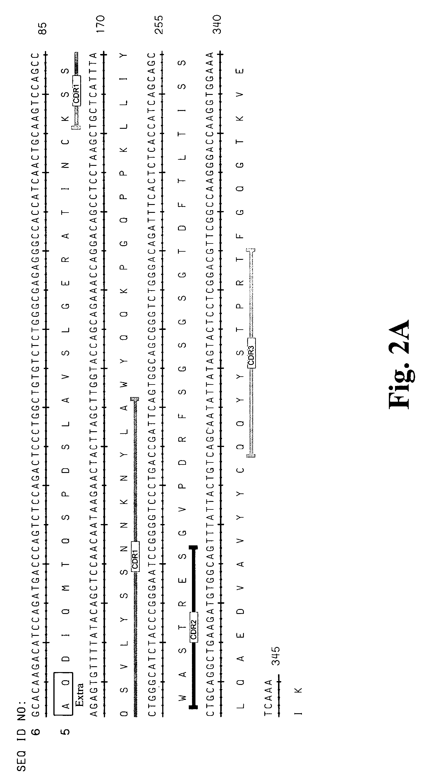
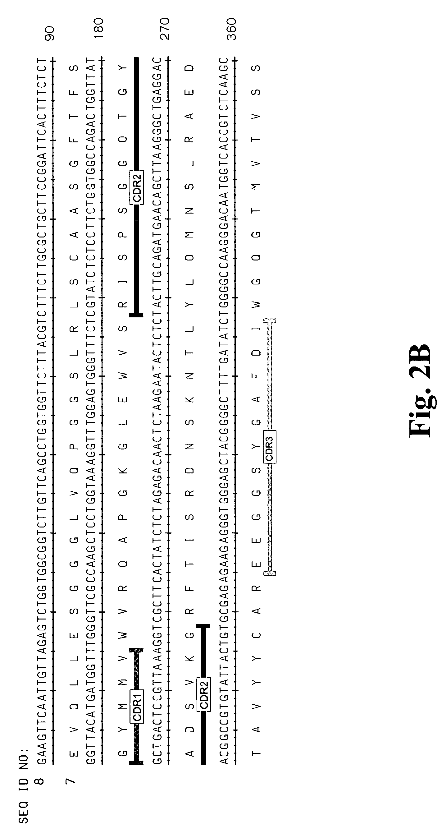
[0310]A large panel of human anti-HMG1 antibodies were isolated from a naïve human Fab phage display library by several rounds of panning against human HMG1 (SEQ ID NO: 1 and 2, also see FIG. 1). The clones were then sequenced to eliminate duplicate clones and the Fab fragments were subcloned into an expression vector for the production of full length IgG. The nucleotide and corresponding amino acid sequences of the variable regions of the light and heavy chains of several antibody clones (G2, G4, G9, G 12, G 16, G20, G34, G35, S2, S6, S10, S12, S14, S16, S17 and E11) are provided in the sequence listing (see Table 3 for specific SEQ ID NOS.). FIG. 2 represents the variable regions of the heavy and light chains of several antibody clones (S2, S6, S16 and G4) that have been deposited with the American Type Culture Collection (deposit numbers PTA-6142, PTA-6143, PTA-6259 and PTA-6258, respectively). Al...

example 2

7.2 Example 2

Kinetic Analysis of Human anti-HMG1 Antibodies

[0316]The binding kinetics and specificity of several human anti-HMG1 antibodies was examined using a variety of techniques. In addition, several peptides were used in epitope mapping studies. As summarized in Table 1, these analysis show that the human anti-HMG1 antibodies have differing binding kinetics and specificity. In addition, the data indicate that the anti-HMG1 antibodies bind to a variety of epitopes including the HMG1 B-Box and A-Box.

[0317]7.2.1 Materials and Methods

[0318]Production / Isolation of Recombinant HMG1: Recombinant HMG1 is purified from E. coli as a calmodulin binding protein (CBP) fusion protein (CBP is fused to N-terminal end of HMGB1). E. coli expressing CBP-HMG1 are induced for 2-3 hours and the protein is release by microfluidization in 25 mM Tris-HCl, 150 mM NaCl, 2 mM CaCl2, pH 8.0. The lysed cells are centrifuged at 125,000×g for 1 hour, the filtered supernatant is applied to a calmodulin column...

example 3

7.3 Example 3

Anti-HMGB1 Antibodies Inhibit Cytokine Release from Human PBMC's

[0334]The ability of a panel of human anti-HMG1 antibodies to inhibit HMG1 induced cytokine release from human peripheral blood mononuclear cells (PBMCs) was determined. The effect on the following cytokines were examined, IL-12, IL-1β, TNF-α, and IL-6. A number of anti-HMG1 antibodies are capable of inhibiting the release of one or more of these cytokines induced by HMG1. In addition, it was determined that HMG1 can stimulate the release of NO and that antibodies against HMG1 can inhibit this release. The ability of several human anti-HMG1 antibodies to reduce HMG1 induced cytokine gene expression in mouse macrophages was also demonstrated.

7.3.1 Materials and Methods

[0335]Inhibition of Cytokine Release: Human peripheral blood mononuclear cells (PBMCs) were isolated from the peripheral blood of healthy volunteers by a density gradient centrifugation. Freshly drawn heparinized whole blood was mixed with two ...


PUM

PropertyMeasurementUnit
dissociation constantaaaaaaaaaa
Tmaaaaaaaaaa

Abstract

Compositions and methods are disclosed for inhibiting the release of a proinflammatory cytokine from a vertebrate cell, and for inhibiting an inflammatory cytokine cascade in a patient. The compositions comprise, for example, high affinity antibodies that specifically bind HMG1 and antigenic fragments thereof. The high affinity antibodies of the present invention and pharmaceutical compositions comprising the same are useful for many purposes, for example, as therapeutics against a wide range of inflammatory diseases and disorders such as sepsis, rheumatoid arthritis, peritonitis, Crohn's disease, reperfusion injury, septicemia, endotoxic shock, cystic fibrosis, endocarditis, psoriasis, psoriatic arthritis, arthritis, anaphylactic shock, organ ischemia, reperfusion injury, and allograft rejection. In addition, the high affinity antibodies of the present inventions are useful as diagnostic antibodies.

Description

1. CROSS REFERENCE TO RELATED APPLICATIONS[0001]This application claims the benefit under 35 U.S.C. §119(e) of the following U.S. Provisional Application Nos.: 60 / 620,726 filed Oct. 22, 2004; 60 / 651,512 filed Feb. 10, 2005; 60 / 658,572 filed Mar. 7, 2005; 60 / 662,944 filed Mar. 18, 2005 and 60 / 713,712 filed Sep. 6, 2005. The priority applications are hereby incorporated by reference herein in their entirety for all purposes.2. BACKGROUND OF THE INVENTION[0002]Inflammation is often induced by proinflammatory cytokines, such as tumor necrosis factor (TNF), interleukin (IL)-1α, IL-1β, IL-6, platelet-activating factor (PAF), macrophage migration inhibitory factor (MIF), and other compounds. These proinflammatory cytokines are produced by several different cell types, most importantly immune cells (for example, monocytes, macrophages and neutrophils), but also non-immune cells such as fibroblasts, osteoblasts, smooth muscle cells, epithelial cells, and neurons. These proinflammatory cytoki...

Claims


Application Information

Patent Timeline
Patent Type & Authority Patents(United States)
IPC IPC(8): A61K39/395
CPCC07K16/24A61K2039/505C07K2316/96C07K2319/30C07K2317/55C07K2317/565C07K2317/92C07K2317/21C07K2317/76A61P1/04A61P17/06A61P19/02A61P29/00A61P31/04A61P37/06Y02A50/30A61K39/395C07K16/40
Inventor WU, HERRENALLAN, CHRISTIAN B.GAO, CHANGSHOUAN, LING-LINGKIENER, PETERMAO, SU-YAUCOYLE, ANTHONY
Owner MEDIMMUNE LLC
Why Eureka
Try Eureka