Core sampling biopsy device with short coupled MRI-compatible driver

a biopsy device and driver technology, applied in the field of biopsy devices, can solve the problems of heterogeneous tissue sample weight variation, and achieve the effect of reducing cutter translation speed and rotation speed

Inactive Publication Date: 2009-11-03
DEVICOR MEDICAL PROD
View PDF22 Cites 1233 Cited by
  • Summary
  • Abstract
  • Description
  • Claims
  • Application Information

AI Technical Summary

Problems solved by technology

Therefore the uncoordinated relationship between the cutter rotation speed and translation speed coupled with the inherent poor response of the pneumatically driven cutter can result in variations in the tissue sample weight when sampling heterogeneous tissue due to variations in cutter rotations through the aperture.

Method used


Image

  • Core sampling biopsy device with short coupled MRI-compatible driver
  • Core sampling biopsy device with short coupled MRI-compatible driver
  • Core sampling biopsy device with short coupled MRI-compatible driver

Examples


Embodiment Construction

Pneumatic Biopsy Device

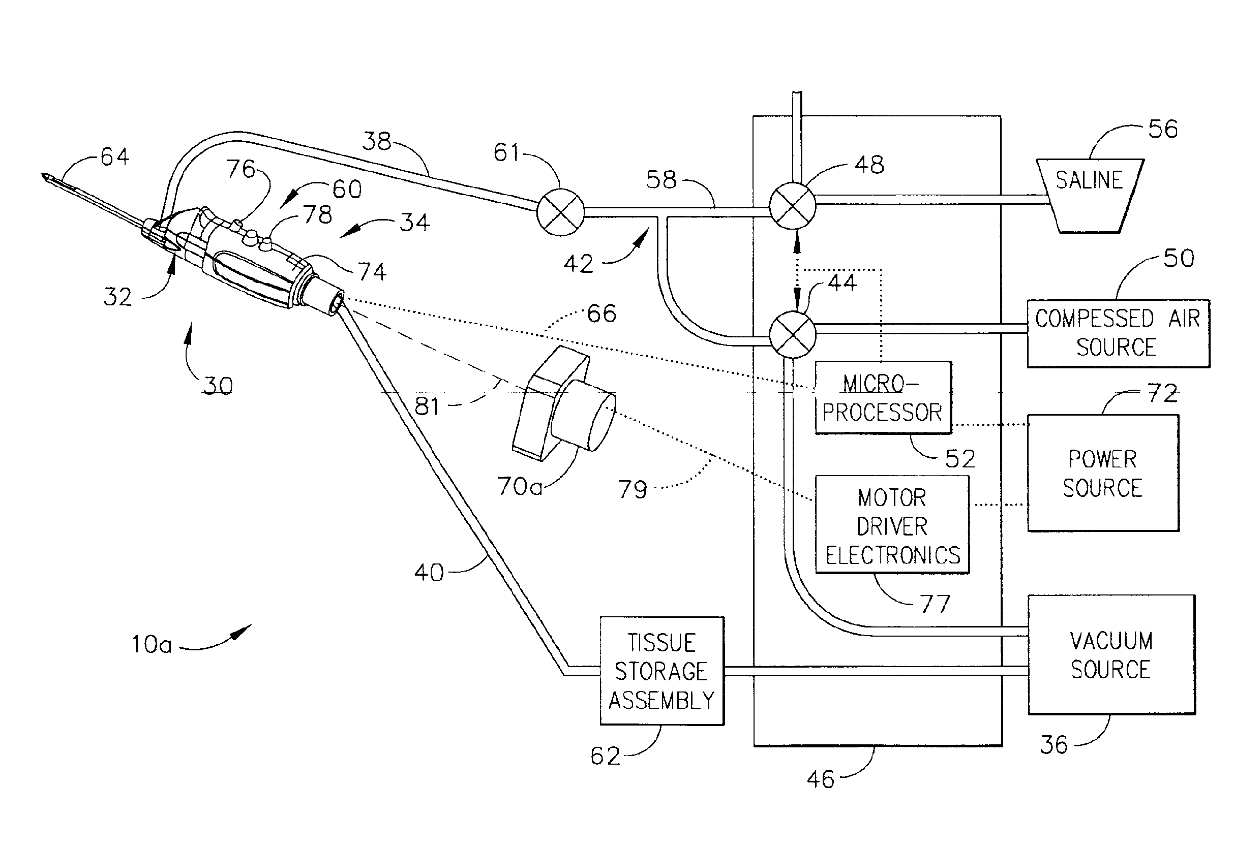
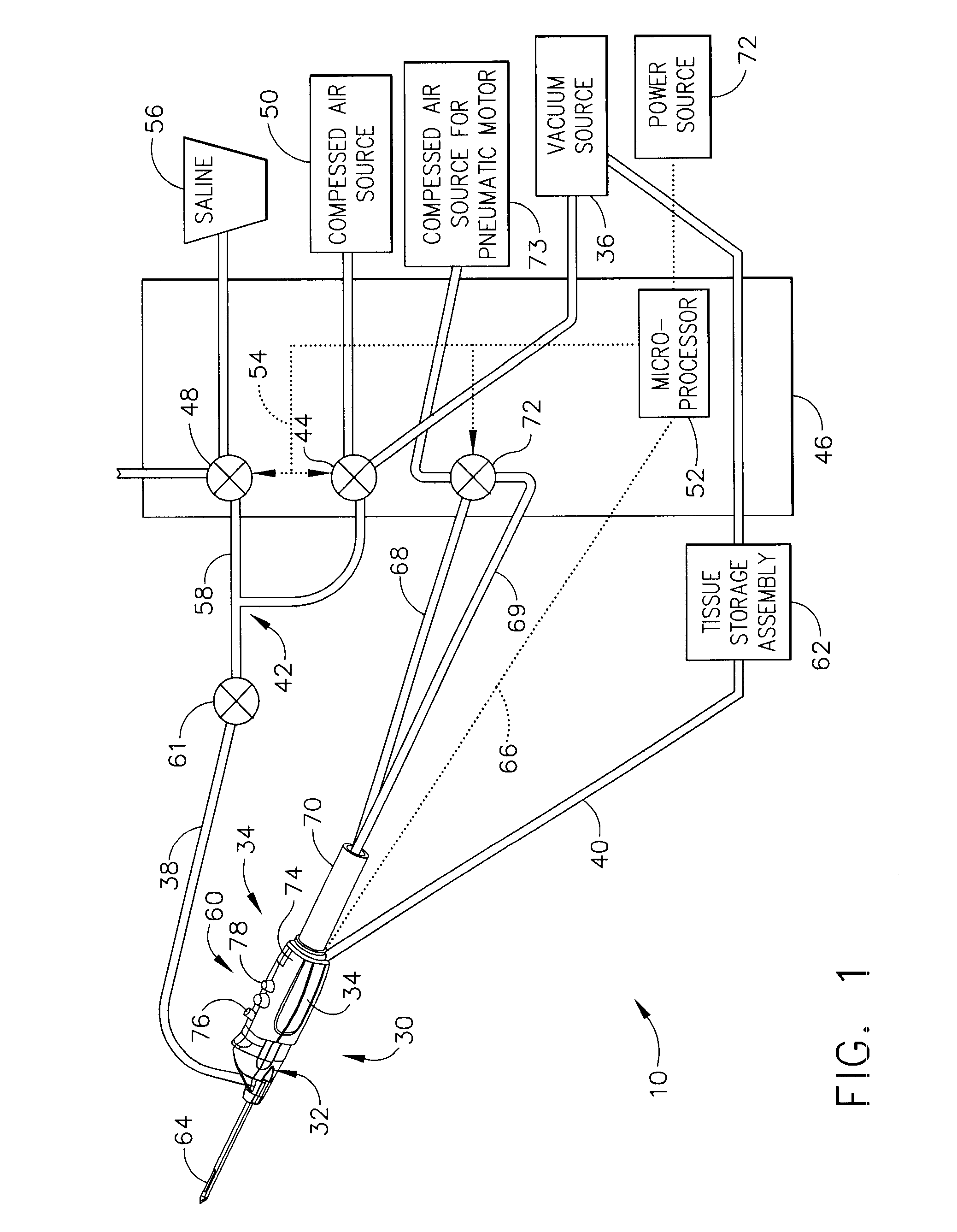
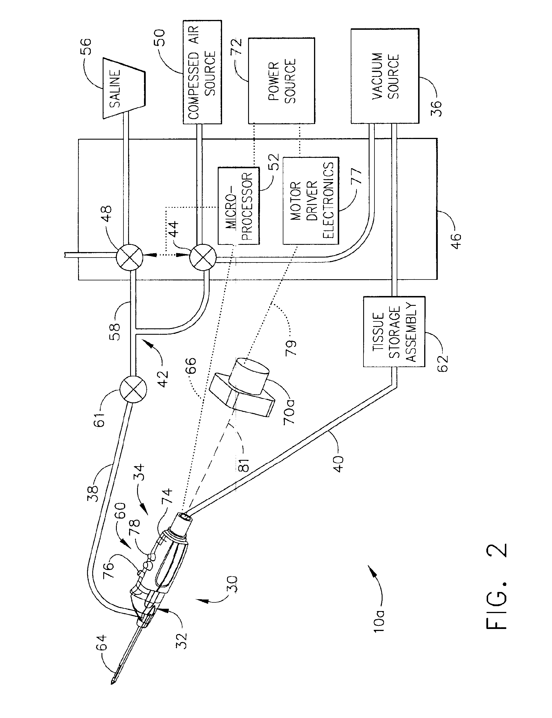
[0023]In FIG. 1, a pneumatic core sampling biopsy system 10 includes a handpiece 30 that may be held comfortably in a single hand, and may be manipulated with a single hand. Handpiece 30 may include a probe assembly 32 and a detachably connected holster 34. Probe assembly 32 may be operatively connected to a vacuum source 36, such as by a first, lateral tube 38 and a second, axial tube 40. First and second tubes 38, 40 may be made from a flexible, transparent or translucent material, such as silicon tubing, PVC tubing or polyethylene tubing. Using a transparent material enables visualization of the matter flowing through tubes 38, 40.

[0024]First tube 38 may include a Y connector 42 for connecting to multiple fluid sources. A first proximal end of Y connector 42 may extend to a first solenoid controlled rotary valve 44 in a control module 46, while the second proximal end of the Y connector 42 may extend to a second solenoid controlled rotary valve 48 in contro...


PUM


Abstract

A core sampling biopsy device is compatible with use in a Magnetic Resonance Imaging (MRI) environment by being driven by either a pneumatic rotary motor or a piezoelectric drive motor. The core sampling biopsy device obtains a tissue sample, such as a breast tissue biopsy sample, for diagnostic or therapeutic purposes. The biopsy device may include an outer cannula having a distal piercing tip, a cutter lumen, a side tissue port communicating with the cutter lumen, and at least one fluid passageway disposed distally of the side tissue port. The inner cutter may be advanced in the cutter lumen past the side tissue port to sever a tissue sample. A cutter drive assembly maintains a fixed gear ratio relationship between a cutter rotation speed and translation speed of the inner cutter regardless of the density of the tissue encountered to yield consistent sample size.

Description

FIELD OF THE INVENTION[0001]The present invention relates in general to biopsy devices, and more particularly to biopsy devices having a cutter for severing tissue.BACKGROUND OF THE INVENTION[0002]The diagnosis and treatment of patients with cancerous tumors, pre-malignant conditions, and other disorders has long been an area of intense investigation. Non-invasive methods for examining tissue include: palpation, X-ray imaging, magnetic resonance imaging (MRI), computed tomography (CT), and ultrasound imaging. When a physician suspects that tissue may contain cancerous cells, a biopsy may be done using either an open procedure or a percutaneous procedure. For an open procedure, a scalpel is used to create a large incision to provide direct visualization of and access to the tissue mass of interest. The entire mass (excisional biopsy) or a part of the mass (incisional biopsy) may then be removed. In percutaneous biopsy procedures, a needle-shaped instrument is inserted through a small...

Claims


Application Information

Patent Timeline
Patent Type & Authority Patents(United States)
IPC IPC(8): A61B10/00
CPCA61B10/0266A61B10/0283A61B2017/00544A61B2017/00539A61B2017/00535
Inventor HIBNER, JOHN A.PREDMORE, KEVIN D.BULLEN, LAWRENCECONTINI, VINCENT J.DVORSKY, JAMES E.NAVIN, ERIC R.
Owner DEVICOR MEDICAL PROD
Why Eureka
Try Eureka