Image-based indicia obfuscation system and method

a technology of indicia and masking system, applied in the field of medical imaging system and technique, can solve the problems of indicia provided in the image itself, sensitive nature of medical diagnostic images, and inability to remove or mask indicia, etc., and achieve the effect of reducing the number of indicia, reducing and improving the accuracy of indicia

Active Publication Date: 2009-12-01
GE MEDICAL SYST INFORMATION TECH
View PDF5 Cites 18 Cited by
  • Summary
  • Abstract
  • Description
  • Claims
  • Application Information

AI Technical Summary

Problems solved by technology

Medical diagnostics images are, however, sensitive in nature.
More problematic, however, are indicia that are provided in the image itself.
However, where pixilated information is encoded into the image itself, removal or masking of the indicia can be extremely time consuming and imprecise.

Method used


Image

  • Image-based indicia obfuscation system and method
  • Image-based indicia obfuscation system and method
  • Image-based indicia obfuscation system and method

Examples


Embodiment Construction

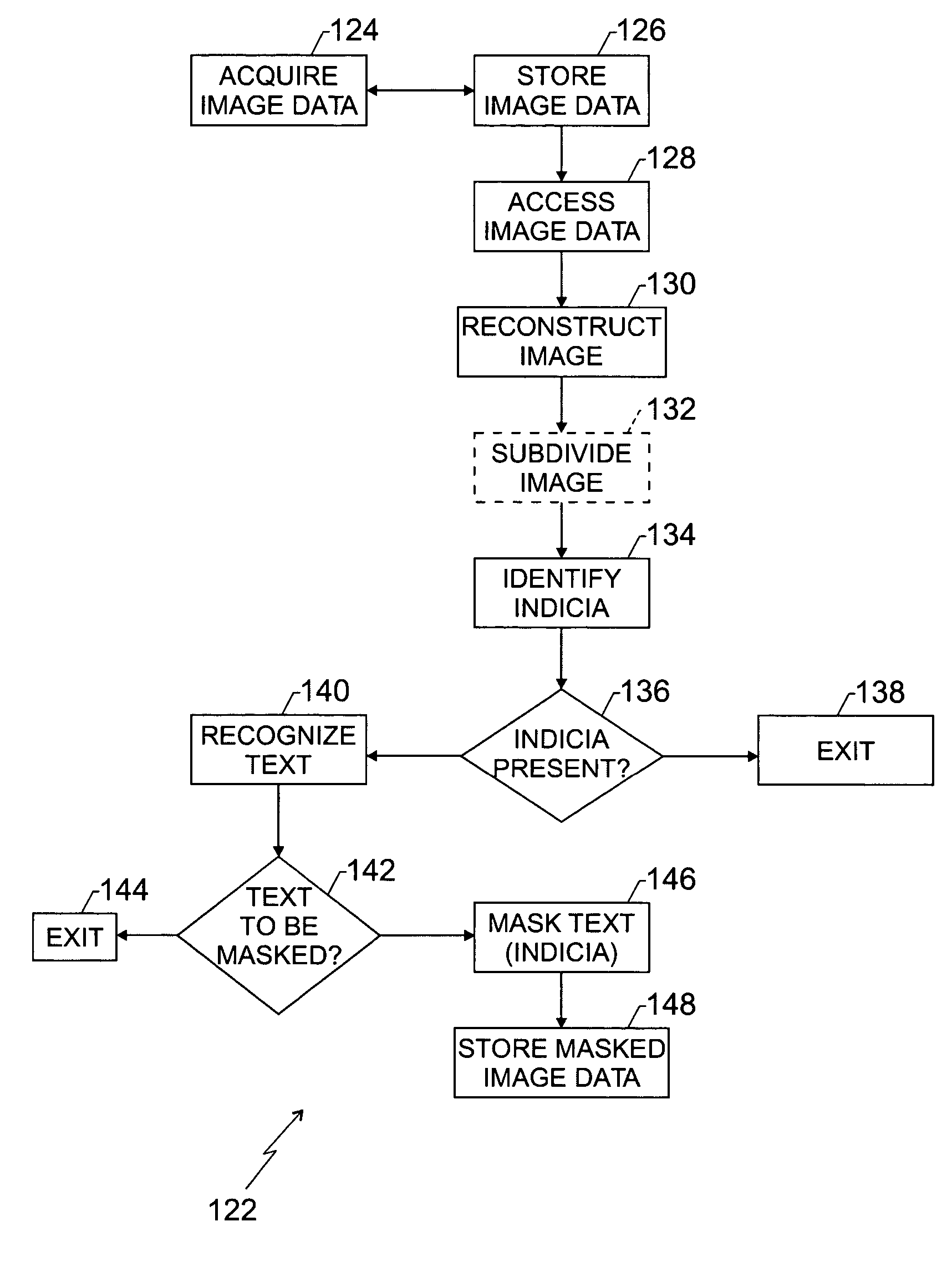
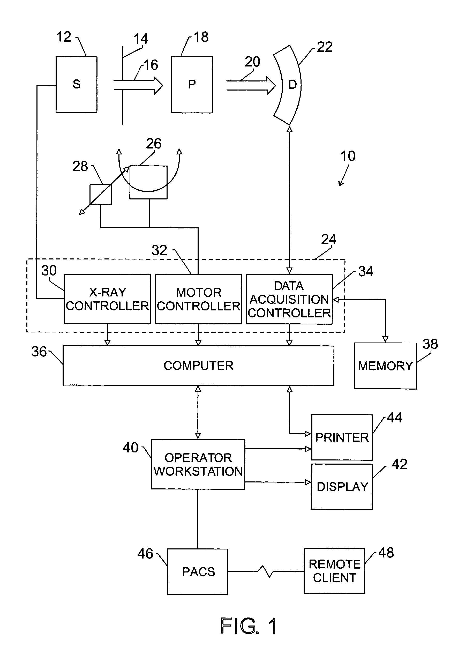
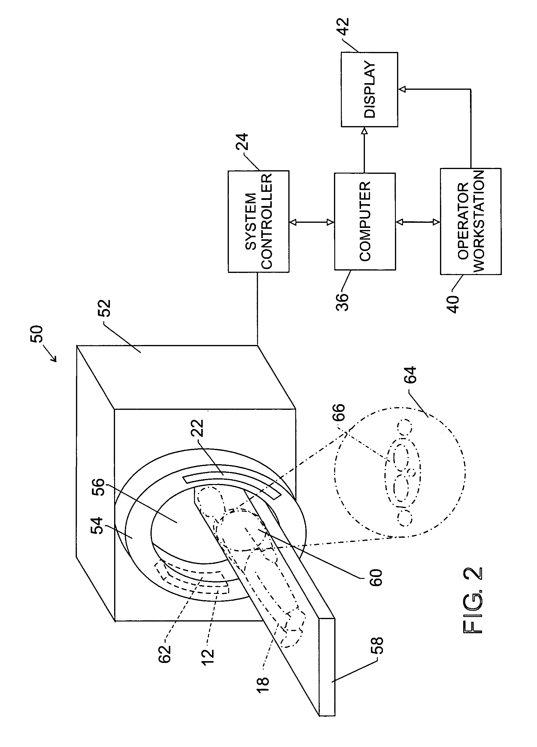
[0017]FIG. 1 illustrates diagrammatically an imaging system 10 for acquiring and processing image data for which navigational images may be generated, as described in detail below. In the illustrated embodiment, system 10 is a computed tomography (CT) system designed both to acquire original image data, and to process the image data for display and analysis while the CT system 10 is described herein as one source of image data for which navigational images may be generated, it should be borne in mind that other imaging modalities may be employed as well, such as MRI systems, X-ray systems, ultrasound systems, PET systems, and so forth.

[0018]In the embodiment illustrated in FIG. 1, imaging system 10 includes a source of X-ray radiation 12 positioned adjacent to a collimator 14. In this exemplary embodiment, the source of X-ray radiation source 12 is typically an X-ray tube. Collimator 14 permits a stream of radiation 16 to pass into a region in which a subject, such as a human patien...


PUM


Abstract

A technique is provided for obfuscating or masking indicia viewable in a reconstructed image. Image data is acquired and stored and then accessed and the reconstructed image is analyzed to determine whether any indicia are viewable in the image that are in need of masking. Such indicia may include, for example, patient-identifying data. Other information may be left unmasked, such as orientation information, descriptive information, dates, and so forth. The technique permits automated recognition of indicia and altering of image data files so as to render certain sensitive information undecipherable in the processed reconstructed images.

Description

BACKGROUND OF THE INVENTION[0001]The present invention relates generally to medical imaging systems and techniques, and more particularly, to a technique for masking or obfuscating text or other indicia in medical images on an automated basis.[0002]Many techniques are known for generating images in the medical diagnostics contexts. These include X-ray systems, computed tomography systems, magnetic resonance imaging systems, positron emission tomography systems, tomosynthesis systems, to mention just a few. In modern medical imaging, pixilated images are generated in digital formats. The pixilated images comprise rows and columns of pixels that are associated with one another in a matrix to define a useful image when reconstructed. The images typically depict anatomies and features of interest in diagnosing physical conditions, disease states, and so forth.[0003]Medical diagnostics images are, however, sensitive in nature. Many such images not only illustrate the subject's anatomy, b...

Claims


Application Information

Patent Timeline
Patent Type & Authority Patents(United States)
IPC IPC(8): G06K9/00G06F19/00G06K9/46G06T5/00G06T11/60
CPCG06T5/005G06T11/60G06F19/321G06T2207/30004G16H30/20
Inventor MATHEW, PRAKASH PARAYILSAMARA, YASEENYELURI, VIJAYKALYANLAU, DENNY WINGCHUNG
Owner GE MEDICAL SYST INFORMATION TECH
Why Eureka
Try Eureka