
Method of treating arthritis and promoting growth of articular chondrocytes

a technology of chondrocytes and chondrocytes, which is applied in the direction of skeletal/connective tissue cells, peptide/protein ingredients, immunological disorders, etc., can solve the problems of inability to achieve spontaneous recovery, low self-reproduction ability of cartilage, severe destruction of articular bone and cartilage, etc., to inhibit arthritis in osteoarthritis, induce osteoarthritis, and achieve the effect of treating osteoarthritis

Active Publication Date: 2010-01-05
KAZUWA NAKAO +1
View PDF24 Cites 35 Cited by
  • Summary
  • Abstract
  • Description
  • Claims
  • Application Information

AI Technical Summary

Benefits of technology

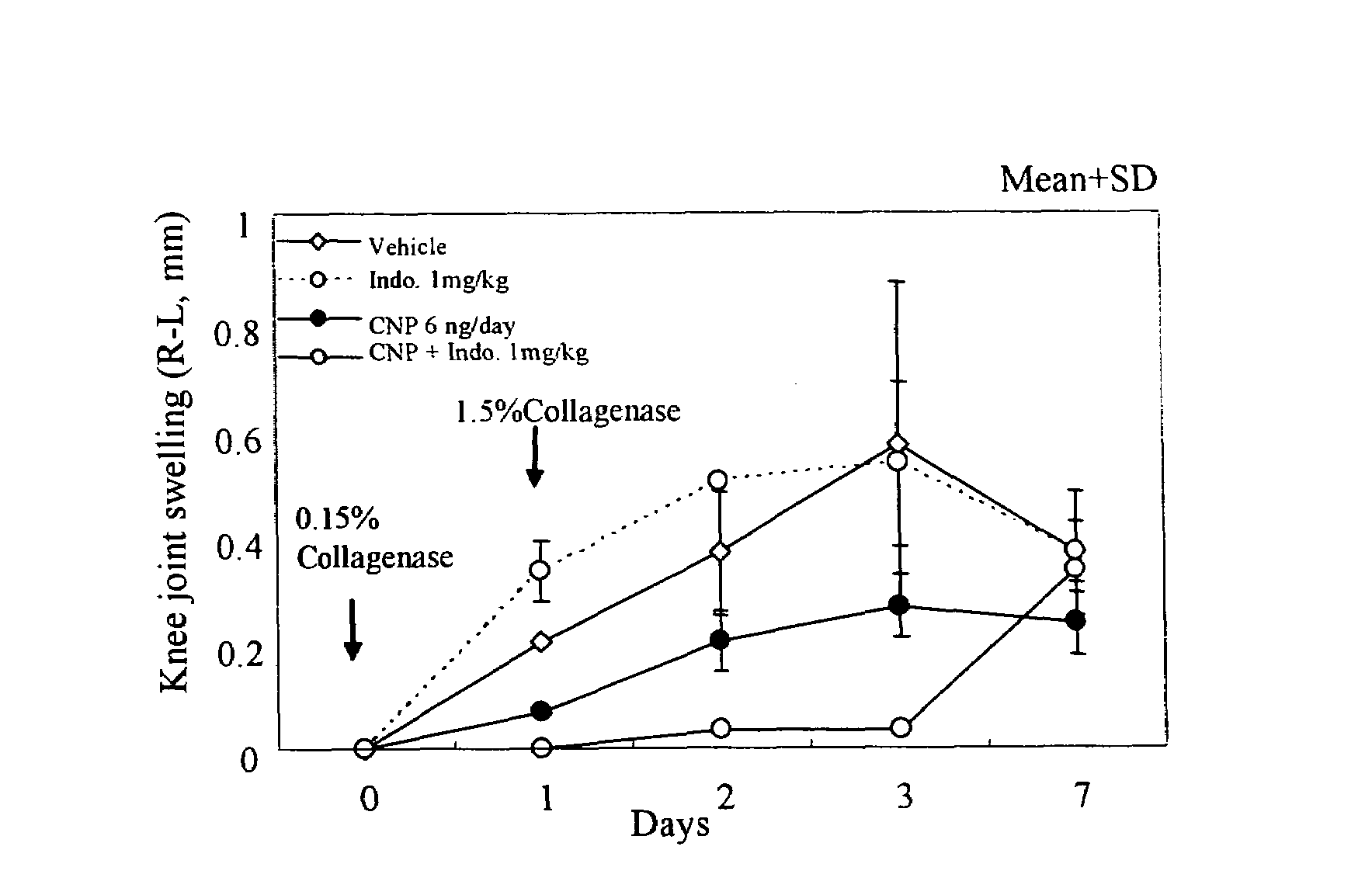
[0017]We prepared a CNP transgenic mouse that overexpresses C-type natriuretic peptide (CNP), which is a kind of guanyl cyclase B (GC-B) activator, to study the effect on the articular cartilage, and the following results were obtained: in the CNP transgenic mouse, the thickness of the articular cartilage and the number of articular chondrocytes increased significantly; in an osteoarthritic model prepared from the CNP transgenic mouse, it was resistant to articular swelling, with the degeneration of the articular cartilage reduced, there were slight changes in synovial cell growth, granulation and inflammatory cell infiltration, and the proteoglycan content in articular cartilage did not decrease, while in an osteoarthritic model prepared from a normal mouse, there were marked changes in synovial cell growth, granulation and inflammatory cell infiltration. From these findings, we have now found that the GC-B activator possesses anti-arthritis effect as well as assimilating action on the articular cartilage.

Problems solved by technology

Rheumatoid arthritis is thought to be an autoimmune disease, accompanied by articular pain, stiffening and swelling, and as the disease progresses, may often lead to the degeneration of the articular cartilage surfaces similar to osteoarthritis, resulting in severe destruction of the articular bone and cartilage.
However, due to the lack of blood vessels distributed in the cartilage tissue, the small number of chondrocytes that are highly differentiated, the small number of cartilage precursor cells, and the slow turnover of the cartilage matrix, the cartilage has too low self-reproduction ability to ensure spontaneous recovery from the decreases in articular cartilage matrix and chondrocytes in osteoarthritis (Novartis Found. Symp. 2003; 249:2-16).
Consequently, no finding has been obtained regarding the action of CNP on arthritis.
Furthermore, although the in vitro activity of CNP to enhance the hypertrophy of articular chondrocytes, which is permanent cartilage, has been reported (J Biol Chem 2003; 278(21): 18824-18832), no finding has been obtained regarding the in vivo action on the articular cartilage in normal animals, or on the degeneration of the articular cartilage or arthritis in osteoarthritis.

Method used


Image

  • Method of treating arthritis and promoting growth of articular chondrocytes
  • Method of treating arthritis and promoting growth of articular chondrocytes
  • Method of treating arthritis and promoting growth of articular chondrocytes

Examples


example 1

Construction of Vector for Preparing CNP Transgenic Mouse

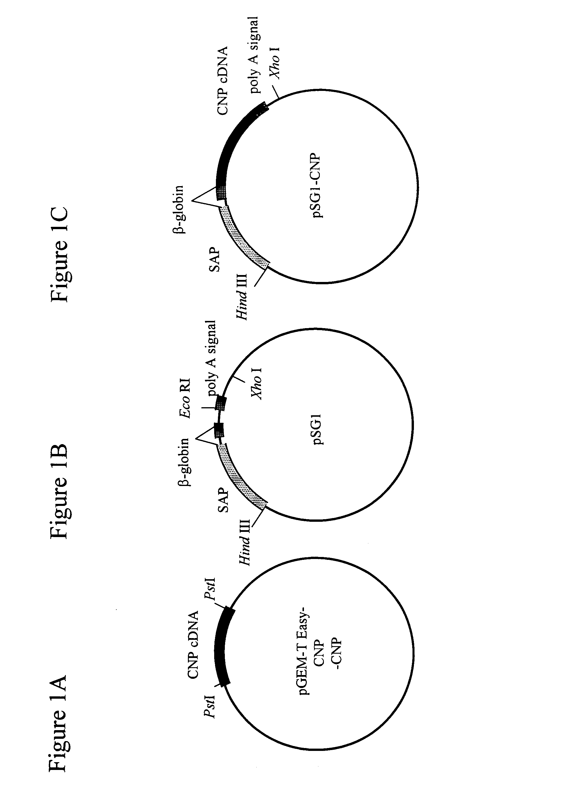
[0209]As shown in FIG. 1A, the murine CNP cDNA (526 bp; FEBS Lett. 276:209-213, 1990) was subcloned into pGEM-T easy vector (Promega), and was then cut with Pst I and blunt-ended to prepare a mouse CNP cDNA. The vector PSG 1 (Promega; FIG. 1B) was cut with EcoRI, blunt-ended and ligated with the murine CNP cDNA, as shown in FIG. 1C, to prepare a SAP-mCNP vector (pSG1-CNP).

example 2

Production of CNP Transgenic Mouse

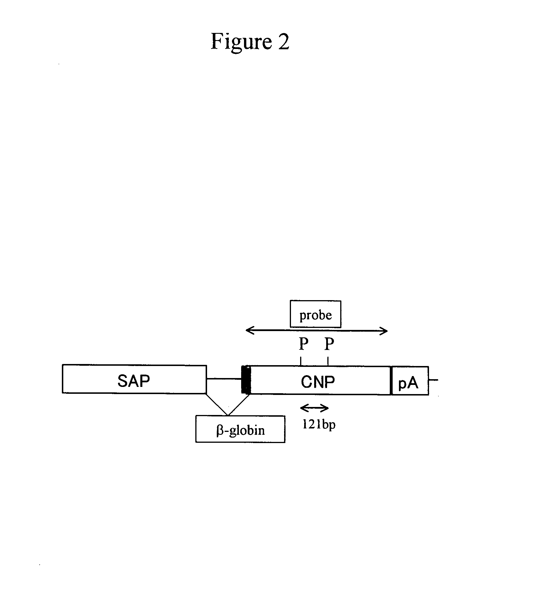
[0210]A DNA fragment for injection was prepared as follows. The SAP-mCNP vector (pSG1-CNP; FIG. 1C) with an inserted CNP gene was first treated with Hind III and Xho I to cut out a fragment (about 2.3 kb) containing the CNP gene. The fragment was then collected using Gel Extraction Kit (QIAGEN), and was diluted with PBS− at a concentration of 3 ng / μl, thereby obtaining the DNA fragment for injection (FIG. 2).

[0211]The mouse egg at pronucleus stage into which the DNA fragment was injected was collected as follows. First, a C57BL / 6 female mouse (Clea Japan, Inc.) was injected intraperitoneally with 5 i.u pregnant mare serum gonadotropin (PMSG), and 48 hours later, with 5 i.u human chorionic gonadotropin (hCG), in order to induce superovulation. This female mouse was crossed with a congeneric male mouse. In the next morning of the crossing, in the female mouse the presence of a plug was confirmed and subsequently the oviduct was perfused to collect a m...

example 3

Genotype Analysis of CNP Transgenic Mouse

[0215]Genotype analysis was performed by Southern blotting according to procedures as described below.

[0216]The tail (about 15 mm) was taken from the 3-week old mouse and treated with proteinase K (at 55° C., with shaking at 100 rpm over day and night) to obtain a lysis solution. The obtained solution was then subjected to an automated nucleic acid separator (KURABO NA-1000; Kurabo, Japan) to prepare genomic DNA. The genomic DNA (15 μg) was treated with Pvu II (200 U), then with phenol-chloroform to remove the restriction enzyme, and was precipitated with ethanol to collect the DNA. The obtained DNA was dissolved in 25 μL of TE and subjected to electrophoresis on 0.7% agarose gel (at 50V constant voltage), then the gel was treated with 0.25M HCl solution for 15 minutes to cleave the DNA, washed with water, and blotted overnight onto a nylon membrane in 0.4M NaOH solution. Thereafter, the DNA on the membrane was fixed by the UV crosslink metho...


PUM

PropertyMeasurementUnit
temperatureaaaaaaaaaa
concentrationaaaaaaaaaa
constant voltageaaaaaaaaaa

Abstract

This invention provides a new therapeutic or prophylactic agent for arthritis such as osteoarthritis. Specifically, it provides a therapeutic or prophylactic agent for arthritis such as osteoarthritis, or an agent for promoting the growth of articular chondrocyte, comprising a guanyl cyclase B (GC-B) activator as an active ingredient; or a method for inhibiting arthritis or for promoting the growth of articular chondrocyte by activating GC-B; or a method for screening an agent for promoting the growth of articular chondrocyte or an agent capable of treating arthritis using the GC-B activity as an indication.

Description

[0001]This application is the national phase of PCT International Application No. PCT / JP05 / 06831 filed on Mar. 31, 2005 under 35 U.S.C. § 371. This application also claims priority under 35 U.S.C. 119(a) of Patent Application No. 2004 / 107924 filed in Japan on Mar. 31, 2004. The entire contents of each of the above-identified applications are hereby incorporated by reference.TECHNICAL FIELD[0002]The present invention relates to a therapeutic or prophylactic agent for arthritis, particularly osteoarthritis and similar types of arthritic diseases, or an agent for promoting the growth of articular chondrocytes, or a method for inhibiting the arthritis or a method for promoting the growth of articular chondrocyte, using a guanyl cyclase B (hereinafter referred to as “GC-B”) activator. The present invention further relates to a method for screening of a therapeutic agent for arthritis or an articular chondrocyte growth promoter using GC-B activity as an indication.BACKGROUND ART[0003]Arth...

Claims


Application Information

Patent Timeline
Patent Type & Authority Patents(United States)
IPC IPC(8): A61K38/22A61K38/10C07K14/575C07K7/08A61K38/00A61K45/00A61K45/06A61P17/00A61P17/06A61P19/02A61P19/06A61P19/10A61P29/00A61P31/04A61P31/06A61P31/10A61P31/12A61P37/02A61P43/00C12N5/07C12N5/077
CPCA61K38/2242A61P17/00A61P17/06A61P19/02A61P19/06A61P19/10A61P29/00A61P31/04A61P31/06A61P31/10A61P31/12A61P37/02A61P43/00A61K38/16A61K45/06
Inventor NAKAO, KAZUWAKITAMURA, HIDETOMO
Owner KAZUWA NAKAO
Learn More