MiRNA molecules isolated from human embryonic stem cell
a human embryonic stem cell and mirna technology, applied in the field of new drugs, can solve the problems of inability to isolate mirnas, laborious and skill-intensive procedures for maintaining and expanding human embryonic stem cells, and inability to produce mirnas
- Summary
- Abstract
- Description
- Claims
- Application Information
AI Technical Summary
Benefits of technology
Problems solved by technology
Method used
Image
Examples
reference example 1
[0071]Culture of Embryonic Stem Cells
[0072]Human embryonic stem cells, SNU-hES3 (Seoul National University, Korea) and MIZ-hES1 (Mizmedi hospital, Korea) were maintained in DMEM / F12 (Gibco BRL) supplemented with 20% (v / v) serum replacements (Gibco BRL), penicillin (100 IU / ml, Gibco BRL) and streptomycin (100 μg / ml, Gibco BRL), 0.1 mM nonessential amino acids (NAA, Gibco BRL), 0.1 mM Mercaptoethanol (Sigma) and 4 ng / ml basic FGF (R&D). Media were changed daily. Human embryonic stem cell colonies were cultured on a feeder layer of mouse STO (ATCC CRL-1503) cells pre-treated with mitomycin C (Sigma) and were manually detached and transferred onto new STO feeders every 5-6 days. HS-3 mouse embryonic stem cells (Postech) were grown under standard condition (Evans and Kaufman, Nature, 292: 154-156, 1981).
reference example 2
[0073]Differentiation of Human Embryonic Stem Cells
[0074]To prepare embryoid bodies (EBs), whole colonies of human embryonic stem cells were detached by glass pipette, transferred onto petri-dishes coated with pluronic F-127 (Sigma), and incubated for 10 days. The media for EB were identical to the media for human embryonic stem cell except that it lacked bFGF. Every two days, media were changed using a pipette. To further differentiate EBs made from SNU-hES3, they were plated onto tissue culture plates coated with poly-L-ornithin (0.01% (v / v)) / fibronectin (5 g / ml (w / v)). Cells were further incubated for 5 days in N2 supplement medium containing 20 ng / ml bFGF and the medium was changed daily. Confluent cells were manually detached, and then pipetted using yellow tips and transferred onto new plates coated with poly-L-omithin / fibronectin. Cells were cultured for 5 days in N2 medium containing 20 ng / ml bFGF. When the cells reached confluency, they were trypsinized and split 2:1 or 3:1...
example 1
[0075]miRNAs Cloning from Human Embryonic Stem Cells
[0076] cDNA Library Construction from Human Embryonic Stem Cells and Culture Thereof
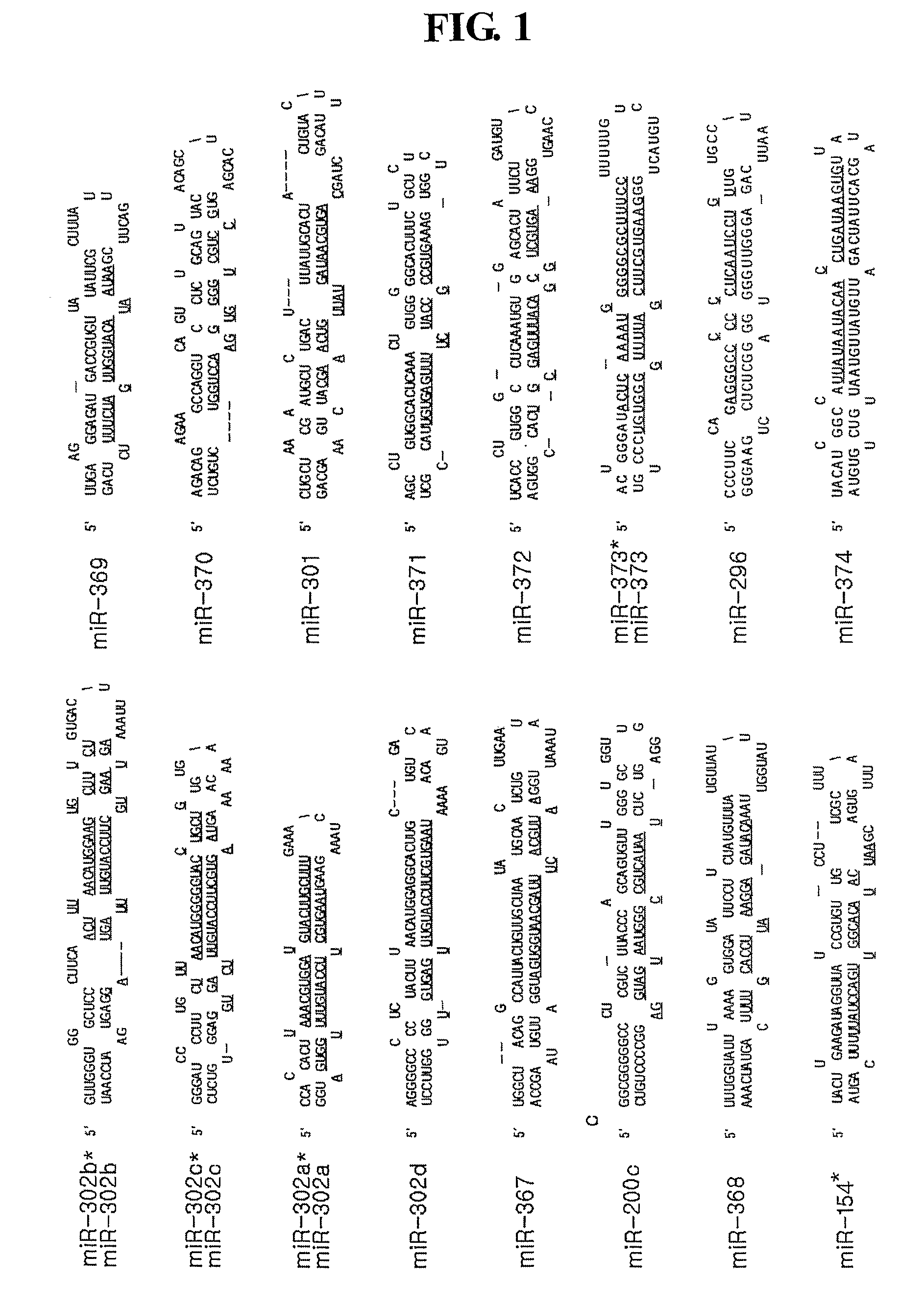
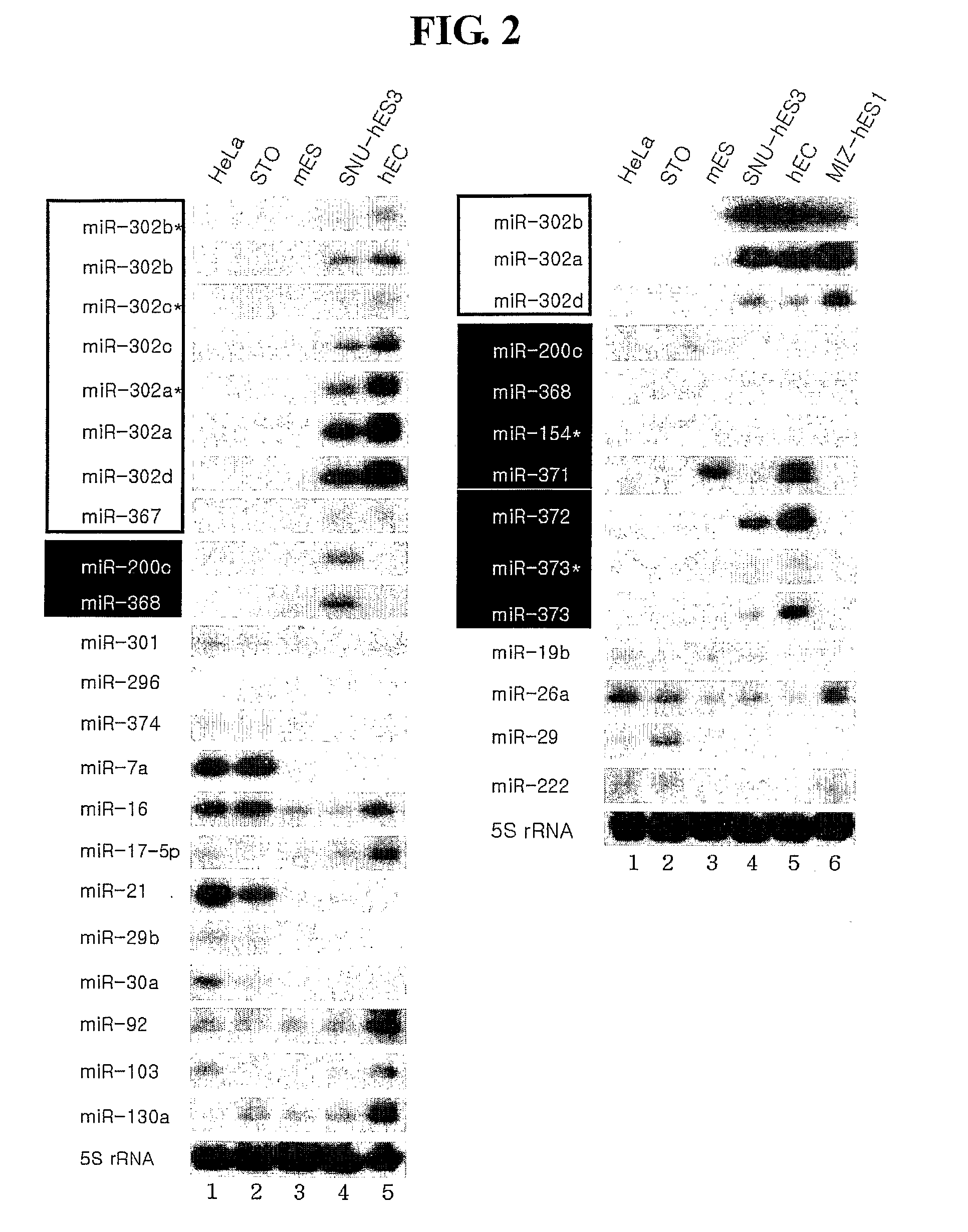
[0077]To identify miRNAs expressed in human embryonic stem cells, two independent cDNA libraries were constructed. Total RNA was prepared from each cell line with TRIzol reagent (Gibco BRL). The cDNA libraries were then constructed by directional cloning method using size fractionated RNA (17-26 nt) from undifferentiated human embryonic stem cells (SNU-hES-3, registered at the Korea Stem Cell Research Center) (Lagos-Quintana et al. Science, 294: 853-858, 2001). To validate the undifferentiating status of the human embryonic stem cell, SNU-hES3, the steady-state level of Oct4 mRNA was determined by RT-PCR. The RT-PCR was performed by following method: the first-strand cDNA from the indicated cells was synthesized with SUPERSCRIPT (Gibco BRL) using 2-5 μg of total RNA. The PCR was performed using primers of SEQ ID NO: 37 and 38. To assess the undiffer...
PUM
| Property | Measurement | Unit |
|---|---|---|
| nucleic acid | aaaaa | aaaaa |
| pharmaceutical composition | aaaaa | aaaaa |
| population-doubling time | aaaaa | aaaaa |
Abstract
Description
Claims
Application Information
 - R&D
- Intellectual Property
- Life Sciences
- Materials
- Tech Scout
- Unparalleled Data Quality
- Higher Quality Content
- 60% Fewer Hallucinations
Browse by: Latest US Patents, China's latest patents, Technical Efficacy Thesaurus, Application Domain, Technology Topic, Popular Technical Reports.
© 2025 PatSnap. All rights reserved.Legal|Privacy policy|Modern Slavery Act Transparency Statement|Sitemap|About US| Contact US: help@patsnap.com


