
Solid-state NMR method for screening cell membrane protein binding drug candidates

a cell membrane protein and solid-state nmr technology, applied in the field of drug testing, can solve the problems of affecting the tertiary structure of membrane proteins, affecting the resolution and sensitivity of nmr methods, and affecting the stability of membrane proteins,

Inactive Publication Date: 2010-03-16
FLORIDA STATE UNIV RES FOUND INC
View PDF0 Cites 1 Cited by
  • Summary
  • Abstract
  • Description
  • Claims
  • Application Information

AI Technical Summary

Benefits of technology

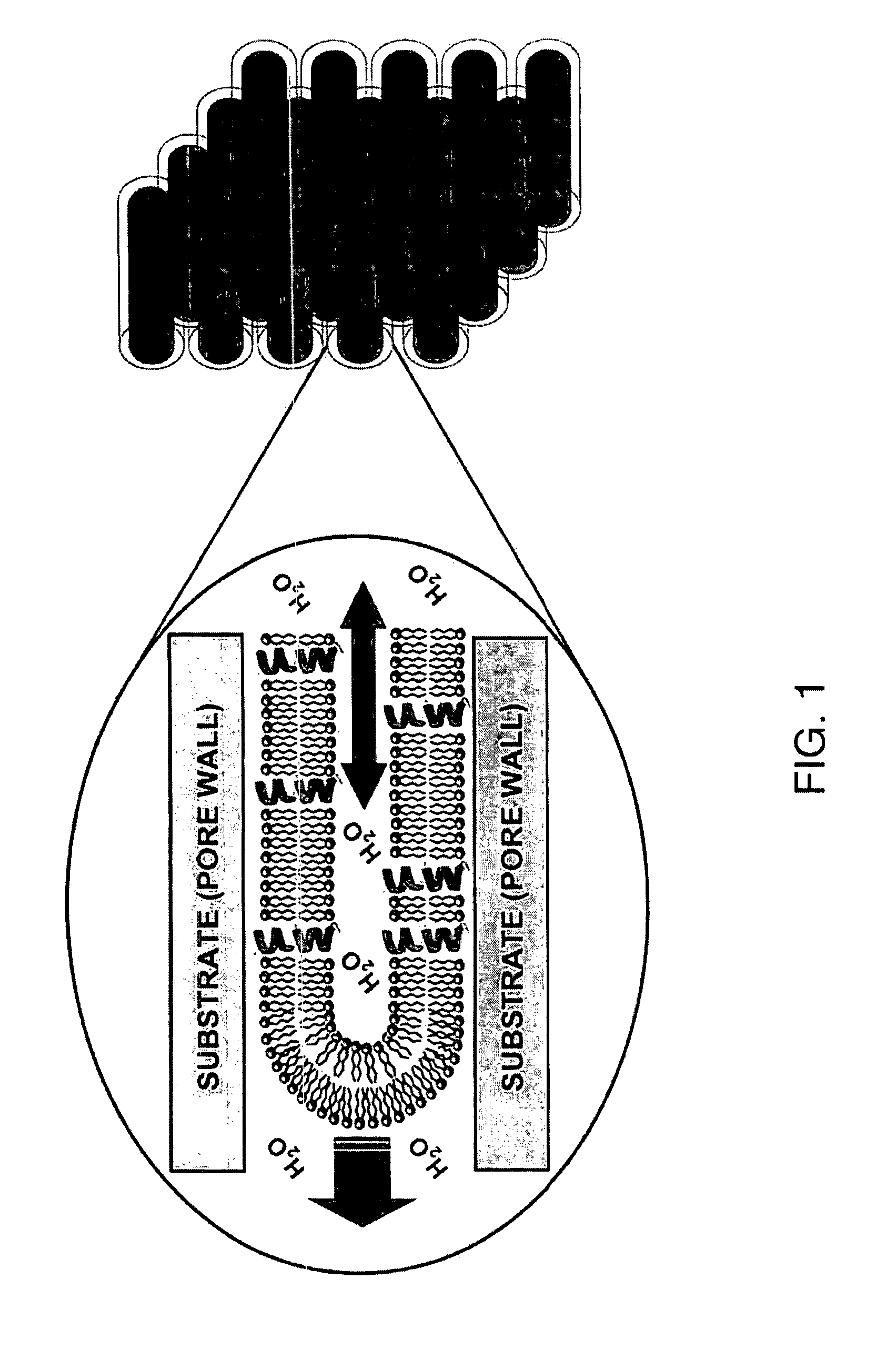
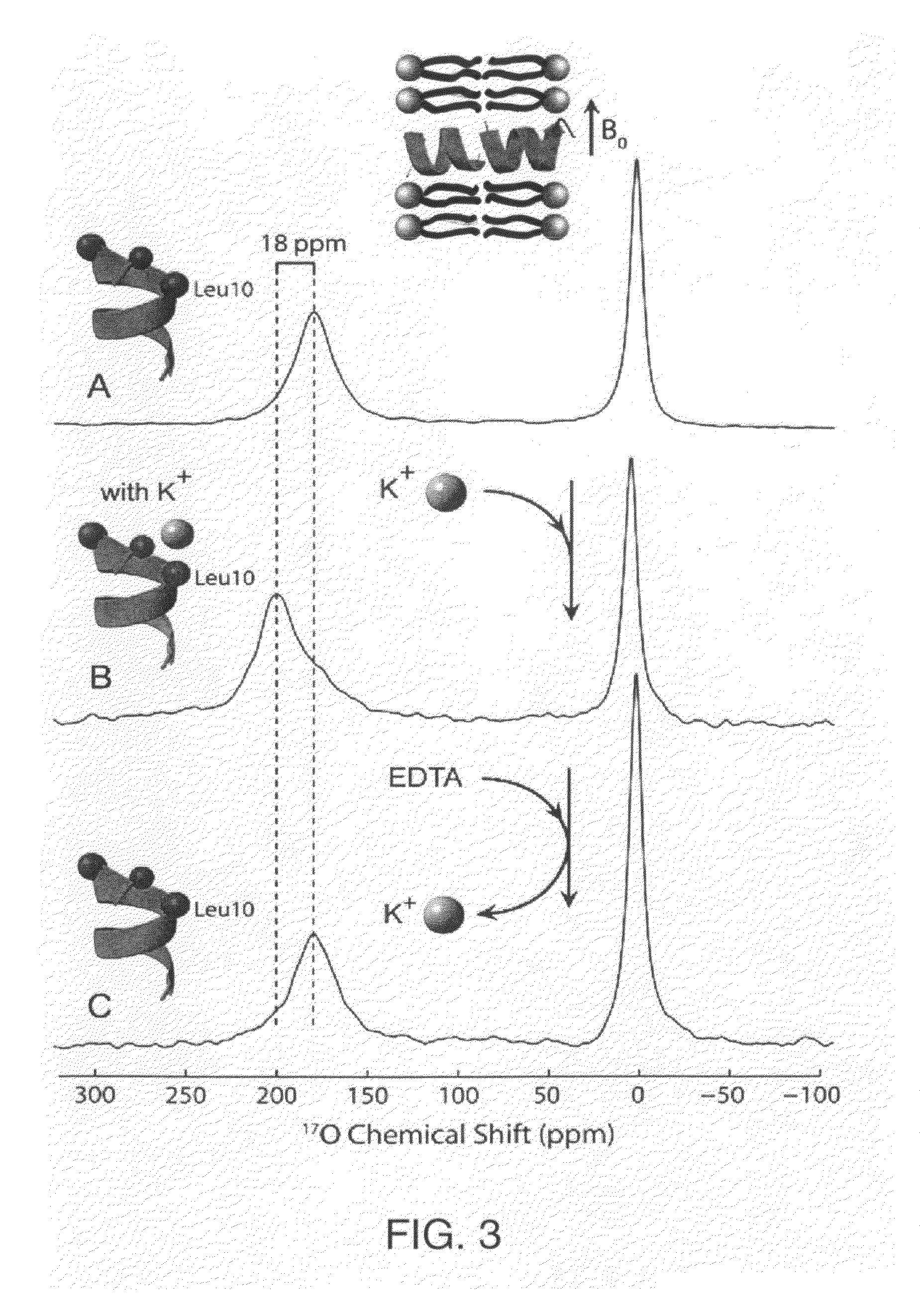
"The present invention provides a method for detecting the binding of a ligand to a cell membrane protein using solid state NMR spectroscopy. The method involves forming lipid bilayers containing the membrane protein in nanopores of an anodic aluminum oxide substrate and treating them with a candidate ligand. The treated preparation is then compared to an untreated preparation using solid-state NMR analysis. A shift in the NMR spectrum of the treated membrane protein indicates binding by the candidate ligand. This method can be used to screen drug candidates for their binding affinity to cell membrane proteins."

Problems solved by technology

Although modern high-resolution NMR spectroscopy offers unique opportunities for structure-function studies of many water-soluble proteins, membrane proteins are generally less amendable to solution NMR methods.
Molecular tumbling of proteins in lipid bilayer systems is insufficient to average out anisotropic magnetic interactions, thus causing broad NMR lines that lead to inevitable loss of resolution and sensitivity.
Unfortunately, surrounding some proteins with detergent molecules is known to destabilize functional conformational states and / or perturb the membrane protein tertiary structure (1).
However, longitudinal orientation of the protein in this spherical coordinate system defined by the bilayer normal remains undetermined, and the anisotropy resulting from observations over the longitudinal angle can still lead to line broadening.
Although several other lipid compositions for bicelles have been reported in the literature, including mixtures that maintain alignment at both acidic and basic pH values (9) and extended temperature range (10), the choice of lipid composition for anisotropic bicelles that would undergo magnetic field alignment remains limited in comparison with the mechanical (or substrate) alignment method.
One significant drawback of the substrate-alignment method derives from the somewhat limited ability to modify the sample hydration, pH, ionic strength, and concentration of molecules in the aqueous phase.
Consequently, it is virtually impossible to expose the same sample, without lengthy sample regeneration and realignment, to a variety of water-soluble agents that could be used to trigger changes in membrane protein conformation or function.
These limitations of the planar substrate-aligned lipid bilayer samples prevent many applications of high-resolution solid-state NMR methods for detailed structure-function studies of membrane proteins.

Method used


Image

  • Solid-state NMR method for screening cell membrane protein binding drug candidates
  • Solid-state NMR method for screening cell membrane protein binding drug candidates
  • Solid-state NMR method for screening cell membrane protein binding drug candidates

Examples


Embodiment Construction

[0029]The present invention will now be described more fully hereinafter with reference to the accompanying drawings, in which preferred embodiments of the invention are shown. Unless otherwise defined, all technical and scientific terms used herein have the same meaning as commonly understood by one of ordinary skill in the art to which this invention pertains. Although methods and materials similar or equivalent to those described herein can be used in the practice or testing of the present invention, suitable methods and materials are described below. Any publications, patent applications, patents, or other references mentioned herein are incorporated by reference in their entirety. In case of conflict, the present specification, including any definitions, will control. In addition, the materials, methods and examples given are illustrative in nature only and not intended to be limiting. Accordingly, this invention may, however, be embodied in many different forms and should not ...


PUM

PropertyMeasurementUnit
molar ratioaaaaaaaaaa
pore diameteraaaaaaaaaa
surface areaaaaaaaaaaa

Abstract

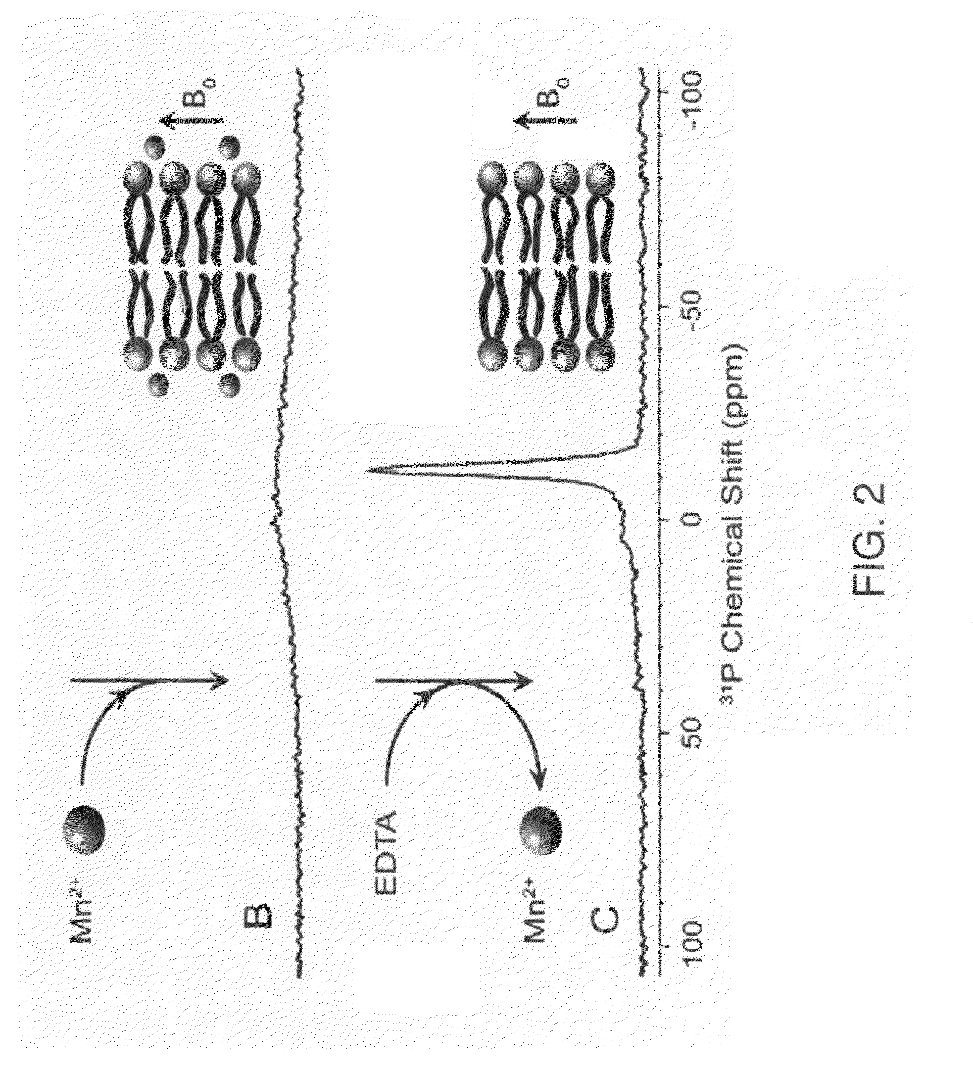
Disclosed is a method for detection of ligand-cell membrane protein binding by solid state NMR spectroscopy. The method starts by forming a lipid bilayer inside nanopores of an anodic aluminum oxide (AAO) substrate, the lipid bilayer containing a membrane protein sample. The AAO substrate is treated with multiple candidate ligands having potential binding affinity for the membrane protein. Solid-state NMR analysis is performed on the treated AAO / lipid preparation so as to generate an NMR spectrum for the treated membrane protein. The solid-state NMR spectrum of the treated membrane protein is compared with the spectrum of the same preparation of membrane protein in the absence of the ligands. It is then determined whether the solid-state NMR spectrum of the treated membrane protein has shifted from the NMR spectrum of the untreated membrane protein, a shift being indicative of protein binding by the candidate ligand.

Description

RELATED APPLICATION[0001]This application claims priority from provisional application Ser. No. 60 / 827,552, which was filed on Sep. 29, 2006, and which is incorporated herein by reference in its entirety.FIELD OF THE INVENTION[0002]The present invention relates to the field of drug testing, and, more particularly, to a method for detecting a binding interaction between a drug and a cell membrane protein target.BACKGROUND OF THE INVENTION[0003]Structure-function studies of proteins impose two major requirements on biophysical spectroscopic methods. First, to succeed in such studies, a spectroscopic method should be capable of providing atomic-scale structural data with minimal perturbation to the system. Second, the protein should be maintained in a functionally relevant conformational state. Ideally, one would like to observe detailed molecular structure while the protein progresses through conformational states associated with normal function.[0004]Although modern high-resolution N...

Claims


Application Information

Patent Timeline
Patent Type & Authority Patents(United States)
IPC IPC(8): G01N33/53
CPCG01N24/08G01N24/088G01N33/554G01N2500/10G01R33/465
Inventor CROSS, TIMOTHYBREY, WILLIAM W.SMIRNOV, ALEXEJCHEKMENEV, EDUARD Y
Owner FLORIDA STATE UNIV RES FOUND INC
Learn More