
Compensating for truncated CT images for use as attenuation maps in emission tomography

a technology of emission tomography and compensation, which is applied in the field of medical diagnostic imaging and to correction of medical images, can solve the problems of insufficient computational power, inability to use iterative techniques, and inability to achieve the effect of sufficient computational power

Inactive Publication Date: 2010-06-15
SIEMENS MEDICAL SOLUTIONS USA INC
View PDF3 Cites 20 Cited by
  • Summary
  • Abstract
  • Description
  • Claims
  • Application Information

AI Technical Summary

Problems solved by technology

However, iterative methods are generally more computationally intensive and more time-consuming than non-iterative methods.
Consequently, in the past iterative techniques were not used in clinical environment as sufficient computational power was cost prohibitive.
According to U.S. Pat. No. 5,376,795, photon attenuation constitutes a major deficiency in diagnosis of heart disease with SPECT and is a major source of error in the measurement of tumor metabolism using radionuclide techniques.

Method used


Image

  • Compensating for truncated CT images for use as attenuation maps in emission tomography
  • Compensating for truncated CT images for use as attenuation maps in emission tomography
  • Compensating for truncated CT images for use as attenuation maps in emission tomography

Examples


Embodiment Construction

[0034]As required, disclosures herein provide detailed embodiments of the present invention; however, the disclosed embodiments are merely exemplary of the invention that may be embodied in various and alternative forms. Therefore, there is no intent that specific structural and functional details should be limiting, but rather the intention is that they provide a basis for the claims and as a representative basis for teaching one skilled in the art to variously employ the present invention.

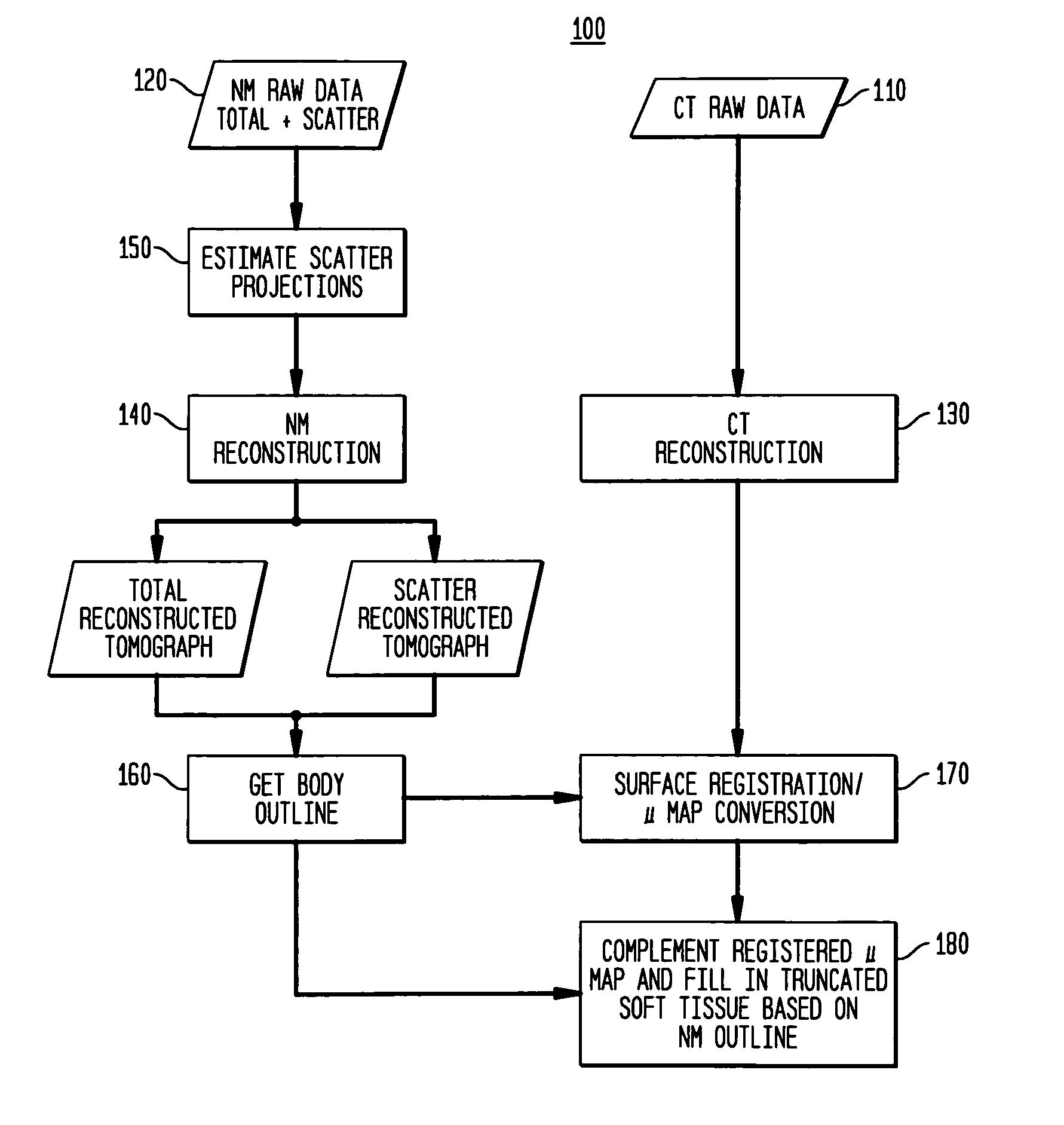
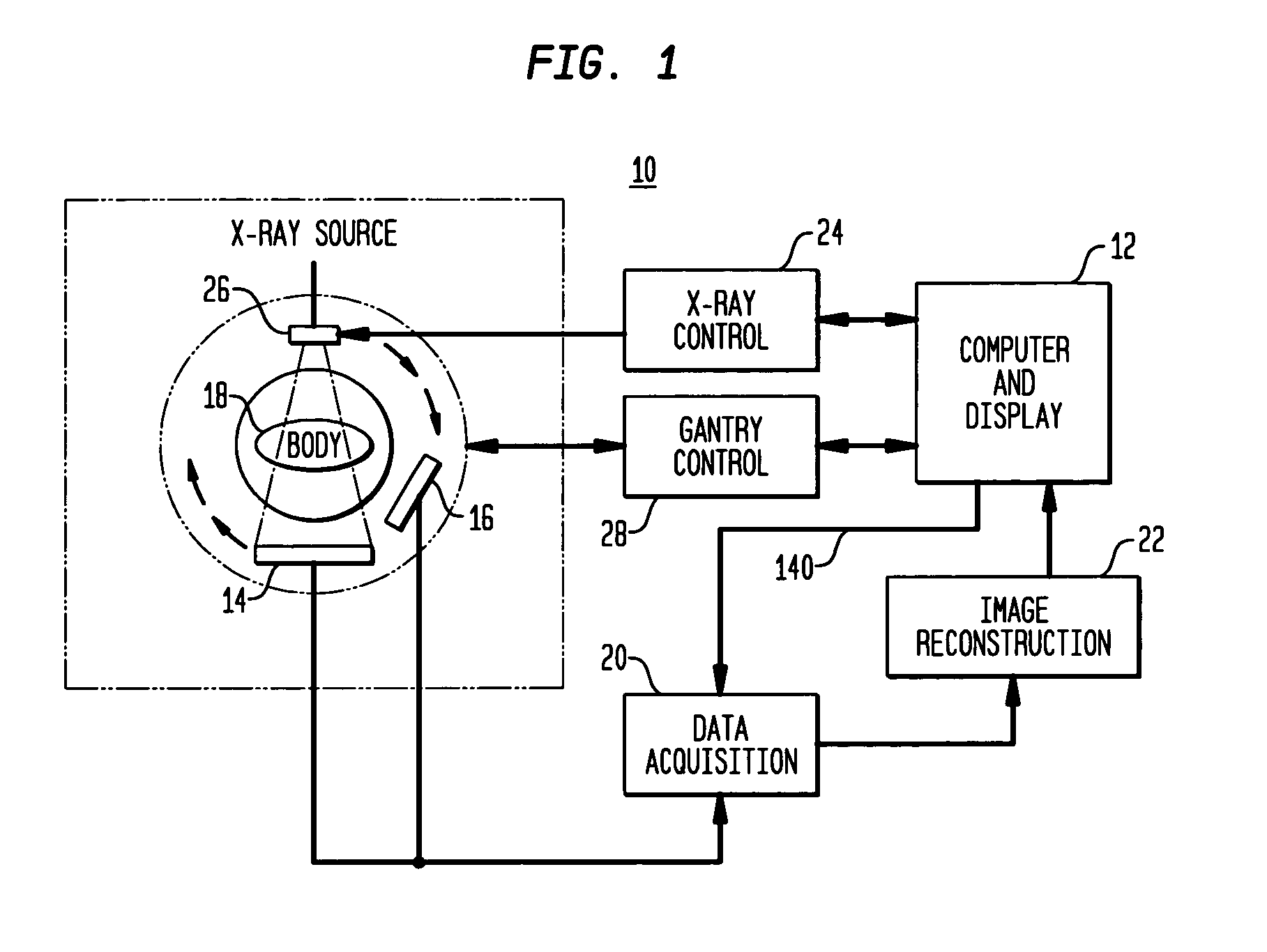
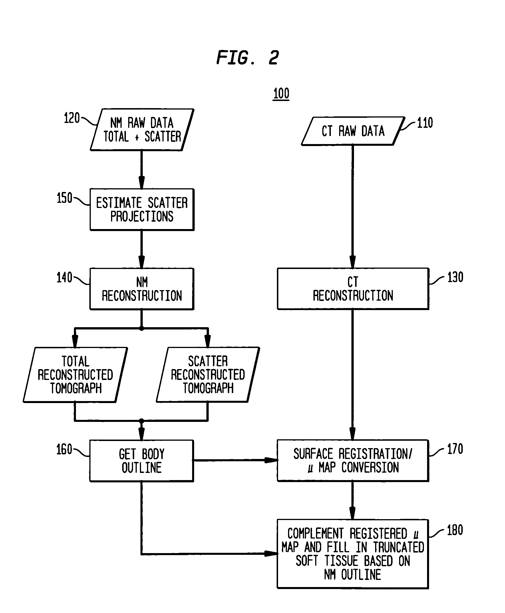
[0035]A process according to the present invention uses an imaging system as a means to reveal the presence of tumors or other defects in the organs or tissues of a patient who exhibits symptoms of an undesirable condition. The imaging system first requires that the patient adopt a position for collection of data from the organ or area of tissue under study, also referred to herein as the imaged object. Data collection proceeds using a dual modality technique that may include either simultaneous ...


PUM


Abstract

A process for obtaining an attenuation map from a truncated transmission scan of an imaged object, by compensating for missing emission data as a result of truncation by using non-truncated emission data of the imaged object to derive “fill-in” emission data. The truncation-compensated emission data then is used to generate an attenuation map for correcting a reconstructed emission image for effects of attenuation.

Description

TECHNICAL FIELD[0001]The present invention relates generally to medical diagnostic imaging and to correction of medical images from one imaging modality with image data from another imaging modality. More specifically the invention relates to application of an attenuation map, developed from X-ray computed tomography (CT) transmission data, for attenuation correction of radionuclide emission data from single-photon emission computed tomography (SPECT). In particular, the invention relates to the use of emission image data to compensate for missing data from transmission image data that is used for creating of attenuation correction or “mu” maps.BACKGROUND OF THE INVENTION[0002]In nuclear medicine imaging techniques such as single photon emission computed tomography (SPECT) and positron emission tomography (PET), medical images are regenerated based on radioactive signals, typically in the form of gamma rays, emitted from the body of a patient after the patient has been injected with...

Claims


Application Information

Patent Timeline
Patent Type & Authority Patents(United States)
IPC IPC(8): G01T1/166
CPCG01T1/1611
Inventor VIJA, A HANSREMPEL, TRUDY DAYLE
Owner SIEMENS MEDICAL SOLUTIONS USA INC
Learn More