Method for predicting immune response to neoplastic disease based on mRNA expression profile in neoplastic cells and stimulated leukocytes

a neoplastic cell and expression profile technology, applied in the field of predicting the immune response to neoplastic disease based on the expression profile of mrna in neoplastic cells and stimulating leukocytes, can solve the problem of difficult interpretation of the identification of the tnf superfamily subtype expressed in infiltrating mononuclear cells

Inactive Publication Date: 2010-06-22
RESONAC CORPORATION +1
View PDF29 Cites 16 Cited by
  • Summary
  • Abstract
  • Description
  • Claims
  • Application Information

AI Technical Summary

Benefits of technology

[0013]If a pathological specimen shows IgG deposition around cancer margins, this means that the patient is at least capable of raising cancer-recognizing IgG. If the infiltration of mononuclear leukocytes exists at the margin of the cancer mass, this means that the patient's leukocytes are somehow attracted toward the cancer lesion, probably due to the release of chemotactic factors (C3a, C5a, C567) (see Becker, The relationship of the chemotactic behavior of the complement-derived factors, C3a, C5a, and C567, and a bacterial chemotactic factor to their ability to activate the proesterase 1 of rabbit polymorphonuclear leukocytes, J. Exp. Med. 1972; 135:376-87) during complement activation of cancer-bound IgG. The type of TNF-R superfamily subtypes expressed on the surface of cancer cells can be identified by, for example, immunostaining (if antibodies are available), in-situ hybridization, or in-situ PCR (sequences are available from GenBank). Identification of the TNF superfamily subtypes expressed in infiltrating mononuclear cells is difficult to interpret, because FcγR may not be activated yet. Since leukocytes in peripheral blood are like reserve police officers, a supply of appropriately and fully equipped cytotoxic T-cells in peripheral blood is a critical first step for the battle against cancer. Thus, this method makes it possible to predict anti-cancer immunity in each patient by quantitating the inducibility of the appropriate TNF superfamily subtypes in peripheral blood leukocytes.

Problems solved by technology

Identification of the TNF superfamily subtypes expressed in infiltrating mononuclear cells is difficult to interpret, because FcγR may not be activated yet.

Method used


Image

  • Method for predicting immune response to neoplastic disease based on mRNA expression profile in neoplastic cells and stimulated leukocytes
  • Method for predicting immune response to neoplastic disease based on mRNA expression profile in neoplastic cells and stimulated leukocytes
  • Method for predicting immune response to neoplastic disease based on mRNA expression profile in neoplastic cells and stimulated leukocytes

Examples


embodiment 1

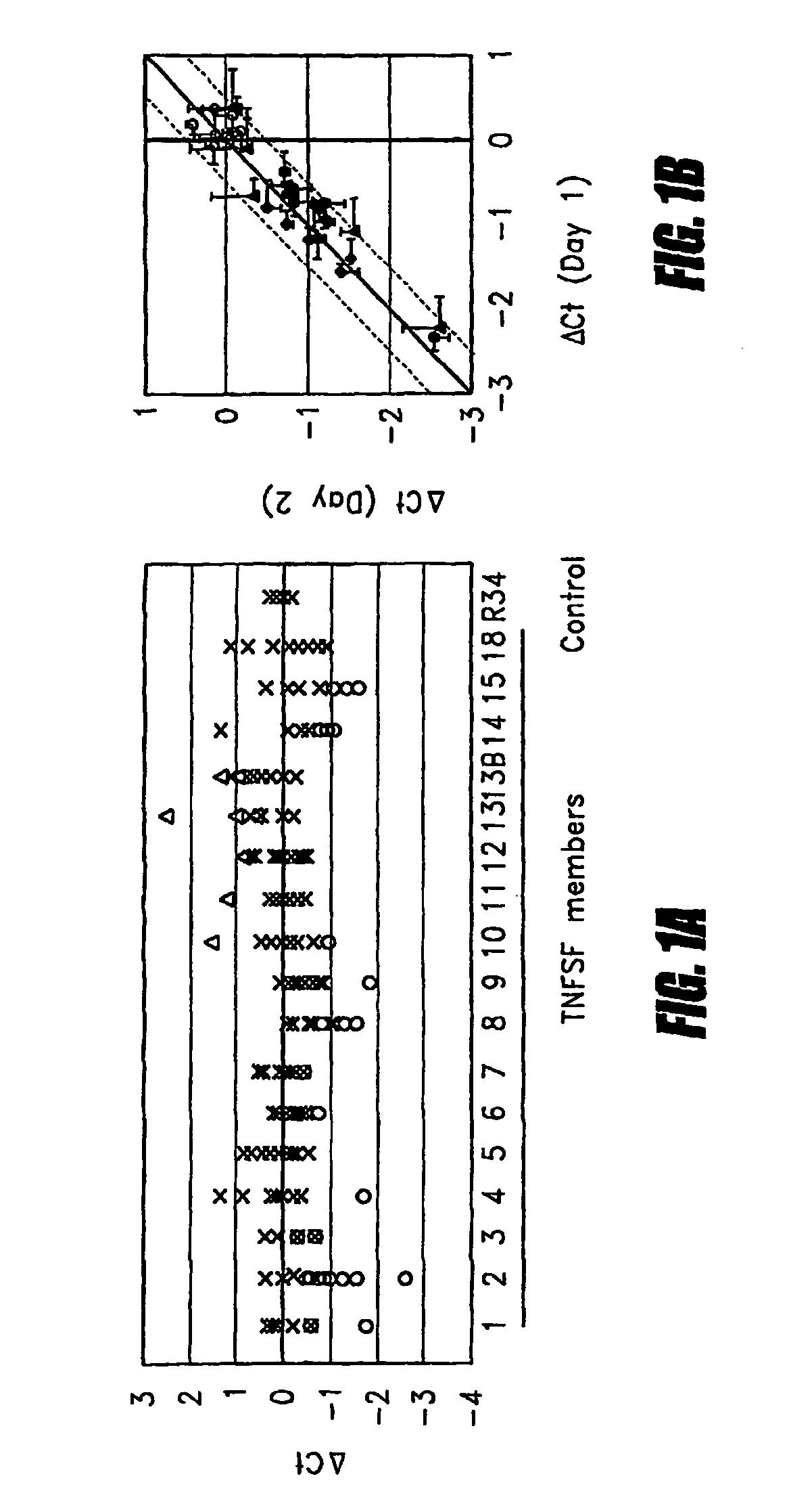
[0039]The mRNA quantitation method described above was employed to quantitate all types of TNF superfamily mRNA, both with and without ex vivo stimulation using heat-aggregated IgG (HAG), which was employed as a model of immune complex stimulation (see Ostreiko et al., Production and characterization of heat-aggregated IgG complexes with pre-determined molecular masses: light-scattering study, Immunol Lett. 1987; 15:311-6). The immune complex is formed when IgG molecules bound to a target cell bind with high avidity to Fc receptors on a leukocyte, causing cross-linking of the Fc receptors and generation of a leukocyte activation signal.

[0040]Triplicate aliquots of 50 μL each of heparinized whole blood was incubated with 200 μg / mL HAG at 37° C. for 2 hours. Then blood samples were applied to 96-well filterplates to trap leukocytes, and cell lysates were transferred to 96-well oligo(dT)-immobilized microplates for isolation of poly(A)+ mRNA as described above. cDNA was synthesized on ...

embodiment 2

[0044]DNA sequences of TNF-R superfamily and TNF superfamily mRNA were downloaded from GenBank, as shown in Table 1, and the primer sequences shown in Table 2 below were designed by Primer Express (Applied Biosystems) and HYBsimulator (RNAture). These sequences were designed at common regions among multiple transcripts if such variants exist. No amplification happened without cDNA (no primer dimer), and a single peak was detected at melting curve analysis.

[0045]

TABLE 2Primer sequences usedTarget mRNAForwardReverseTNFRSF-1ACCTGCCAGGAGAAACAGAACAGGAGACACACTCGTTTTCTCTTAGAATNFRSF-1BCAAGCCAGCTCCACAATGGTGACCGAAAGGCACATTCCTTNFRSF-3CCTCCCGGGCTCTCTACACTCATGGGTGATAAATTGGTTCCTTNFRSF-4ACGACGTGGTCAGCTCCAAGCGGCAGACTGTGTCCTGTGTTNFRSF-5GGCCAAGAAGCCAACCAATAGAAGATCGTCGGGAAAATTGATTNFRSF-6TGGCATCAACTTCATGGAAAGAGCAAGAGTACAAAGATTGGCTTTTTTNFRSF-7CTGCAGAGCCTTGTCGTTACAGGCTCCGGTTTTCGGTAATCCTNFRSF-8GGTTGAGGCAGCAAACAGATGGCCTGGTGGTTAAGGTCTGATGTNFRSF-9CGTCGACTGCGTTGCTCTTTTCTGCCCCGTTAACAACAGTNFRSF-10ATGAGGACAATGCT...

embodiment 3

[0053]In this embodiment, as in Embodiment 1, heat-aggregated human IgG (HAG), which is used widely as a model of the immune complex as found in ADCC, was used to stimulate the Fc receptors of leukocytes in whole blood. In brief, human IgG (Sigma) was suspended in PBS at 20 mg / mL and heated at 63° C. for 20 min. Sixty μL of whole blood was stimulated at 37° C. for only 2-4 hours in triplicate with 200 μg / mL HAG or control phosphate-buffered saline (PBS). The dose of HAG and incubation time were determined by preliminary analysis, as shown in FIGS. 5 and 6.

[0054]FIG. 5A shows the results of an analysis of the dose response. Triplicate aliquots of 60 μL each of heparinized whole blood were mixed with various concentrations of human IgG (∘), or HAG (•) and incubated at 37° C. for 2 hours. FIG. 5B shows the results of an analysis of kinetics. Triplicate aliquots of 60 μL each of heparinized whole blood was mixed with PBS (∘), or 200 mg / mL HAG (•) and incubated at 37° C. for 0-12 hours. ...


PUM

PropertyMeasurementUnit
pHaaaaaaaaaa
pHaaaaaaaaaa
volumeaaaaaaaaaa

Abstract

Tumor necrosis factor (TNF) is capable of inducing apoptosis by interacting with specific TNF receptors on the surface of cancer cells. Because multiple members of TNF ligand and receptor are present within each superfamily, over 300 different ligand-receptor combinations exist. Activated blood leukocytes produce TNF as part of the immune response to cancer, as well as producing chemokines to attract other leukocytes to the site. A method is disclosed of detecting significant induction of a variety of TNF superfamily subtype and chemokine mRNAs in blood leukocytes when whole blood is exposed to heat-aggregated IgG or anti-T cell receptor antibodies as a model of immune system interactions. Substantial individual-to-individual variation is observed in TNF subtypes and chemokines induced. Since peripheral blood leukocytes are the supply of anti-cancer immune cells, the quantitation of ex vivo inducibility of appropriate TNF ligands and chemokines in blood will be useful in individualized cancer immunotherapy. If the tumor mass is small, such as with early invisible metastatic lesions, appropriate TNF assaults may be sufficient to prevent relapse.

Description

RELATED APPLICATIONS[0001]This application claims priority to U.S. Provisional Application Nos. 60 / 688,744 filed on Jun. 8, 2005 and 60 / 735,508 filed on Nov. 11, 2005.BACKGROUND OF THE INVENTION[0002]1. Field of the Invention[0003]The present invention relates to a method for predicting the immune response in a mammal to neoplastic disease based on the expression profile of tumor necrosis factor receptor (TNF-R) superfamily mRNA in neoplastic tissue, and of tumor necrosis factor (TNF) superfamily mRNA in circulating leukocytes. In the method, samples of neoplastic tissue are obtained and the TNF-R superfamily subtype mRNAs expressed in those tissues are assessed. Furthermore, whole blood of the mammal is subjected to a stimulus that activates T-cells in the blood and the TNF superfamily subtype mRNAs that exhibit a significant change in expression level in response to the stimulus are identified. Individuals that exhibit a change in expression level in TNF superfamily subtypes that ...

Claims


Application Information

Patent Timeline
Patent Type & Authority Patents(United States)
IPC IPC(8): C12Q1/68G01N33/48G01N33/574
CPCC12Q1/6886G01N33/574G01N33/505C12Q1/6806C12Q2600/158C12Q2600/118
Inventor MITSUHASHI, MASATO
Owner RESONAC CORPORATION
Why Eureka
Try Eureka