Tissue sample serial capturing biopsy device

a biopsy device and tissue sample technology, applied in the field of biopsy devices, can solve the problems of high cost and high level of trauma to the patient, open biopsy carries a relatively higher risk of infection and bleeding, and is difficult to read future mammograms

Inactive Publication Date: 2010-07-13
DEVICOR MEDICAL PROD
View PDF46 Cites 57 Cited by
  • Summary
  • Abstract
  • Description
  • Claims
  • Application Information

AI Technical Summary

Benefits of technology

[0011]The present invention addresses these and other problems of the prior art by providing a biopsy device that has a probe cannula that is inserted into tissue to obtain a core biopsy sample by translating a cutter with the probe cannula. A pneumatic pressure differential is used to draw a severed tissue sample proximally from the probe cannula into a proximal stacking tube that provides confirmation of successful sample collection, preserves the sequential order of the samples, allows multiple samples to be taken without reinserting the probe cannula, provides a convenient holder for on-site scans for calcifications, and avoids a biohazard as the samples are taken to a pathology assessment.

Problems solved by technology

An open biopsy is a surgical procedure that is usually done as an outpatient procedure in a hospital or a surgical center, involving both high cost and a high level of trauma to the patient.
Open biopsy carries a relatively higher risk of infection and bleeding than does percutaneous biopsy, and the disfigurement that sometimes results from an open biopsy may make it difficult to read future mammograms.
Further, the aesthetic considerations of the patient make open biopsy even less appealing due to the risk of disfigurement.
Given that a high percentage of biopsies show that the suspicious tissue mass is not cancerous, the downsides of the open biopsy procedure render this method inappropriate in many cases.
However, a shortcoming of FNA is that only a small number of cells are obtained through the procedure, rendering it relatively less useful in analyzing the suspicious tissue and making an assessment of the progression of the cancer less simple if the sample is found to be malignant.
With a long probe, the rate of sample taking is limited not only by the time required to rotate or reposition the probe but also by the time needed to translate the cutter.

Method used


Image

  • Tissue sample serial capturing biopsy device
  • Tissue sample serial capturing biopsy device
  • Tissue sample serial capturing biopsy device

Examples


Embodiment Construction

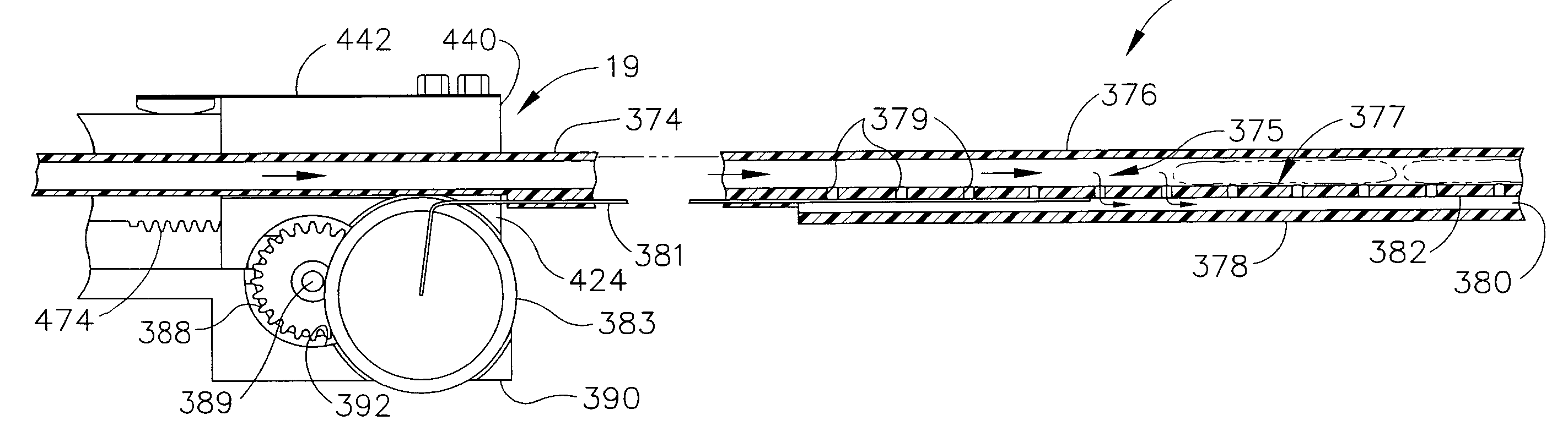
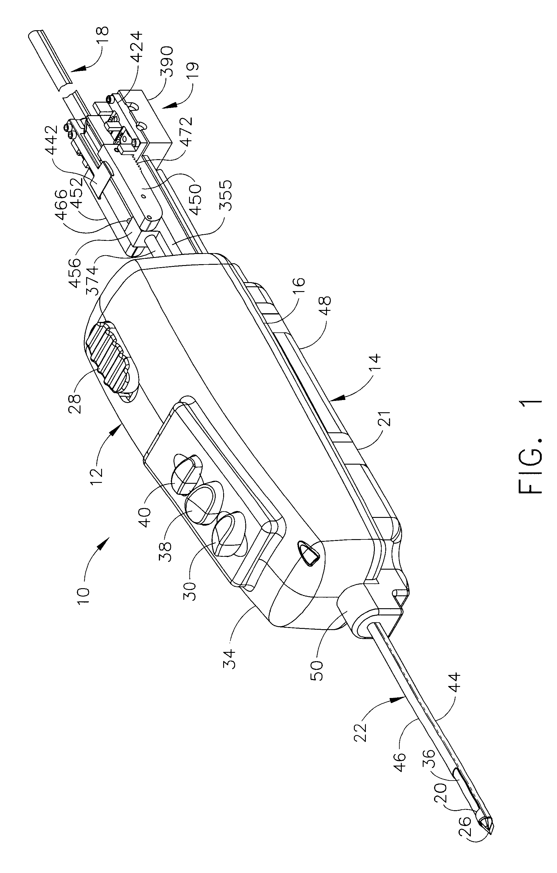
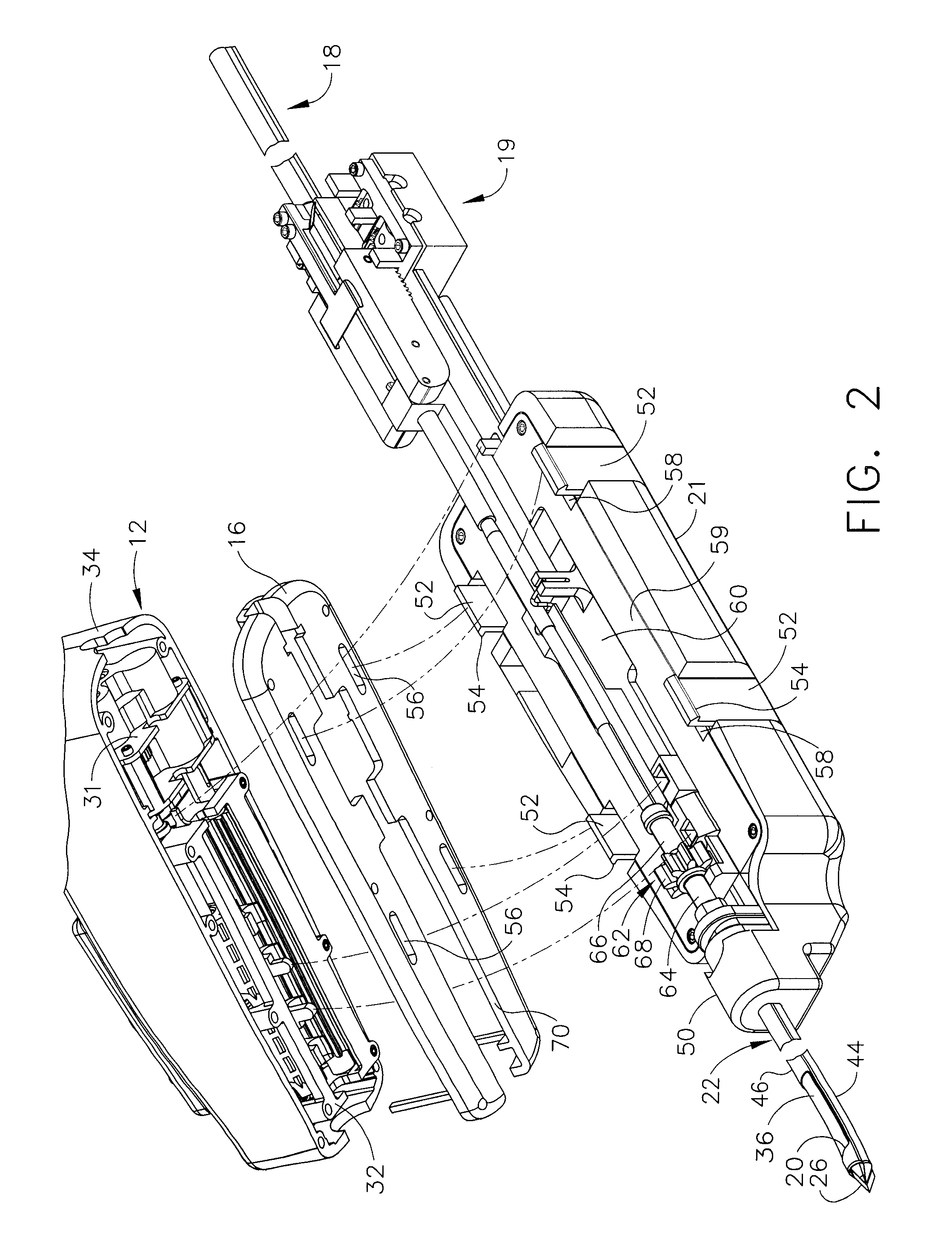
[0044]Turning to the Drawings, wherein like numerals denote like components throughout the several views, in FIGS. 1-2, a biopsy device 10 includes a reusable handpiece 12, and a disposable probe assembly 14. A lower handle tray 16 is disassembled from upper portions of the reusable handpiece 12 to expose portions that operably engage the disposable probe assembly 14. A replaceable serial tissue stacking assembly 18 is prepared to receive the next tissue sample by a tape indexing assembly 19 attached to a hand-held distal portion 21 of the disposable probe assembly 14 that mounts to and is actuated by the reusable handpiece 12. Tissue that is drawn by vacuum assistance into a side aperture 20 of a probe cannula 22 of the disposable probe assembly 14 is severed by a DC motor 24 (FIG. 3) in the reusable handpiece 12 that also powers rotation and staging of the serial tissue stacking assembly 18 to serially stack and store the tissue samples in the order received.

[0045]With particular ...


PUM


Abstract

A biopsy device and method are provided for obtaining a tissue sample, such as a breast tissue biopsy sample. The biopsy device includes a disposable probe assembly with an outer cannula having a distal piercing tip, a cutter lumen, and a cutter tube that rotates and translates past a side aperture in the outer cannula to sever a tissue sample. The biopsy device also includes a reusable handpiece with an integral motor and power source to make a convenient, untethered control for use with ultrasonic imaging. The reusable handpiece incorporates a probe oscillation mode to assist when inserting the distal piercing tip into tissue. The motor also actuates an attached tape indexing mechanism in coordination with movement of the cutter tube to provide sequentially stored tissue samples in a replaceable serial stacking assembly.

Description

CROSS REFERENCE TO RELATED APPLICATIONS[0001]The present application is a continuation-in-part of the co-pending and commonly-owned U.S. patent application Ser. No. 10 / 953,834, “BIOPSY APPARATUS AND METHOD” to Hibner et al., filed 29 Sep. 2004 (U.S. Pat. Publ. No. 2006 / 0074345), the disclosure of which is hereby incorporated by reference in its entirety.FIELD OF THE INVENTION[0002]The present invention relates in general to biopsy devices, and more particularly to biopsy devices having a cutter for severing tissue, and even more particularly to biopsy devices for multiple sampling with a probe remaining inserted.BACKGROUND OF THE INVENTION[0003]When a suspicious tissue mass is discovered in a patient's breast through examination, ultrasound, MRI, X-ray imaging or the like, it is often necessary to perform a biopsy procedure to remove one or more samples of that tissue in order to determine whether the mass contains cancerous cells. A biopsy may be performed using an open or percutan...

Claims


Application Information

Patent Timeline
Patent Type & Authority Patents(United States)
IPC IPC(8): A61B10/00
CPCA61B10/0275A61B10/0283A61B2010/0225A61B2010/0208
Inventor HIBNER, JOHN A.
Owner DEVICOR MEDICAL PROD
Why Eureka
Try Eureka