

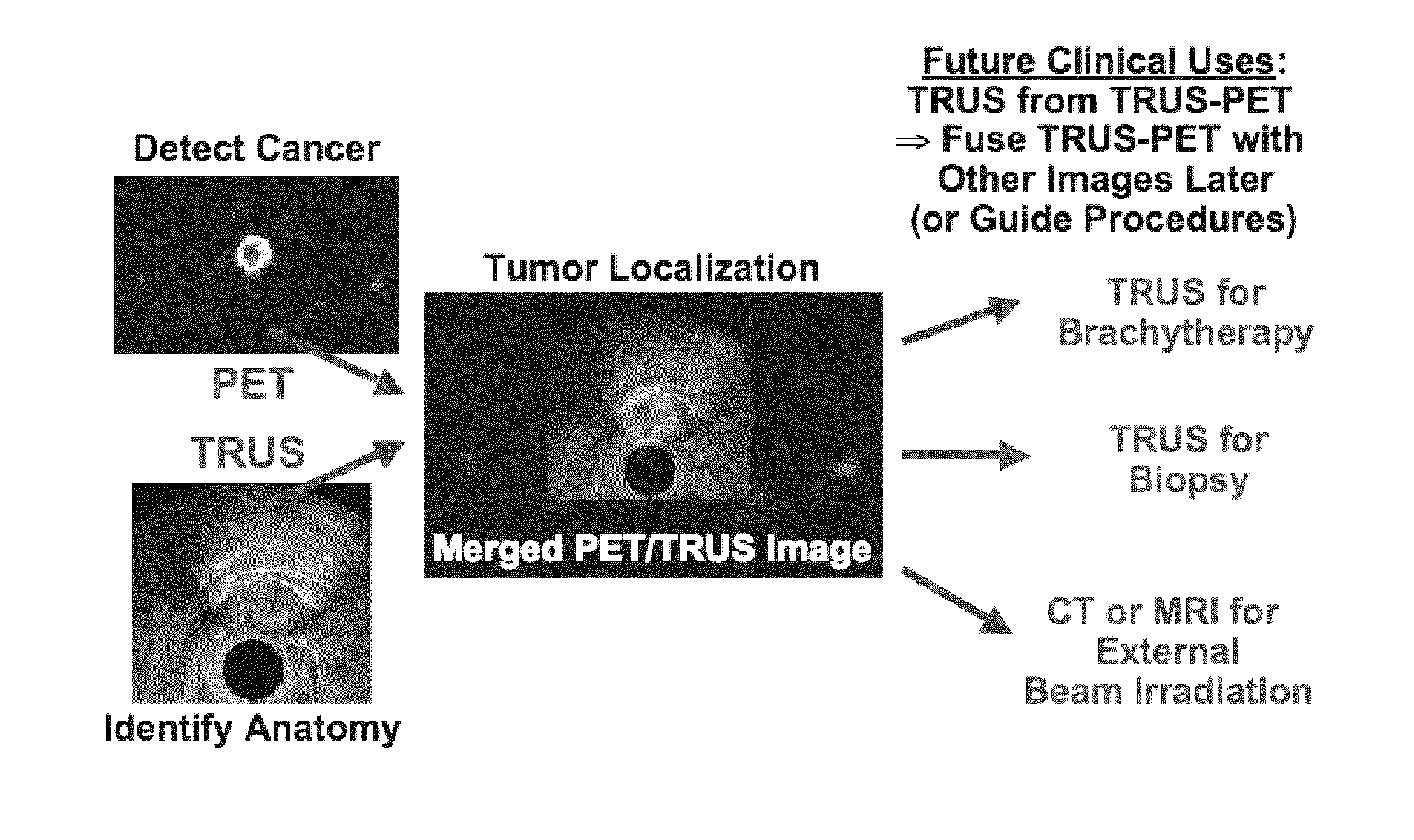
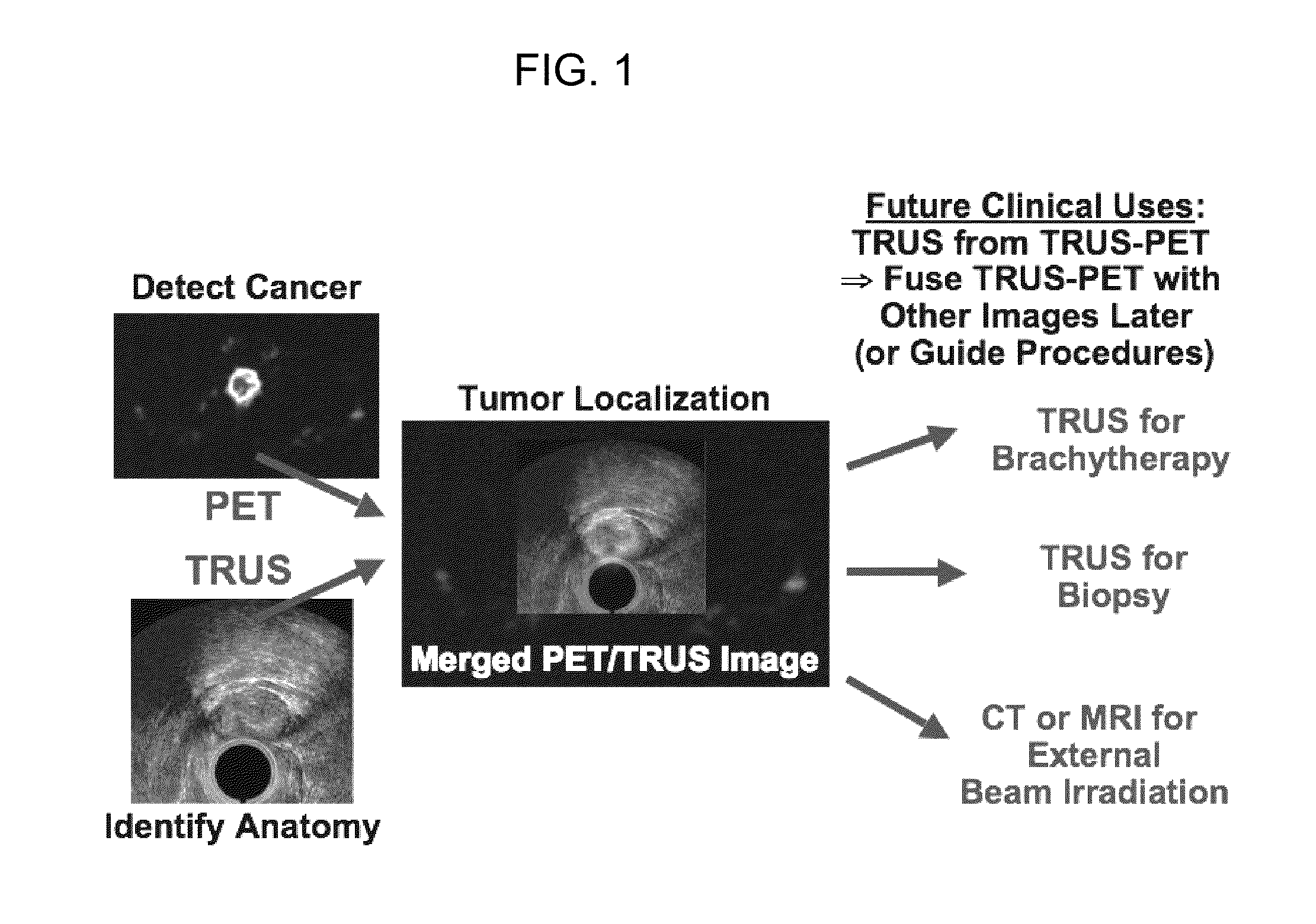
Multi-Modality Phantoms and Methods for Co-registration of Dual PET-Transrectal Ultrasound Prostate Imaging
a transrectal ultrasound and multi-modal technology, applied in the field of phantoms for multi-modal imaging, can solve the problems of high false-positive rate of prostate specific antigen and digital rectal exam screening in general clinical practice, high difficulty in acquisition and interpretation of prostascintTM images, and high difficulty in co-registration. , to achieve the effect of accurately superimposing the pet and accurate co-registration
- Summary
- Abstract
- Description
- Claims
- Application Information
AI Technical Summary
Benefits of technology
Problems solved by technology
Method used
Image

Examples
example 1
PET-TRUS Prostate Scanner and Imaging
[0068]LBNL Prostate-Optimized PET Scanner
[0069]LBNL has built a high performance positron emission tomograph optimized to image the prostate [32-34]. Coincidence imaging of positron emitters is achieved using a pair of external curved detector banks with the patient centered between them. The two banks form an incomplete elliptical ring of detectors with a 45 cm minor axis and a 70 cm major axis, which reduces the distance between the detectors and patient. FIG. 3 shows the transaxial and sagittal views of the scanner. Each bank consists of two axial rows of 20 ECAT HR+PET block detector modules for a total of 80 detectors per scanner; thus the scanner uses about one-quarter the number of detectors as an EXACT HR or HR+ scanner. The ECAT HR+ block detectors are three attenuation lengths thick for good detection efficiency with narrow detector elements (i.e., 8×8 arrays of 4.4×4.1×30 mm3 BGO crystals) to achieve good spatial resolution. The indivi...
example 2
PET-TRUS Prostate Positioning
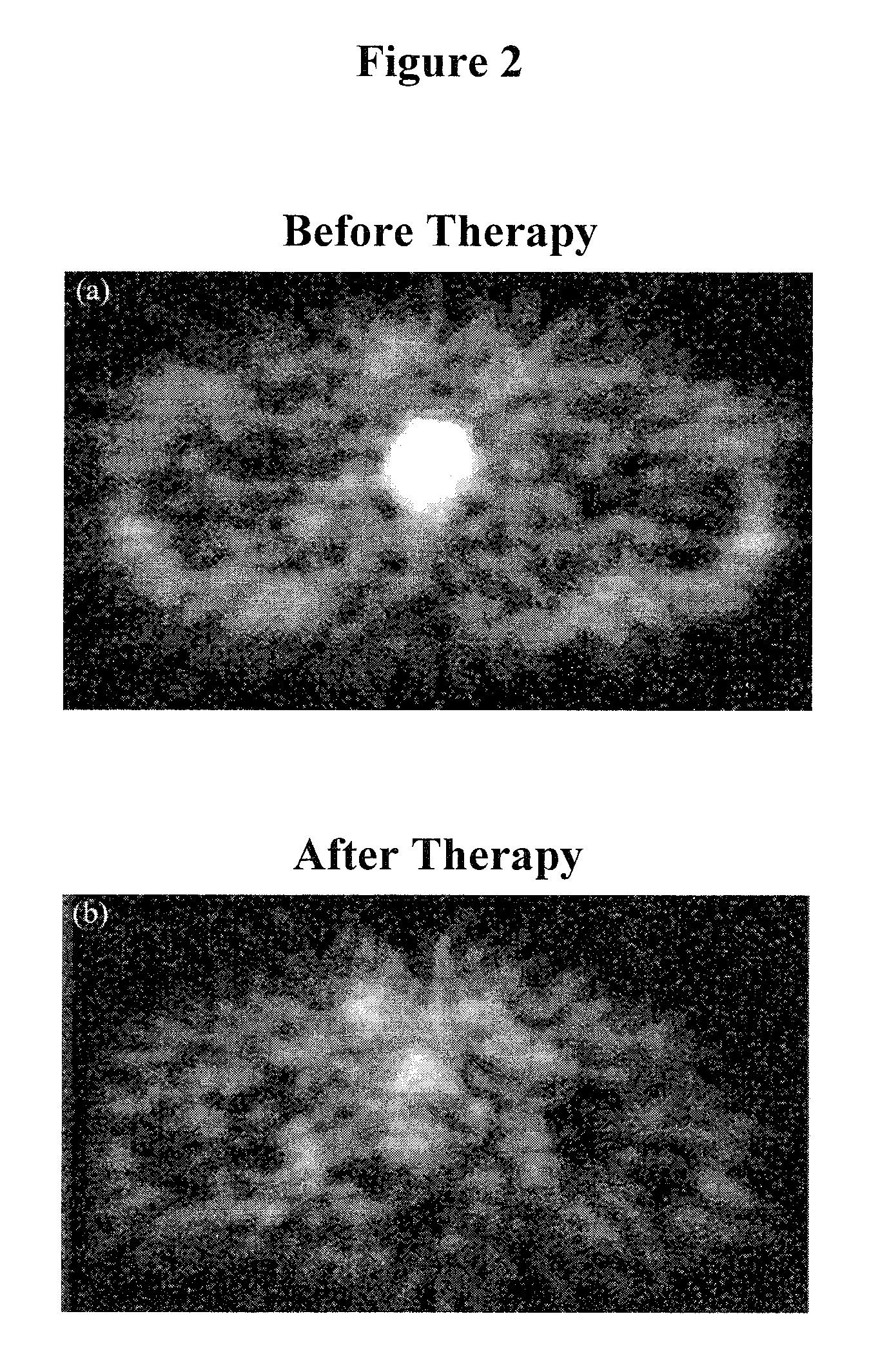
[0073]We used the LBNL prostate-optimized PET scanner, shown in FIG. 3, to acquire 3D volumetric PET images [J. S. Huber, W. S. Choong, W. W. Moses, J. Qi, J. Hu, et al., “Initial Results of a Positron Tomograph for Prostate Imaging,”IEEE Trans Nucl Sci, NS-53, pp. 2653-2659 (2006)]. We also use a commercial TRUS imaging system to acquire a series of 2D TRUS images, then reconstruct them to visualize a 3D TRUS image of the prostate region (see FIG. 4). We have mechanically modified this TRUS equipment to work when mounted onto a common patient table in conjunction with the PET scanner (see FIG. 5b). The TRUS probe is rigidly attached to the modified TRUS stepper that allows calibrated linear displacement along its axis. A point source holder (with two 68Ge point sources) is attached to the TRUS stepper. The TRUS probe-stepper-point source holder unit is mounted onto a moveable TRUS stabilizer arm that is rigidly attached to the patient table. The stabili...
example 3
Co-Registration and PET-TRUS Prostate Phantoms
Custom PET-US Phantom
[0085]We have developed a method to co-register PET and TRUS images and validate this method using a custom TRUS-PET prostate phantom. We construct and use a TRUS-PET prostate phantom with structures that simulate the acoustical properties for TRUS and 511 keV activity concentrations for PET. We use agar-gelatin-based tissue mimicking materials (TMMs) that are mixed with radioactive water solutions [J. S. Huber, Q. Peng, and W. W. Moses, “Multi-Modality Phantom Development,”IEEE Nuclear Science Symposium Conference Record 2007, vol. 4, pp. 2944-2948, (Edited by B. Yu), Honolulu, Hi., 2007]. When developing the procedures for the agar-gelatin-based phantom construction, we first used non-radioactive water to test ultrasound properties. We then used short-lived 18F radioactive (110 minutes half-life) water solutions, since 18F is readily available from our in-house cyclotron and no radioactive waste is generated by the...
PUM
 Abstract
Description
Claims
Application Information