Ultrasonic and nuclear magnetic resonance image fusion transluminal registration device and method

A nuclear magnetic resonance image and ultrasound technology, applied in medical science, sensors, catheters, etc., can solve the problems of prostate cancer biopsy, limited application range, and limited applicable population, so as to avoid damage, use limitations, and a wide range of applications , well tolerated by patients

Inactive Publication Date: 2011-09-14
XIN HUA HOSPITAL AFFILIATED TO SHANGHAI JIAO TONG UNIV SCHOOL OF MEDICINE
View PDF5 Cites 16 Cited by
  • Summary
  • Abstract
  • Description
  • Claims
  • Application Information

AI Technical Summary

Problems solved by technology

[0007] 1. The radioactive particles implanted in the prostate for prostate brachytherapy are used as image registration markers. They must be placed before imaging through invasive techniques, which will cause varying degrees of damage to patients, and the applicable population is limited to prostate cancer patients. Patients with brachytherapy are not conducive to large-scale clinical application;
[0008] 2. To use the contour features and pixel features of the prostate, calcifications in the prostate, anatomical landmarks such as the internal opening of the urethra, the edge contours of the prostate and its transition zone, or related lesions in the prostate as internal markers for image registration, the operator needs to be aware of these In addition, due to the pressure formed by the bladder and rectum with different filling levels, the patient's position, the inner diameter of the rectal ultrasound probe and MRI rectal coil are inconsistent, and the biopsy needle during the intervention These factors will lead to different degrees of displacement and deformation of the prostate. At this time, the registration method relying on the geometric shape of the prostate and local points, lines, surfaces, and surface contour features will have a large error.
The utility model has a complex structure, and can only perform registration processing on in vitro images, and has a limited range of application, and cannot perform accurate biopsy on prostate cancer, etc.
[0011] Since transrectal ultrasonography-guided prostate biopsy and pathological diagnosis are the gold standard for clinical preoperative diagnosis of prostate cancer, there is an urgent need for a method that can be clearly identified by other imaging examinations (such as magnetic resonance imaging) without conventional ultrasonography. An image fusion and registration device for accurate biopsy of the target prostate cancer lesion, but there is no report on this device

Method used


Image

  • Ultrasonic and nuclear magnetic resonance image fusion transluminal registration device and method
  • Ultrasonic and nuclear magnetic resonance image fusion transluminal registration device and method
  • Ultrasonic and nuclear magnetic resonance image fusion transluminal registration device and method

Examples


Embodiment 1

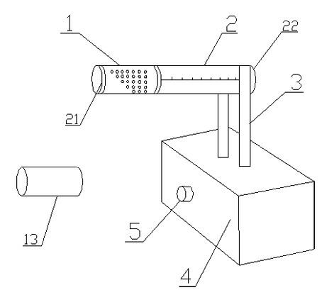
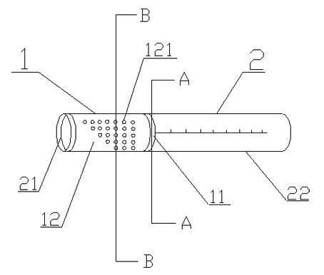
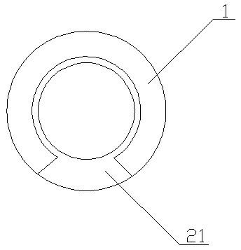
[0049] Please refer to figure 1 , figure 1 It is a schematic structural diagram of an intracavity registration device for ultrasound and nuclear magnetic resonance image fusion of the present invention. The intracavity registration device for ultrasound and nuclear magnetic resonance image fusion is provided with a bracket 2, the bracket 2 is a hollow cylinder, and the bracket 2 is divided into heads part 21 and tail part 22, the inner and outer diameters of the cross-section of the head 21 are consistent with the inner and outer diameters of the tail part 22, the outer surface of the head 21 is provided with a carrier 1, the carrier 1 is provided with a thin envelope 13, and the thin envelope 13 can cover on carrier 1. Please refer to figure 2 , figure 2 It is a schematic structural diagram of the carrier and bracket structure of an ultrasound and nuclear magnetic resonance image fusion intracavity registration device of the present invention. The carrier 1 is divided in...

Embodiment 2

[0055] 1. Marker preparation materials

[0056] Ferric oxide powder, agar, distilled water

[0057] 2. Marker preparation method

[0058] Weigh 0.1g of ferric oxide and 100g of agar (mass ratio: 1:1000) and add distilled water to make a 500ml mixed solution. When the mixed solution is heated to a liquid gel state, mix well, and then put the mixed solution while hot. The marker is poured into the blind cylindrical hole 121 to be shaped, wait for it to cool and solidify, and it is ready for use.

Embodiment 3

[0060] 1. Marker preparation materials

[0061] Ferric oxide powder, agar, distilled water

[0062] 2. Marker preparation method

[0063] Weigh 0.2g of ferric oxide and 100g of agar (mass ratio: 1:500) and add distilled water to make a 500ml mixed solution. When the mixed solution is heated to a liquid gel state, mix well, and then put the mixed solution while hot. The marker is poured into the blind cylindrical hole 121 to be shaped, wait for it to cool and solidify, and it is ready for use.


PUM


Abstract

The invention relates to an ultrasonic and nuclear magnetic resonance image fusion transluminal registration device, which is provided with a carrier, a support, a regulating device and a base, wherein the carrier is arranged on the support; the support is connected with the base through the regulating device; and markers arranged in the carrier. The invention also provides an ultrasonic and nuclear magnetic resonance image fusion transluminal registration method. The invention has the advantages that: the device and the method are simple, convenient, accurate and quick in prostate transrectal ultrasonic and nuclear magnetic resonance image registration, and only needs to register layers, which have the same number of target markers, in two modes; the device and the method avoid injuring patients and avoids implanting the markers in the bodies of the patients and injuries and use limitation thus caused; and the volume of the device is small, the diameter of the device is equal to thatof a transrectal ultrasonic probe, and the tolerance and acceptability of the patients are high.

Description

technical field [0001] The invention relates to the field of image registration, in particular to an ultrasonic and nuclear magnetic resonance image fusion intracavity registration device and method thereof. Background technique [0002] In the image registration fusion of prostate magnetic resonance imaging (MRI) and transrectal ultrasound imaging (TRUS) to guide prostate biopsy, the existing image registration techniques mainly include: [0003] 1. Using radioactive particles implanted in the prostate in prostate brachytherapy as image registration markers; [0004] 2. Use the contour features and pixel features of the prostate itself, the calcification in the prostate, the internal urethral opening and other anatomical landmarks, the edge contour of the prostate and its transition zone, or the relevant lesions in the prostate as internal markers for image registration; [0005] 3. An electromagnetic tracer positioning system consisting of a magnetic field generator, an u...

Claims


Application Information

Patent Timeline
Patent Type & Authority Applications(China)
IPC IPC(8): A61B5/055A61B8/12
Inventor 陈亚青戚庭月
Owner XIN HUA HOSPITAL AFFILIATED TO SHANGHAI JIAO TONG UNIV SCHOOL OF MEDICINE
Why Eureka
Try Eureka