Increased T-cell tumor infiltration and eradication of metastases by mutant light

a t-cell tumor and mutant light technology, applied in the direction of viruses, peptide/protein ingredients, fusion polypeptides, etc., can solve the problems of tumor rejection, tumor volume reduction, and tumor removal surgery boosting the immune response to tumors, so as to achieve the effect of reducing tumor volum

Inactive Publication Date: 2010-10-12
UNIVERSITY OF CHICAGO
View PDF21 Cites 7 Cited by
  • Summary
  • Abstract
  • Description
  • Claims
  • Application Information

AI Technical Summary

Benefits of technology

[0018]A novel method to treat tumors (solid tumors in particular) is to create lymphoid-like microenvironments that express chemokines, adhesion molecules, and costimulatory molecules required for priming naïve T cells and expanding activated T cells by the use of mutant LIGHT molecules. Broader T cells are generated against tumors. Adenoviral vectors that include mutant LIGHT encoding sequences, are effective against tumors and metastasis. Tumor volume was reduced in vivo when vectors delivered mutant LIGHT to tumors as compared to tumors injected with control vectors.
[0028]Cancer metastasis is reduced by stimulation of cytotoxic T-lymphocytes, and / or by stimulation of production of chemokines, adhesion molecules, and costimulatory molecules for priming naïve T-cells.

Problems solved by technology

Experiments in animal models as well as clinical studies indicate that the immune system can recognize and kill individual tumor cells, but a host cannot generally eradicate established solid tumors.
There may be several explanations for the failure of the host to respond effectively to established tumors: 1) lack of early T cell priming due to poor direct or indirect presentation in lymphoid tissues because of an inadequate number of tumor cells (especially those of non-hemopoietic origin) migrating to the lymphoid tissue; 2) inadequate numbers of immune cells migrating to tumor sites due to biological barriers around tumor tissues; 3) exhausted or short-lived activated antigen-specific T cells that fail to combat tumor growth due to limited repertoires; 4) unresponsiveness or ignorance of T cells to tumors; 5) an inhibitory microenvironment or lack of stimulation inside tumors to activate the immune system.
After tumor growth has been established, however, the therapeutic vaccinations usually fail to reject tumors.
Surgical debulk of tumor does not boost the immune response to tumors.
Furthermore, it was reported that even the expression of a strong antigen on tumor cells was insufficient in promoting the rejection of an established tumor, despite the presence of excessive numbers of antigen-specific T cells in the lymphoid tissues.
Lack of T cells priming and / or infiltrating an established tumor is one of the major obstacles for either natural or therapeutic approaches against antigenic cancers.
In addition, insufficient expression of costimulatory molecules inside tumor tissues may fail to activate infiltrating T cells and result in the anergy of tumor-reactive T cells.
The mechanisms for priming tumor-specific T cells has been vigorously debated and so far remains inconclusive.
Lack of efficient priming of naïve T cells in draining lymphoid tissues and the inability to expand tumor-specific T cells within tumors prevent the eradication of cancer.
Despite these advances, success has been limited, and optimal treatment of metastatic disease continues to pose a significant challenge in cancer therapy.
However, the naturally existing T cell responses against malignancies in human are often not sufficient to cause regression of the tumors, primary or metastases.
From an immunological perspective, present clinical strategies hinder the immune defense against malignancies and further diminish the effectiveness of immunotherapy.
In addition, currently utilized adjuvant treatment with chemotherapy and radiation therapy that is meant to kill residual tumor cells may in fact impair anti-tumor immune responses by destroying or inhibiting T cells.
A challenge in developing an effective immunotherapy is to devise an approach to increase the number or enhance the function of circulating tumor-specific T cells that may detect and destroy microscopic metastatic cells before they become clinically meaningful.
Non-mutant LIGHT is not expressed on the surface of tumors and does not induce effective anti-tumor activity.

Method used


Image

  • Increased T-cell tumor infiltration and eradication of metastases by mutant light
  • Increased T-cell tumor infiltration and eradication of metastases by mutant light
  • Increased T-cell tumor infiltration and eradication of metastases by mutant light

Examples


example 1

Mutant LIGHT Expressed Inside the Tumor Augmented Host Resistance More than 500 Times

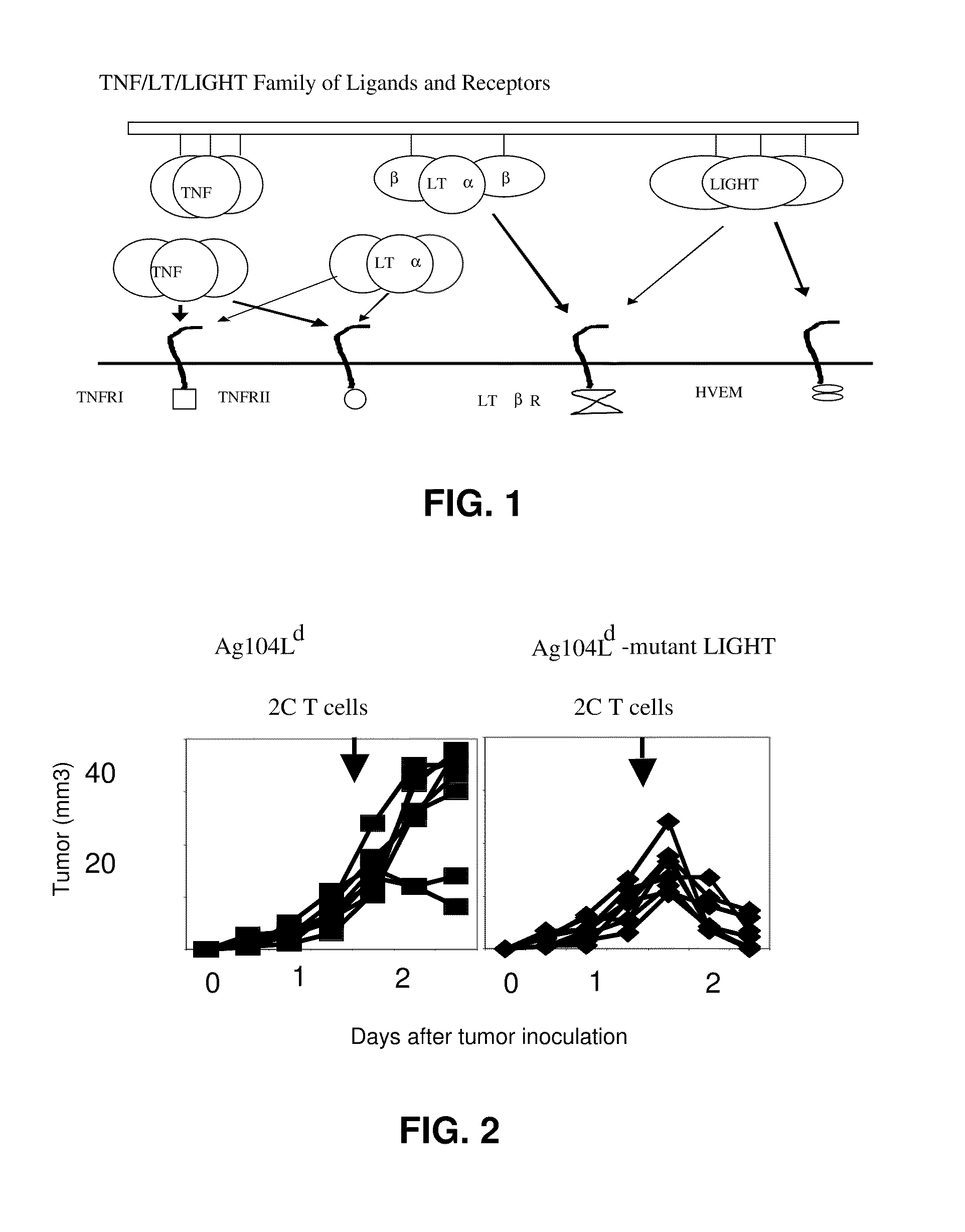
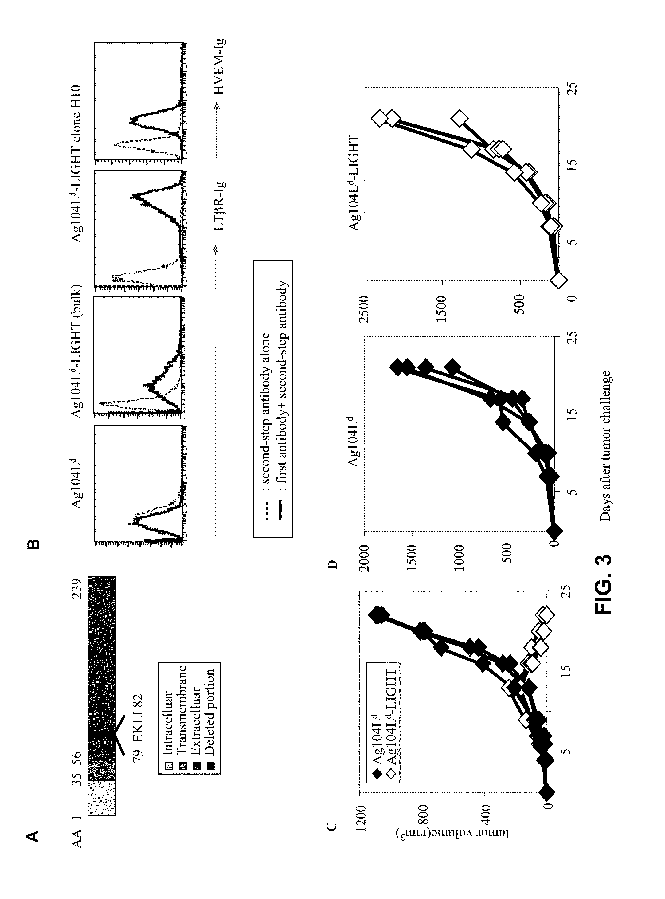
[0096]Fibrosarcoma Ag104Ld was highly tumorigenic and grew out 100% (enlarged tumor growth) when 104 cells were injected into recipient mice C3B6F1 subcutaneously (TABLE 1). It has been reported that 2C T cell receptor (TCR) transgenic mice, which were filled with T cells against antigen Ld expressed on the tumor, failed to eradicate it even after rejection of skin graft containing the same antigen. How to direct tumor-specific T cells into the tumor and activate them at the tumor sites seems to be one critical hurdle for rejection, as well as immunotherapy of cancer clinically. Mutant LIGHT expressed in the tumor environment may break the tolerance by attracting and activating T cells inside the tumor via LTβR and HVEM, respectively, leading to tumor rejection (FIG. 2). To demonstrate this, mutant LIGHT was expressed on this tumor cell line by retroviral transduction utilizing retroviral vector MFG...

example 2

Mutant LIGHT-Mediated Tumor Environment has More Infiltrating CD8+ T Cells

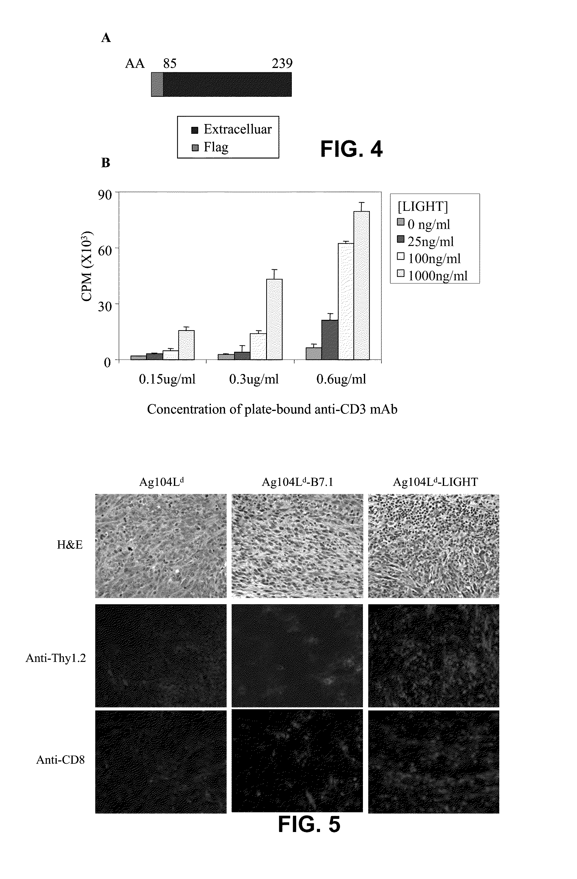
[0097]To investigate the possible mechanisms underlying mutant LIGHT-mediated tumor rejection, 5×10⁶ mutant LIGHT-expressing Ag104Ld or the same number of parental tumor cells were injected subcutaneously to the C3B6F1 mice. Ten to fourteen days after tumor inoculation, before mutant LIGHT-expressing tumors were rejected, tumor tissues were collected. HE-staining of the tumor tissues showed large amount of infiltrating lymphocytes (FIG. 5) while the parental tumors showed very little infiltration (FIG. 5). Immunofluorescent staining confirmed that among the infiltrating lymphocytes, large amount of Thy1.2+ T cells (FIG. 5), especially CD8+ T cells were present inside Mutant LIGHT-expressing tumors (FIG. 5).

example 3

Modified Extracellular Domain of LIGHT is Sufficient to Co-Stimulate T Cells

[0098]In mutant LIGHT, four amino acids corresponding to a proteolytic site in the extracellular domain, very close to transmembrane domain of the molecule were deleted (FIG. 3A). The mutation in the mutant LIGHT molecule affects its co-stimulatory effect. Recombinant mutant LIGHT protein was made that only contains amino acids 85 to 239, a shortened form of extracellular domain, with a flag peptide to facilitate purification (recombinant mutant LIGHT) (FIG. 4A). The modified extracellular domain of mutant LIGHT was sufficient to co-stimulate T cells. For this test, an in vitro co-stimulation assay with plate-bound recombinant mutant LIGHT was used to stimulate purified mouse T cells in the presence of an immobilized monoclonal antibody against CD3 at a sub-optimal dose. Immobilized recombinant mutant LIGHT strongly stimulated a proliferation of purified mouse T cells in a dose-dependent manner in the presen...


PUM

PropertyMeasurementUnit
timeaaaaaaaaaa
thickaaaaaaaaaa
concentrationsaaaaaaaaaa

Abstract

Mutant LIGHT expressed in a tumor environment elicited high levels of chemokines and adhesion molecules, accompanied by massive infiltration of naïve T lymphocytes. Methods and compositions to elicit immune responses against tumors including tumor volume reduction and eradication of metastasis using mutant LIGHT are disclosed.

Description

[0001]This application is a continuation-in-part of U.S. Ser. No. 10 / 865,623, filed Jun. 10, 2004, which claims priority from U.S. Ser. No. 60 / 477,733 filed Jun. 11, 2003 and U.S. Ser. No. 60 / 478,126 filed Jun. 12, 2003.[0002]The government has rights in the invention due to partial support from NIH R01 HD 37104, R01-062026, and R01-DK58897.BACKGROUND OF THE DISCLOSURE[0003]The paucity of activated T cells infiltrating established tumors in immunocompetent hosts helps to explain the inability of hosts to dispose of tumors. Experiments in animal models as well as clinical studies indicate that the immune system can recognize and kill individual tumor cells, but a host cannot generally eradicate established solid tumors. There may be several explanations for the failure of the host to respond effectively to established tumors: 1) lack of early T cell priming due to poor direct or indirect presentation in lymphoid tissues because of an inadequate number of tumor cells (especially those...

Claims


Application Information

Patent Timeline
Patent Type & Authority Patents(United States)
IPC IPC(8): A61K38/00A61K39/00A61K35/12
CPCA61K39/0011C07K14/525C12N7/00A61K2039/5152A61K2039/5156A61K2039/5256A61K2039/53C07K2319/43C12N2710/10043C12N2799/022A61P35/04A61P37/04A61K39/001102
Inventor FU, YANG-XIN
Owner UNIVERSITY OF CHICAGO
Why Eureka
Try Eureka