Method for processing slice images

a slice image and image processing technology, applied in the field of image processing, can solve the problems of difficult tracking of the basal and apical regions of the ventricles using short-axis images, image smoother, small spatial details lost, etc., and achieve the effect of breaking the heart image processing

Inactive Publication Date: 2011-01-25
VALTION TEKNILLINEN TUTKIMUSKESKUS
View PDF5 Cites 48 Cited by
  • Summary
  • Abstract
  • Description
  • Claims
  • Application Information

AI Technical Summary

Benefits of technology

[0011]An object of this invention to improve the accuracy of quantitative information extracted from slice images.
[0016]The method for reducing misalignment errors between image slices is mainly characterized in that, the misalignment errors are reduced by registering slices of an image set to slices from at least one other image set.
[0020]Simultaneous tracking of short-axis (SA) and long-axis (LA) images broadens the heart image processing. The LA images provide comprehensive information especially on tracking the movement of the basal and apical regions of the ventricles in the heart's long-axis direction.
[0021]The image sets that are acquired at least from short-axis and long-axis contribute differently on various regions in the algorithm. For example, the information on the apex of the heart can be extracted from long-axis images as the information on the medial parts of the heart can be retrieved from short-axis images. The simultaneous tracking of two image orientations allows to track more precisely the basal and apical movement of the ventricles. In addition, the motion of the atria can be tracked.

Problems solved by technology

The problem in increasing the slice thickness is that the images become smoother and small spatial details are lost.
Breathing is a major source of movement artifacts in cardiac imaging, as the heart's own movement is handled by the ECG gating.
If the phase of the breathing cycle is not similar during all acquisitions, slices from different image series will be misaligned relative to each other, and a volume built from the image series does not represent the real anatomy of the subject.
Because the slice thickness is much higher than the pixel size in slice level, the tracking of the basal and apical regions of the ventricles using short-axis images is difficult.

Method used


Image

  • Method for processing slice images
  • Method for processing slice images
  • Method for processing slice images

Examples


Embodiment Construction

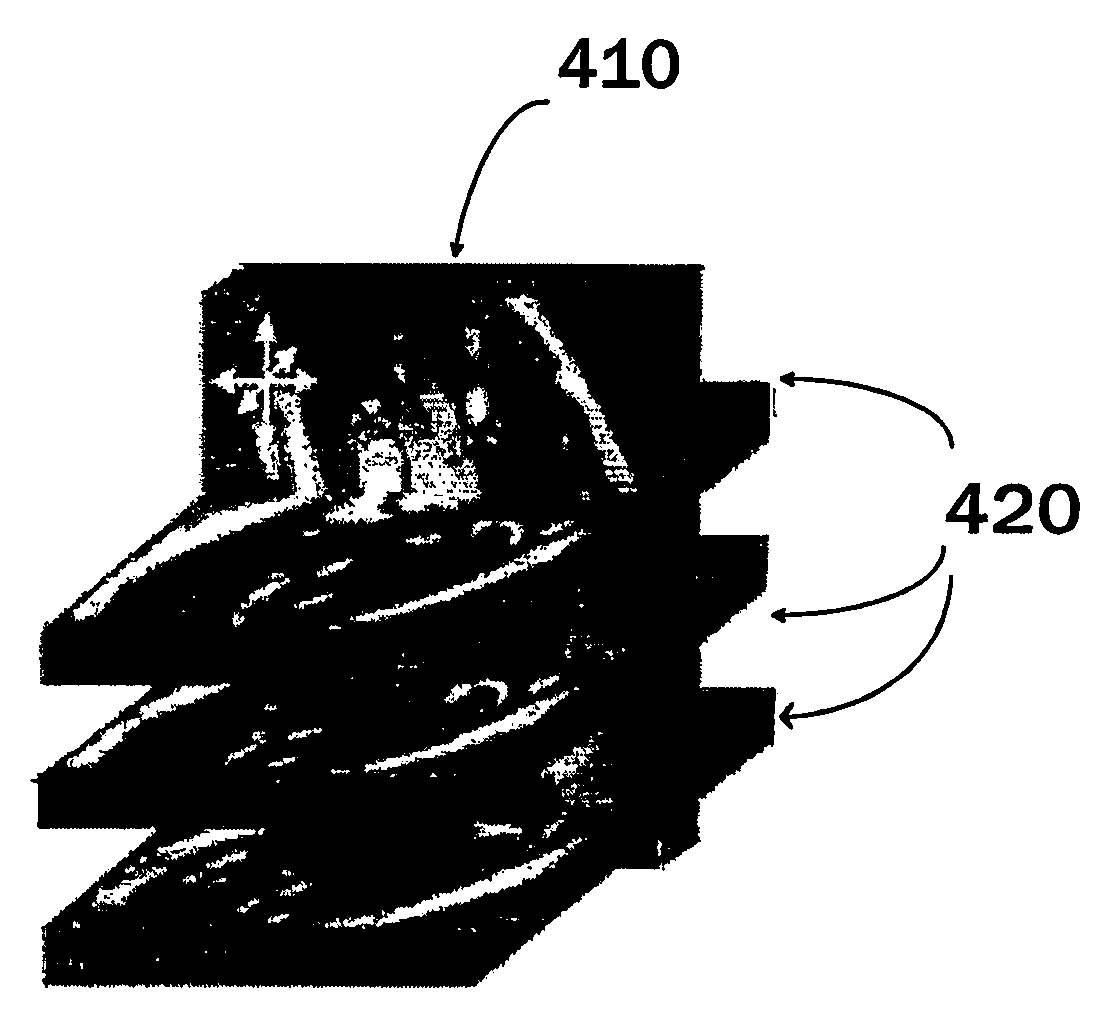
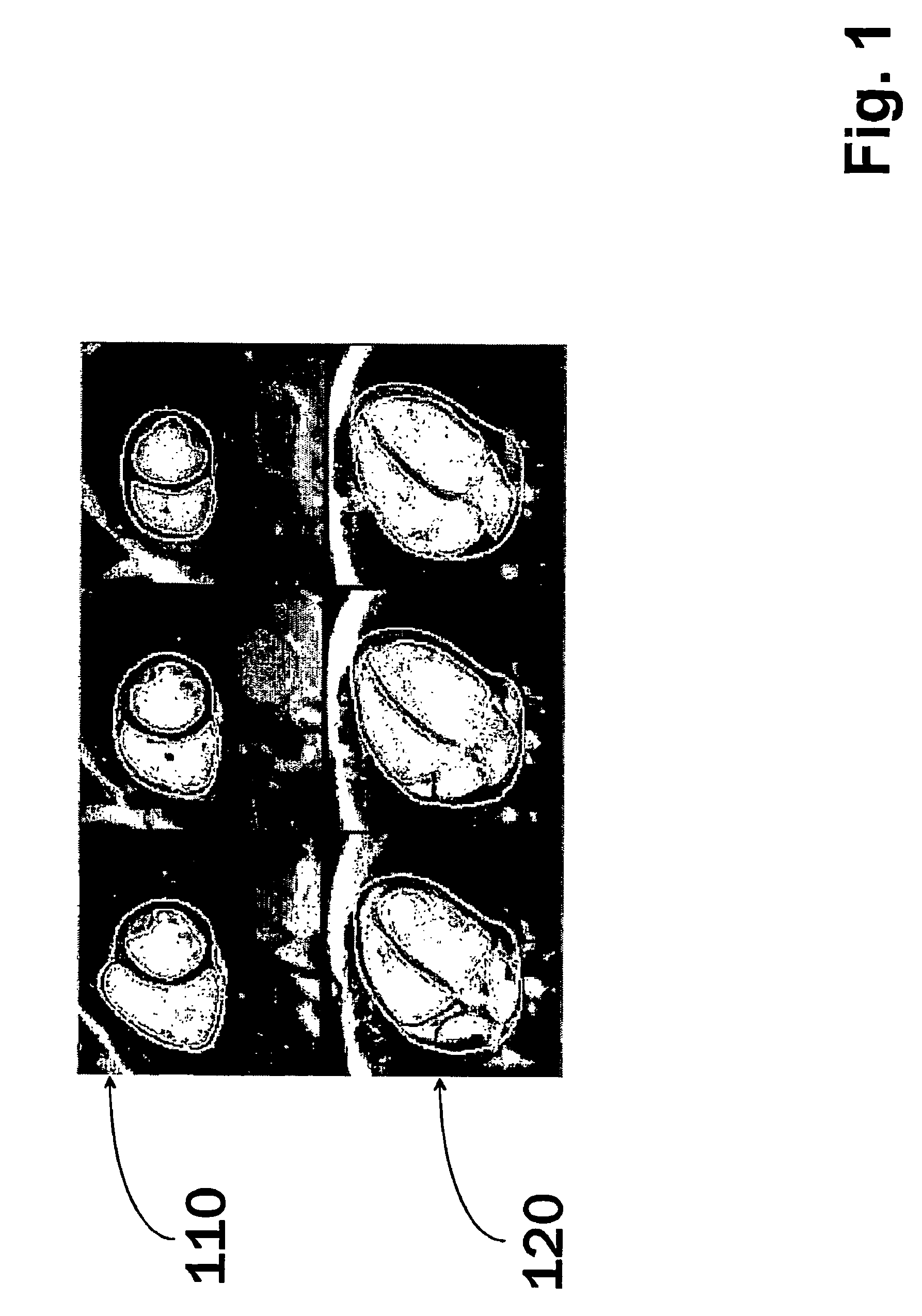
[0032]In this example a method is used for cardiac analysis of a subject, where during scanning (i.e. imaging sessions) the subject, a short-axis (SA) and a long-axis (LA) image volumes are acquired using a known imaging protocol adopted for cardiac subjects e.g. magnetic resonance imaging or other imaging system producing slice images of different levels of the region of interest. Image sets in this description refers to such an image sets that are formed of image slices. The set can be, for example, a stack of image slices or a set of sector slices. What is common here, is that the image set comprises image slices that are recorded from different locations of the region of interest. In addition to cardiac applications, the method can be applied to other regions of interests as well as to other application areas, such in applications related to confocal microscopy, where slice-like images are also used.

[0033]In this example, SA images contain ventricles from valve level until the l...


PUM


Abstract

A method for processing slice images wherein information indicative of at least one quantitative measure is formed from image sets. The image sets are acquired from at least two imaging directions. At least two of the image sets relating to the imaging directions are used for forming the information. A method for reducing misalignments, a module, a system and a computer program product for implementing the method.

Description

CROSS-REFERENCE TO RELATED APPLICATIONS[0001]This application claims priority to Finnish patent application 20035205 filed 12 Nov. 2003 and is the national phase under 35 U.S.C. §371 of PCT / FI2004 / 050164.FIELD OF THE INVENTION[0002]This invention relates generally to a image processing and particularly to such an image processing where image sets are acquired from at least two different directions.BACKGROUND OF THE INVENTION[0003]Since cardiovascular disease is the most common cause of death in the Western countries, there is a strong need to diagnose and to quantify cardiac diseases early enough. The cardiac imaging techniques have improved considerably during the last years providing nowadays detailed anatomic and functional information on the heart. In addition, the automated analysis of the cardiac images has also been intensively developed.[0004]Segmentation of cardiac images is a prerequisite for obtaining many cardiac indices, such as left ventricular volume and ejection frac...

Claims


Application Information

Patent Timeline
Patent Type & Authority Patents(United States)
IPC IPC(8): G06K9/00A61B5/00A61B5/05A61B6/00A61BA61B5/055G06T5/00G06T7/00G06T7/20G06T7/60
CPCG06T7/0038G06T7/602G06T7/0012G06T7/0034G06T2207/30048Y10S128/922G06T7/35G06T7/38G06T7/62
Inventor LOTJONEN, JYRKI
Owner VALTION TEKNILLINEN TUTKIMUSKESKUS
Why Eureka
Try Eureka