
Device for evaluating a position of balance for the human body

Active Publication Date: 2011-06-07
WARSAW ORTHOPEDIC INC
View PDF7 Cites 9 Cited by
  • Summary
  • Abstract
  • Description
  • Claims
  • Application Information

AI Technical Summary

Benefits of technology

The problem addressed by the present invention is that of designing a new system avoiding the drawbacks of prior art systems, in particular by eliminating all sources of nuisance and errors that can stem from the use of a mobile vertical plumbline, and by making totally transparent to the user the transfer of the position of the global axis of gravity of the patient onto the radiographic image.
Another problem addressed by the invention is that of improving the accuracy and reliability of the radiographic images obtained in this way, to enable high-resolution interpretation of the position of the global axis of gravity relative to the skeleton of the patient.
The image is therefore produced by electronic means situated entirely outside the space occupied by the patient when taking the radiographic image, and totally insensitive to the risks of being displaced by movements of the patient. Moreover, the accuracy of the image obtained in this way is significantly improved, since plumbline position errors are entirely eliminated.
In a preferred embodiment, further improving accuracy, the program includes a calibration sequence, adapted for scanning the radiographic image of a calibration object with radio-opaque markers, and calculating said geometrical data from known positions and dimensions of the radio-opaque markers of said calibration object, and storing the data in the memory. This solves the problem of uncertainty related to the unknown position of the source of X-rays, which is generally incorporated into an X-ray head whose interior cannot be seen. By positioning the calibration object appropriately and in a marked way on the patient support means, and taking a radiographic image of the calibration object when so positioned, all the geometrical data necessary, i.e. the relative positions of the source, the patient support means and the target plate, is determined in one operation, enabling subsequent accurate calculation of the position of the image of the global axis of gravity relative to the radiographic image of the bones of the patient.
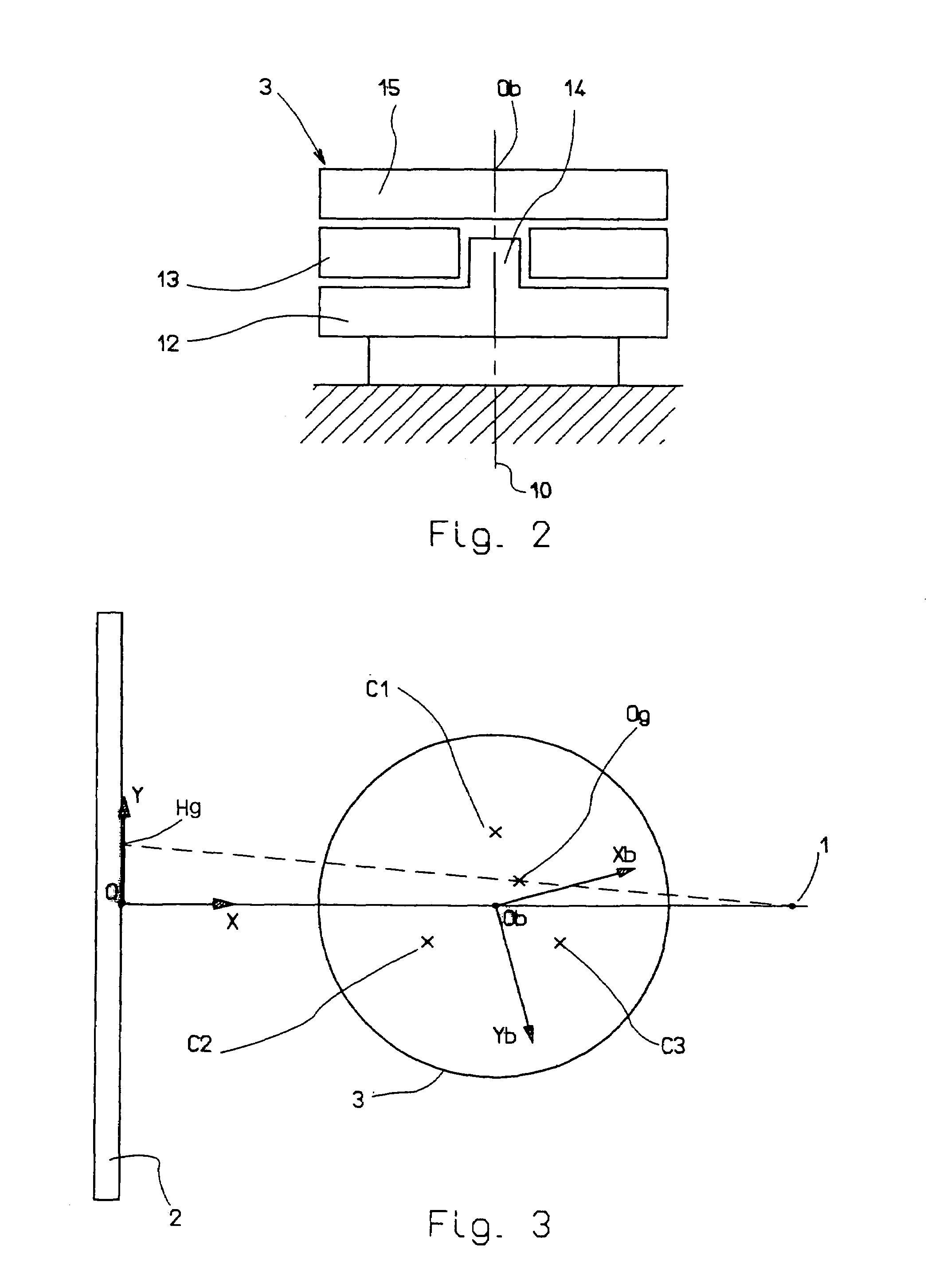
To improve accuracy, the fixed system of axes preferably has its center in the vertical plane passing through the source of X-rays and perpendicular to the support means for the target plate sensitive to X-rays.

Problems solved by technology

Analyzing radiographic images is difficult, and despite the great experience of practitioners, this analysis does not take sufficiently reliable and complete account of the causes of some spinal afflictions.
In some cases, the radiographic images lead to diagnostic errors.
The above kind of device nevertheless has a number of drawbacks that prevent its effective use in practice.
First of all, the precision and reliability of the position of the global axis of gravity in the radiographic image are unsatisfactory.
Most importantly, the mechanical system with a mobile carriage and a vertical plumbline is a nuisance and a source of errors because of its bulkiness, the risks of the plumbline snagging, and the attention that must necessarily be paid to the time of movement of the plumbline support carriage.
Another drawback results generally from the variable and uncontrolled position of the patient on the patient support means, which degrades the readability of the radiographic image.

Method used


Image

  • Device for evaluating a position of balance for the human body
  • Device for evaluating a position of balance for the human body
  • Device for evaluating a position of balance for the human body

Examples


Embodiment Construction

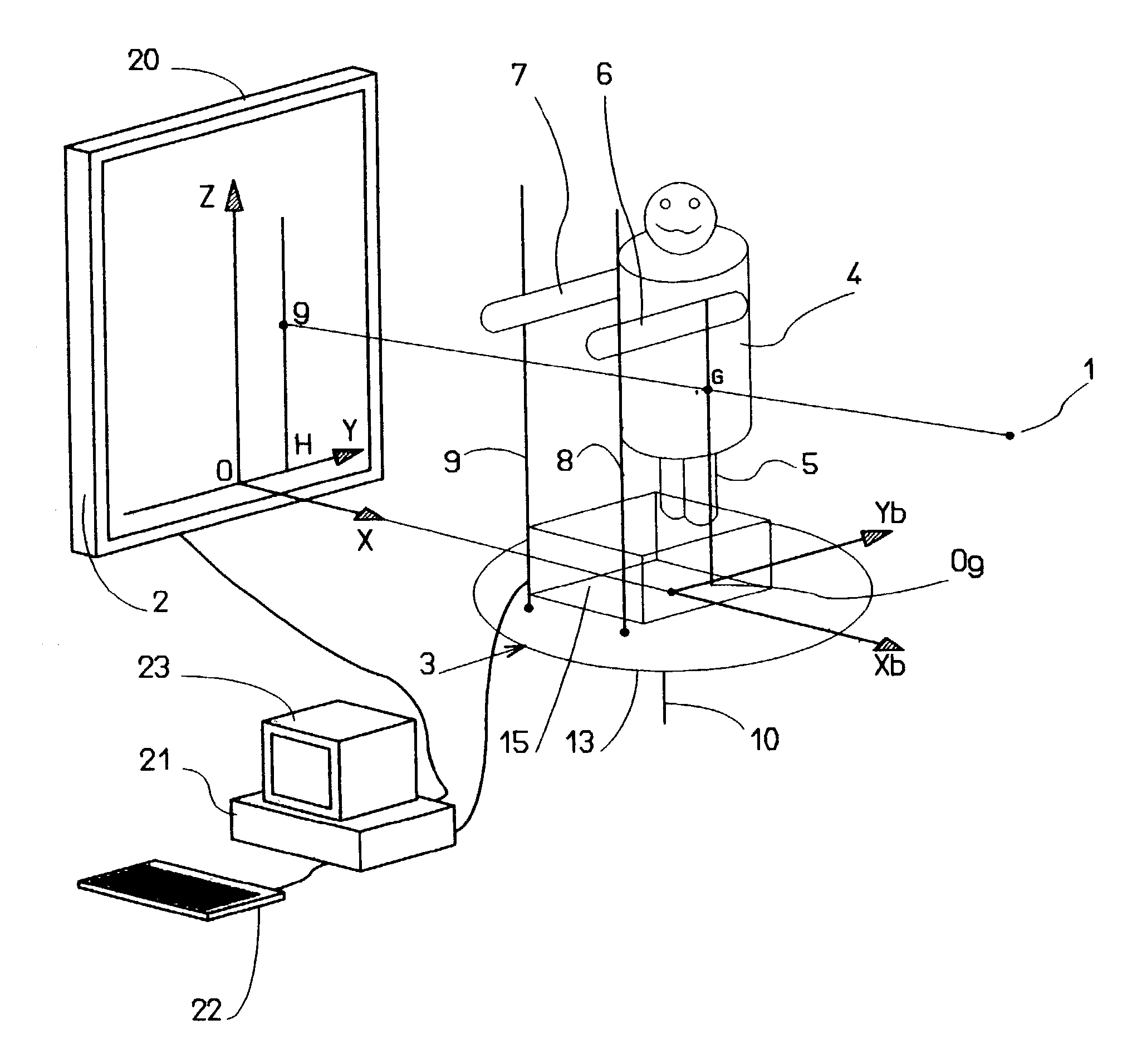
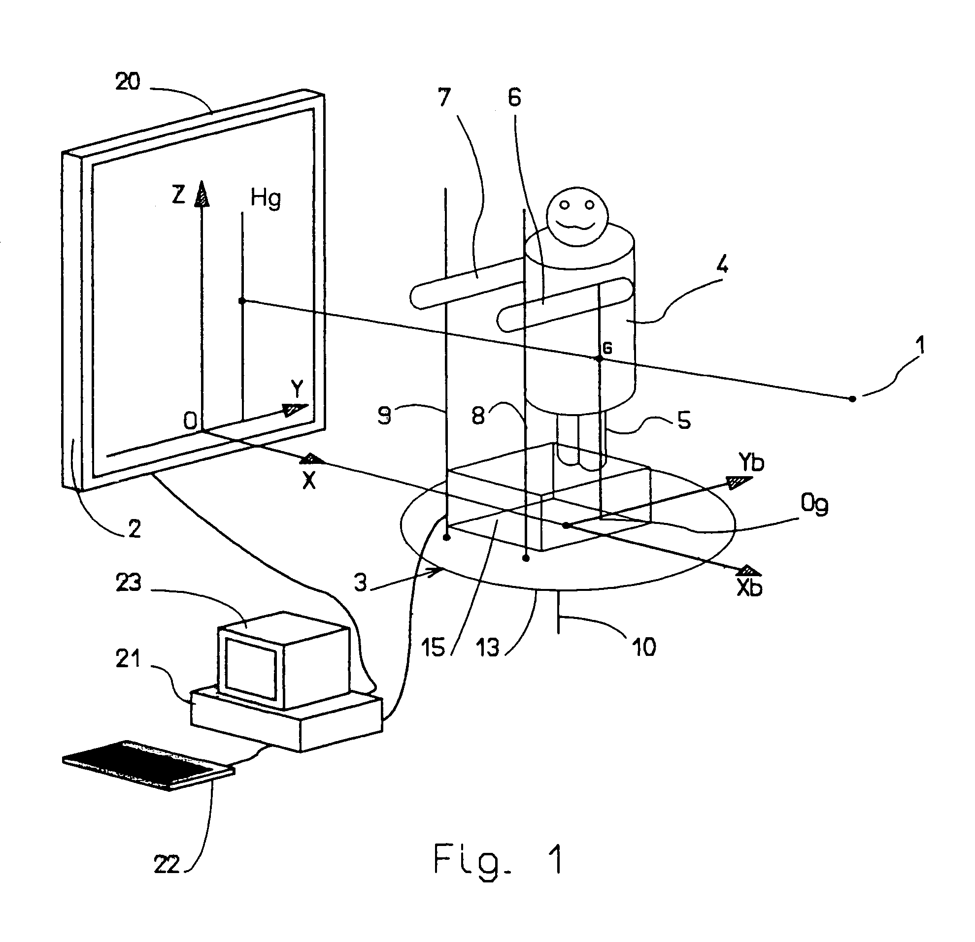
In the embodiment of the system shown in FIGS. 1 and 3, the system includes a source 1 of X-rays, support means for supporting a target plate 2 sensitive to X-rays and oriented vertically at a particular horizontal distance from the source 1 of X-rays, and patient support means 3 adapted to support a patient 4 in a fixed position between the source 1 of X-rays and the support means for the target plate 2. The system according to the invention is an installation for taking radiographic images. In other words, the source 1 of X-rays, and the support means for the target plate 2 can have a structure like those habitually used in medical radiographic image installations.

In accordance with the invention, the patient support means 3 include means for detecting the horizontal position of the global axis of gravity 5 of the patient 4 (the vertical axis passing through the center of gravity G of the patient), i.e. its coordinates in the horizontal plane, during operation of the source 1 of X...


PUM


Abstract

According to the invention, the patient (4) is placed upon a patient support (3) between an X-ray source (1) and a support for the target plate (2) sensitive to the X-rays. The patient support (3) is connected to a device for detecting the horizontal position of the global axis of gravity (5) of the patient when the X-ray source (1) is in operation. The patient (4) can, for example, remain standing. The inventive device can superimpose an image (Hg) of the global axis of gravity (5) on the digital radiographic image of the patient (4).

Description

TECHNICAL FIELD OF THE INVENTIONThe present invention concerns a device for evaluating an equilibrium position of the human body, in particular an equilibrium position of the spinal column.The diagnosis of back pain is generally based on interpreting one or more radiographic images, produced by a machine including a source of X-rays, support means for a target plate sensitive to X-rays, and patient support means adapted to support a patient in a fixed position between the source of X-rays and the target plate support means. The practitioner must interpret the radiographic images to deduce therefrom the probable causes of the patient's back pain.Analyzing radiographic images is difficult, and despite the great experience of practitioners, this analysis does not take sufficiently reliable and complete account of the causes of some spinal afflictions. In some cases, the radiographic images lead to diagnostic errors.To effect a more comprehensive analysis of the human skeleton, with the...

Claims


Application Information

Patent Timeline
IPC IPC(8): A61B5/103A61B5/11G01B21/00A61B6/00
CPCA61B5/4023A61B6/0457A61B6/04A61B6/505A61B6/0487
Inventor BERTHONNAUD, ERICDIMNET, JOANNESROUSSOULY, PIERRE
Owner WARSAW ORTHOPEDIC INC
Learn More