
Method and apparatus for determining autofluorescence of skin tissue

Inactive Publication Date: 2011-06-21
DIAGNOPTICS HLDG
View PDF13 Cites 19 Cited by
  • Summary
  • Abstract
  • Description
  • Claims
  • Application Information

AI Technical Summary

Benefits of technology

[0012]By irradiating a portion of the skin tissue in vivo and carrying out the irradiation noninvasively, a measurement of the autofluorescence of the skin tissue is obtained which can serve as a measure for the AGE content (and possibly also for a potential other underlying explanation for the relation between autofluorescence and diabetes mellitus or possible other disorders, such as renal or hepatic deficiency), without requiring that skin samples be taken and processed to form a homogenized suspension.
[0013]This is despite the fact that in irradiating the skin a part of the radiation is reflected on the skin surface, another part of the radiation, though it penetrates the skin, is absorbed there or, after being scattered in the tissue, exits again, and thus only a small part of the radiation directed at the skin leads to fluorescent radiation. Incidentally, of the fluorescent radiation too, a portion is absorbed in the skin, while the skin tissue in vivo has not been brought into a normalized, homogenized condition as is the case in the known measuring technique. The method and the apparatus according to the present invention nonetheless enable determination of the weak fluorescence from the intact, in vivo skin tissue, which can serve as a measure for AGE contents.

Problems solved by technology

However, that involves the identification of abnormal differences in autofluorescence between different portions of the skin as a result of diseases of the skin, in particular as a result of cancer.

Method used


Image

  • Method and apparatus for determining autofluorescence of skin tissue
  • Method and apparatus for determining autofluorescence of skin tissue
  • Method and apparatus for determining autofluorescence of skin tissue

Examples


Embodiment Construction

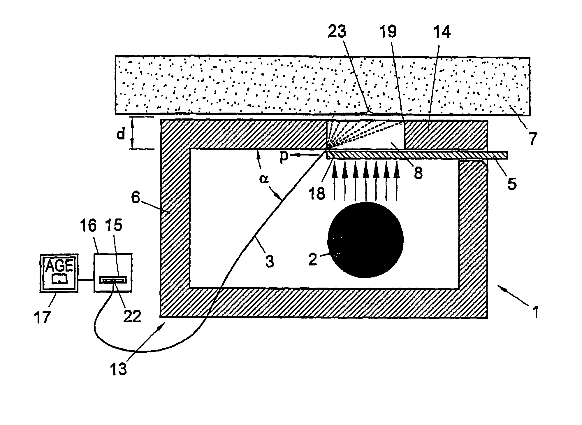
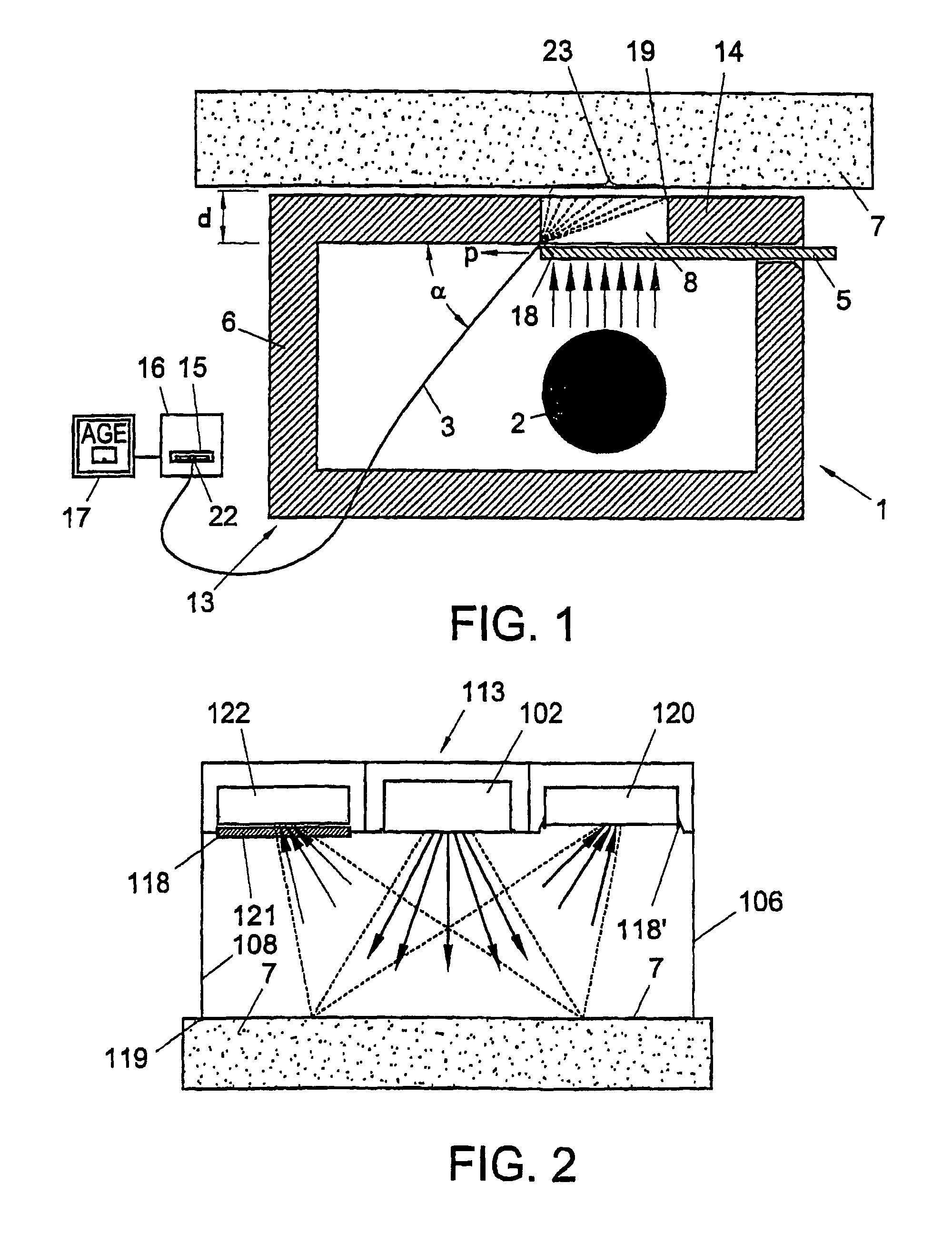
[0021]The measuring system 1 shown in FIG. 1, for measuring an AGE content in a tissue of a patient, constitutes a currently most preferred exemplary embodiment of the invention. The measuring system 1 according to this example comprises a measuring unit 13 having as a light source a fluorescent lamp in the form of a blacklight fluorescent tube 2, which is arranged within a supporting structure in the form of a light-shielding casing 6. The casing 6 has a contact surface 14 which is placed against the skin 7. An opening in the contact surface 14 forms an irradiation window 8 through which a portion of the surface of the skin 7 located behind that irradiation window 8 and adjacent to the window opening, can be irradiated.

[0022]To provide that, of the radiation generated by the fluorescent tube, only UV light in the desired wavelength range reaches the skin 7, there is placed, according to this example, a filter 5 in front of the irradiation window 8. Such filters may be adapted, for ...


PUM


Abstract

For determining autofluorescence of a clinically normal skin issue (7) of a patient, the tissue (7) is irradiated with electromagnetic radiation. An amount of fluorescent radiation emitted by the tissue (7) in response to the irradiation is measured and, in response thereto, a signal is generated which represents a determined autofluorescence in agreement with the measured amount of electromagnetic radiation. Because the tissue (7) is skin tissue (7) in vivo and irradiation is performed noninvasively, a method which is very simple to apply for determining autofluorescence is obtained. An apparatus especially adapted for use in this method is also described.

Description

[0001]This application is a continuation application of U.S. patent application Ser. No. 10 / 089,575, which was the U.S. national phase patent application of PCT / NL99 / 00607, filed Sep. 30, 1999.[0002]This application was published in English on Apr. 5, 2001 as International Publication Number WO 01 / 22869.TECHNICAL FIELD[0003]This invention relates to a method and an apparatus for determining an autofluorescence value of clinically healthy skin tissue. It is supposed that autofluorescence of clinically healthy skin tissue can serve as an indication for an AGE content in a tissue of a patient. This involves measuring the extent to which a portion of the tissue exhibits fluorescence upon excitation with light and / or with radiation in a wavelength region near the visible region.BACKGROUND OF THE PRIOR ART[0004]Studies have shown there is a relation between the occurrence of complications in diabetes mellitus patients and the amount of native fluorescence, or autofluorescence, of the eye ...

Claims


Application Information

Patent Timeline
IPC IPC(8): A61B5/00A61B5/1455A61B5/103
CPCA61B5/0059A61B5/444A61B5/441A61B5/0071
Inventor SMIT, ANDRIES JANGRAAFF, REINDERTOOMEN, PETRUS HENDRICUS N.JAGER, JAN
Owner DIAGNOPTICS HLDG
Learn More