Method and apparatus for diagnosing conditions using tissue color

a tissue color and diagnostic method technology, applied in the field of tissue color diagnostic methods and apparatuses, can solve the problems of difficult calibration of colors observed, difficult to attach quantitative measurements, and high cost of known medical instruments, so as to facilitate and improve the diagnosis of patient conditions, reduce color balance, and improve reliability

Inactive Publication Date: 2011-08-30
CHIO SHIU SHIN
View PDF13 Cites 34 Cited by
  • Summary
  • Abstract
  • Description
  • Claims
  • Application Information

AI Technical Summary

Benefits of technology

[0025]One feature of the present invention is that an electronic visual image of a patient tissue is employed for performing the analysis. The use of an electronic visual image has the advantage of producing a record of the patient tissue that is useable both for archival purposes, and that is capable of being reviewed by other persons at other times. This compares advantageously with the known prior art wherein the color of a sample of tissue is compared by comparing the tissue to a color chart using the technicians “eyes”. As will be appreciated, as the color of tissue is likely to change over time (especially when the tissue is living tissue such as skin, etc.), the ability of an analysis on the tissue to be performed at a later date is reduced significantly, if not impaired totally.
[0028]This feature has the advantage of making a diagnosis of the patient's condition both easier and more reliable. Reliability is enhanced because by standardizing the color of the tissue against a known color sample, the influence of the variations from true color induced in the image by such things as lighting, color balance, and camera programming can be reduced significantly and hopefully, virtually eliminated. Not only does this permit the color of the instant sample to be judged more accurately, but also allows the color of the particular sample (and hence the condition of the patient) to be compared more reliably against other tissue samples taken from other patients, and also other samples taken from the same patient. This advantage becomes especially valuable when one is tracking the progress of a disease. For example, one might seek to monitor the progress of a patient's recovery from a severe sunburn by monitoring the color of the patient's skin during the recovery process.
[0031]This feature has the advantage of enabling the medical practitioner to not only determine the color of a particular tissue sample of interest, but also to determine the size of a particular tissue sample of interest. Determining the size of a tissue sample is often important when tracking the size of skin abnormalities, such as moles or discolorations, because the changes in the size of such moles and discolorations are often indicative of an advancing disease state that may impact the aggressiveness with which the disease state or condition is treated by the medical practitioner.

Problems solved by technology

One of the difficulties with using color identification is that it is difficult to calibrate the colors observed, so that one can easily compare observed colors against either a known standard, or against subsequent observations.
Although sophisticated scientific instruments exist that measure color spectrums of material, most of such known medical instruments are expensive and require a special skill to operate.
Unfortunately, any such match relies to a large extent on the subjective observations of the viewer, along with being the type of observation to which it is very difficult to attach any quantitative measurements.
As will be discussed below, the difficulties that currently exist in capturing “true color” images make color-based diagnoses that are made from color images (rather than the live tissue), very problematic and unreliable.
The upshot of the foregoing discussion is that it is very difficult, if not almost impossible, to achieve standardized colors when using conventional photography equipment.
Since it is very difficult to standardize the color of a particular tissue sample in an image, it is difficult to use an image to analyze the color of the tissue sample to make an accurate diagnosis of the condition of the tissue sample, or more importantly, the condition of the patient from whom the tissue sample was taken.
Further, because of the inconsistency of the way in which colors are reproduced with conventional photograph equipment, it is difficult to track the progress of the condition by tracking changes of the color of a tissue sample shown in the image.
It is difficult to track color changes, because a lack of standardization makes it difficult to determine whether a change in the apparent color of a tissue sample has resulted from an actual change in the color of the tissue sample, or rather, resulted from a change in the conditions under which the picture was taken, or a change in which the color was reproduced on the image.
Unfortunately, these CCD chips currently in use are not inherently better than film in capturing “true color”.
One reason that the CCD chips are no better than film is that CCD chips, when capturing an image, are still subject to the vagaries induced by the ambient light in which the image is being captured.
Unfortunately, those cameras do not allow an image to be captured without this processing; nor is it easy to reverse engineer this processing, as the algorithms and processing methods employed by the camera companies to so process their images are often maintained by the camera companies as trade secrets.

Method used


Image

  • Method and apparatus for diagnosing conditions using tissue color
  • Method and apparatus for diagnosing conditions using tissue color
  • Method and apparatus for diagnosing conditions using tissue color

Examples


Embodiment Construction

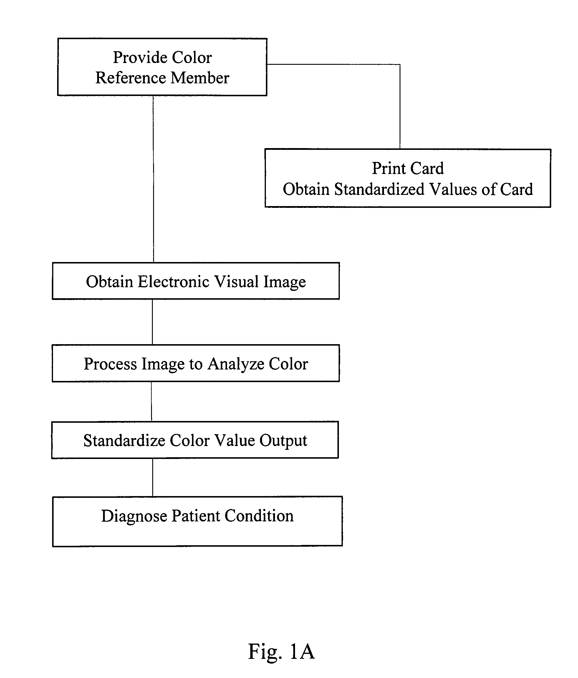
[0042]In FIG. 1, a flow chart that provides an overview of the process of the present invention is shown.

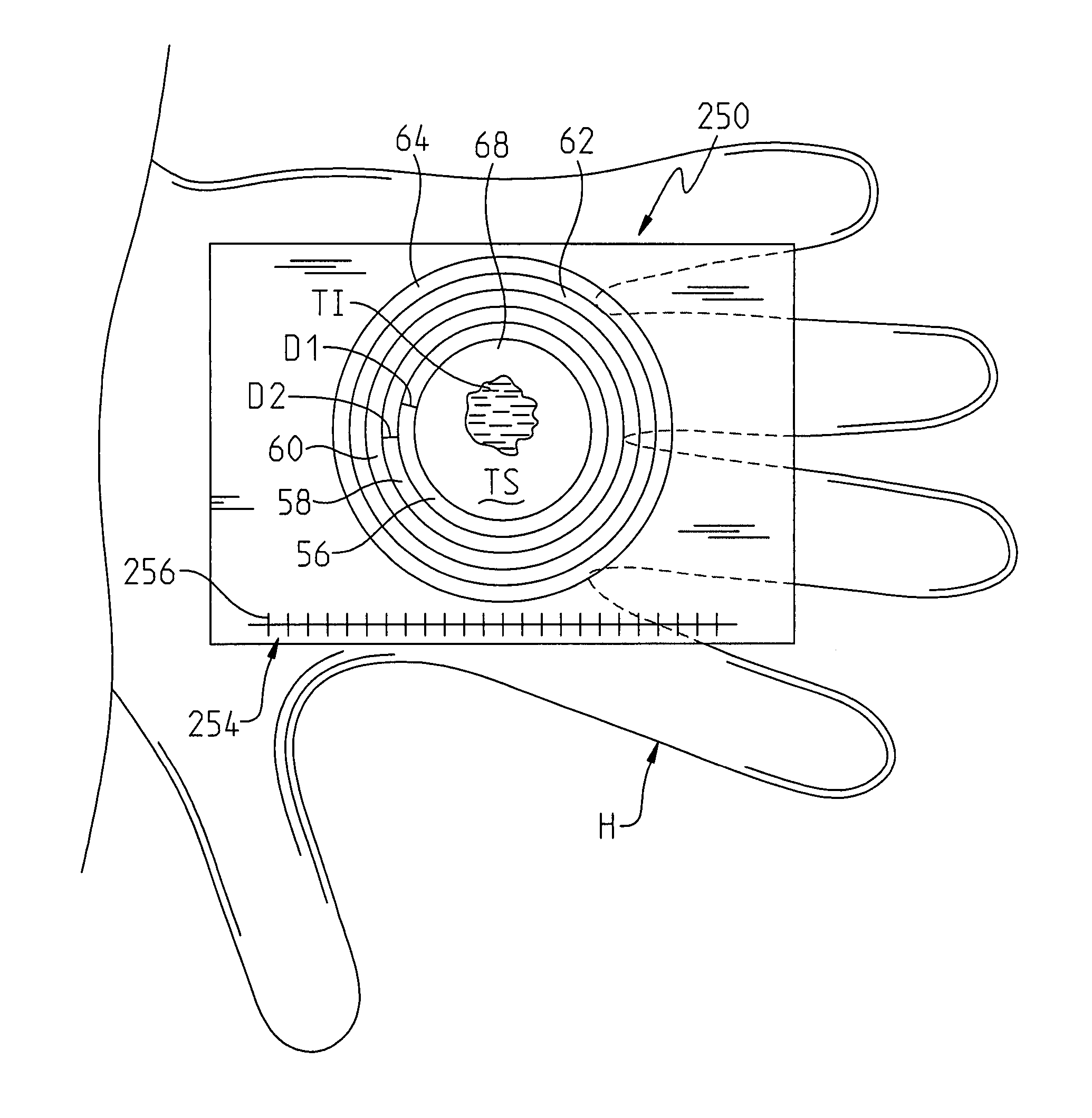
[0043]The first primary step in the process is to provide a color reference member (such as is shown in FIG. 2-6) that includes at least one reference color thereon. Preferably, the color reference member includes a plurality of colors thereon, such as the three colored areas shown in FIG. 1, which actually, includes five colors if one were to include a white area and a black area as “colors”. Alternately, color reference cards having a plurality of other different numbers of colors can be used, such as the six color, color charts shown in FIGS. 4 and 6, that include six colors in addition to black and white, for a total of eight “colors”.

[0044]Prior to the card being used, but after the card is prepared, it may be useful to obtain standardized values of the colors on the card, since the colors on the card are subject to changes from printing to printing. These color values that ...


PUM


Abstract

A method for diagnosing a condition of a patient includes providing a color reference member that includes one reference color thereon. An electronic visual image is obtained of a patient tissue and the at least one reference color on the image. The electronic visual image is processed with the computer by analyzing the colors in the image of the patient tissue and the reference color. This analysis is performed to obtain a standardized color value of the patient tissue. A condition of the patient is diagnosed by using the standardized color value of the patient tissue so obtained.

Description

PRIORITY CLAIM[0001]The present application claims the benefit of priority to Shiu-Shin Chio, U.S. Provisional Application No. 60 / 814,995, that was filed on Jun. 20, 2006, and which is incorporated herein by reference.TECHNICAL FIELD OF THE INVENTION[0002]The present invention relates to a method and apparatus for diagnosing a condition of a patient tissue, and more particularly to a method and apparatus for using standardization techniques for standardizing the color in digital images of tissue to aid in the diagnosis of a patient condition base on the color of the tissue.BACKGROUND OF THE INVENTION[0003]Biological tissue substances are imbued with color that is observable under a visible light. The colors of some substances, such as skin, hair and teeth will often differ widely between individuals, and are used as identifying markers to identify the individual to whom the particular tissue sample belongs.[0004]All colored bio-substances will exhibit the color-based functionalities...

Claims


Application Information

Patent Timeline
Patent Type & Authority Patents(United States)
IPC IPC(8): G06K9/00
CPCA61B5/441G06T7/0012H04N1/6033A61B5/4547
Inventor CHIO, SHIU-SHIN
Owner CHIO SHIU SHIN
Why Eureka
Try Eureka