Implants, tools, and methods for sinus lift and lateral ridge augmentation

a lateral ridge and sinus lift technology, applied in dental implants, dental surgery, medical science, etc., can solve the problems of difficult surgery and replacement of maxillary teeth, and achieve the effect of reducing the risk of perforation of the schneiderian membrane and infection, and enhancing the maxillary alveolar ridg

Inactive Publication Date: 2011-10-04
MAXILLENT
View PDF53 Cites 22 Cited by
  • Summary
  • Abstract
  • Description
  • Claims
  • Application Information

AI Technical Summary

Benefits of technology

[0041]Some embodiments of the present invention provide a self-tapping osseointegrated dental implant and minimally-invasive closed sinus lift techniques for augmenting the maxillary alveolar ridge while reducing the risk of perforating the Schneiderian membrane and of infection. The dental implant is shaped so as to define a lumen therethrough having a distal opening through a distal external surface of a distal portion of the implant. During an implantation procedure, a surgeon simultaneously forms a bore through the maxillary alveolar ridge and advances the implant into the bore by screwing the implant into the ridge. For some applications, while the surgeon screws the implant, a fluid is provided under monitored pressure to the distal implant portion via the lumen. A drop in the pressure is detected as the distal implant end forms an opening through the top of the ridge to below the Schneiderian membrane, thereby bringing the distal opening into fluid communication with a surface of the membrane facing the ridge. Upon detection of the pressure drop, the surgeon ceases screwing the implant to avoid perforating the membrane.
[0043]In some embodiments of the present invention, a proximal end of the lumen of the implant has a lateral opening through a lateral external surface of the implant, and is not open to a proximal external surface of the implant within 2 mm of the proximal-most part of the implant. The implant typically is permanently closed within 3 mm of the proximal-most part. During the implantation procedure, the additional screwing of the implant into the regenerative material advances the lateral external surface of the implant until the lateral opening is positioned entirely within the bore in the ridge and / or within the regenerative material in the cavity between the ridge and the membrane. Such positioning of both ends of the lumen within bone (current or future) substantially reduces the risk of infection, because the proximal end of the implant that is exposed to the oral cavity or gingiva is permanently closed.
[0179]a biodegradable valve coupled to the distal end of the tubular portion, and configured to allow flow through the lumen in a direction from the proximal end to the distal end, and to prevent flow in an opposite direction.
[0247]In an embodiment, the applicator includes an applicator body, which includes a rotatable surface accessible from a proximal end of the applicator, rotation of which rotatable surface with respect to a portion of the applicator body causes the delivery tube to become decoupled from the implant, and the applicator is shaped so as to define a stabilization surface accessible from the proximal end of the applicator, application to which stabilization surface of a stabilizing force stabilizes the implant during rotation of the rotatable surface. For some applications, the rotatable surface and the stabilization surface are configured to facilitate on-axis rotation of the rotatable surface, thereby minimizing any off-axis force that the rotation may cause the apparatus to apply to its surroundings.
[0267]In an embodiment, the applicator includes an applicator body, which includes a rotatable surface accessible from a proximal end of the applicator; decoupling the delivery tube from the implant includes rotating the rotatable surface with respect to a portion of the applicator body; the applicator is shaped so as to define a stabilization surface accessible from the proximal end of the applicator; and rotating the rotatable surface includes stabilizing the implant by applying a stabilizing force to the stabilization surface. For some applications, stabilizing the implant includes removably coupling a stabilization tool to the stabilization surface of the applicator body, and applying the stabilizing force to the stabilization surface using the stabilization tool.

Problems solved by technology

Replacement of the maxillary teeth is often a challenging surgical procedure when the remaining maxillary bone has insufficient height to support the implant.
Bone augmentation procedures are often surgically difficult to perform, and are associated with complications, including infection of the maxillary sinus.
This is often difficult, because of the delicacy of the membrane, and the restricted access afforded by the closed approach.

Method used


Image

  • Implants, tools, and methods for sinus lift and lateral ridge augmentation
  • Implants, tools, and methods for sinus lift and lateral ridge augmentation
  • Implants, tools, and methods for sinus lift and lateral ridge augmentation

Examples


Embodiment Construction

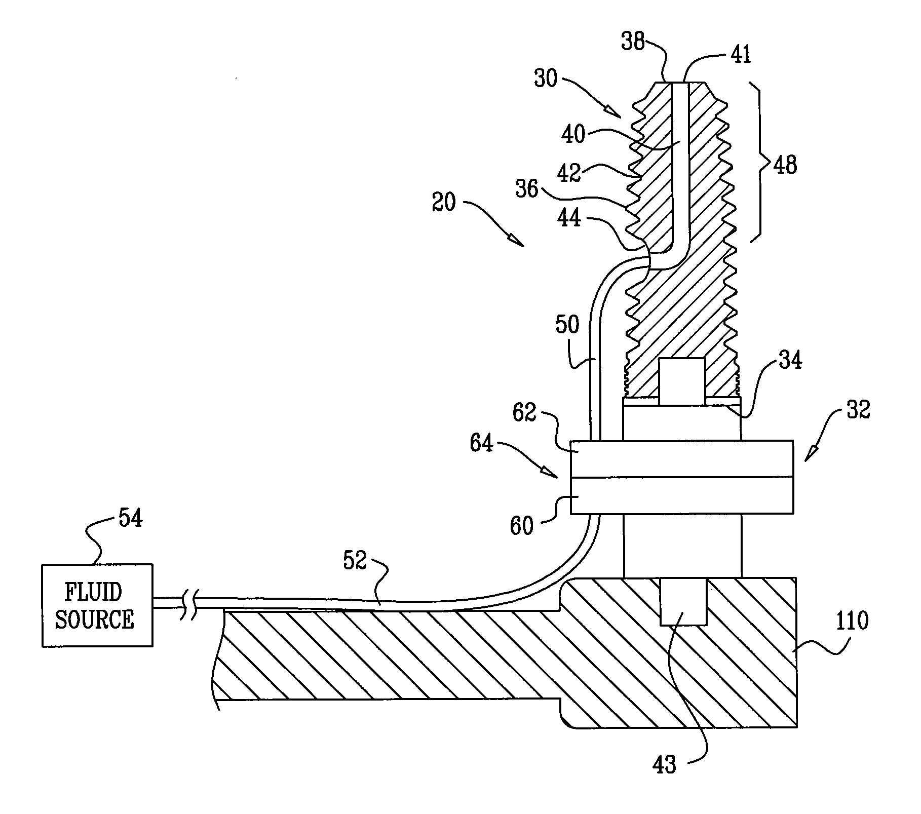
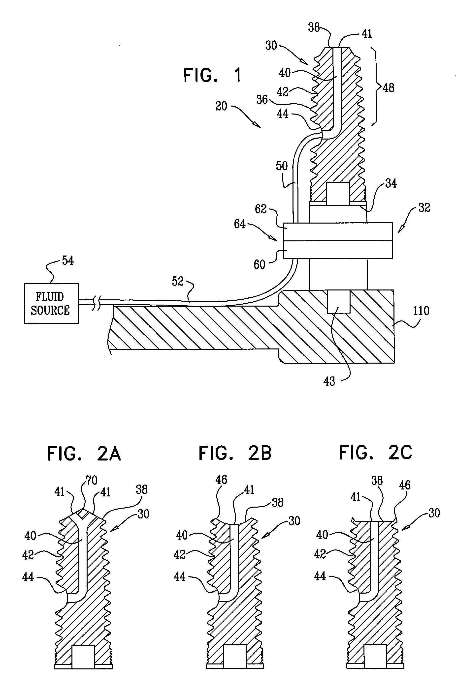
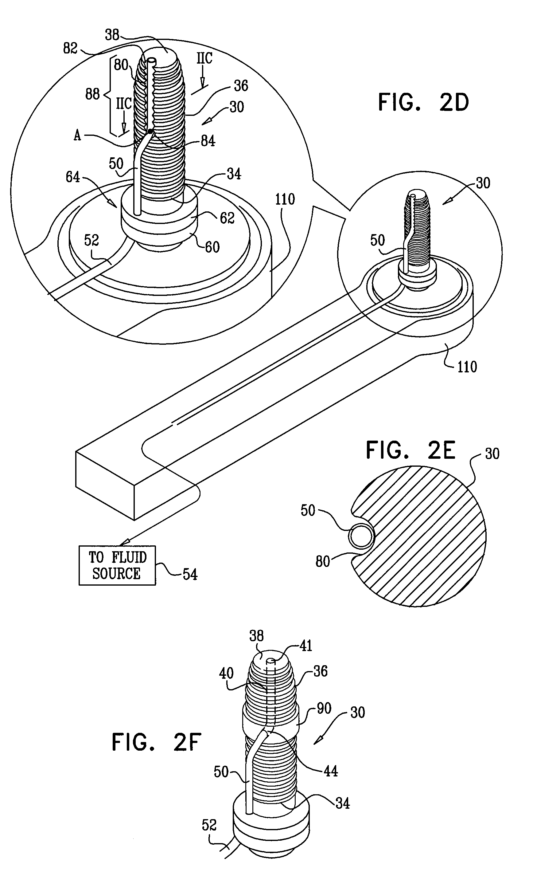
[0357]FIG. 1 is a schematic illustration of a dental implant system 20, in accordance with an embodiment of the present invention. System 20 comprises a dental implant 30, which is typically shaped so as to define a lumen 40 therethrough that is open through a distal opening 41 to a distal portion 48 of the implant that extends from a distal implant end 38 of the implant along up to 50% of a longitudinal length of the implant, such as up to 30% of the length, up to 15% of the length, or up to 5% of the length. For some applications, distal portion 48 has a longitudinal length of up to 6 mm, such as up to 4 mm, or up to 2 mm. As used herein, including in the claims, the “distal” end of the implant is the end that is inserted first into a bone, such as an alveolar ridge, and is sometimes referred to in the art as the apical end, and the “proximal” end of the implant is the end of the implant opposite the distal end, e.g., that faces the oral cavity, and is sometimes referred to in the...


PUM


Abstract

Apparatus is provided that includes a dental implant having a proximal implant end and a lateral external surface. The implant is shaped so as to define a lumen therethrough having a lateral opening through the lateral external surface. The apparatus further includes an applicator, which is removably coupled to the proximal implant end. The applicator includes a delivery tube having a distal tube end that is removably coupled to the implant such that the delivery tube is in fluid communication with the lumen via the lateral opening. Other embodiments are also described.

Description

CROSS-REFERENCE TO RELATED APPLICATIONS[0001]The present application is a continuation-in-part of U.S. patent application Ser. No. 12 / 240,353, filed Sep. 29, 2008, entitled, “Sinus lift implant,” which is assigned to the assignee of the present application and is incorporated herein by reference.FIELD OF THE INVENTION[0002]The present invention relates generally to dental implants and implantation methods, and specifically to minimally-invasive sinus lift implants and implantation methods.BACKGROUND OF THE INVENTION[0003]Osseointegrated dental implants are typically metallic or ceramic screws that are placed in the jawbone for supporting artificial teeth after the loss of natural teeth. Replacement of the maxillary teeth is often a challenging surgical procedure when the remaining maxillary bone has insufficient height to support the implant. One surgical technique for augmenting the maxillary bone includes injecting a regenerative material, such as autogenic, allogeneic, xenogeneic...

Claims


Application Information

Patent Timeline
Patent Type & Authority Patents(United States)
IPC IPC(8): A61C8/00
CPCA61C8/0018A61C19/06A61C8/0092A61C8/0022A61C8/0024A61C8/0039
Inventor BETTER, HADARFOSTICK, GIDEONUCHITEL, ILANGROSS
Owner MAXILLENT
Why Eureka
Try Eureka