Method and apparatus for sealing access

a puncture and apparatus technology, applied in the field of apparatus and a method for sealing a puncture, can solve the problems of increased hospital stay with the associated cost, excessive restriction or interruption of blood flow, and troublesome stemming of blood flow in these patients, so as to reduce the chance of a procedure, the effect of rapid, safe and effective sealing of the punctur

Inactive Publication Date: 2012-02-21
MORRIS INNOVATIVE RES
View PDF133 Cites 25 Cited by
  • Summary
  • Abstract
  • Description
  • Claims
  • Application Information

AI Technical Summary

Benefits of technology

[0009]The present disclosure is directed to an apparatus and method for sealing punctured tubular tissue structures, including arteries and veins, such as punctures which occur during diagnostic and interventional vascular and peripheral catheterizations, or for sealing a puncture in the wall of a body cavity. More specifically, the apparatus and method of the present disclosure employ submucosal tissue or another extracellular matrix-derived tissue or a synthetic bioabsorbable material to seal punctures in tubular tissue structures, such as blood vessels, or in the wall of a body cavity. The submucosal tissue or other extracellular matrix-derived tissue is capable of inducing tissue remodeling at the site of implantation by supporting the growth of connective tissue in vivo, and has the added feature of being tear-resistant so that occlusive material is not introduced into the patient's circulatory system. Also, submucosal tissue or another extracellular matrix-derived tissue has the feature of being resistant to infection, thereby reducing the chances that the procedure will result in systemic infection of the patient.

Problems solved by technology

Therefore, unless the puncture site is closed clinical complications may result leading to increased hospital stays with the associated costs.
Since the control of bleeding in anti-coagulated patients is much more difficult to control, stemming blood flow in these patients can be troublesome.
In particular, when human hand pressure is utilized, it can be uncomfortable for the patient, can result in excessive restriction or interruption of blood flow, and can use costly professional time on the part of the hospital staff.
A potential problem with plugs introduced into the vessel is that particles may break off and float downstream to a point where they may lodge in a smaller vessel, causing an infarct to occur.
Another potential problem with collagen plugs is that there is the potential for the inadvertent insertion of the collagen plug into the lumen of the blood vessel which is hazardous to the patient.
Collagen plugs also can act as a site for platelet aggregation, and, therefore, can cause intraluminal deposition of occlusive material creating the possibility of a thrombosis at the puncture sight.

Method used


Image

  • Method and apparatus for sealing access
  • Method and apparatus for sealing access
  • Method and apparatus for sealing access

Examples


Embodiment Construction

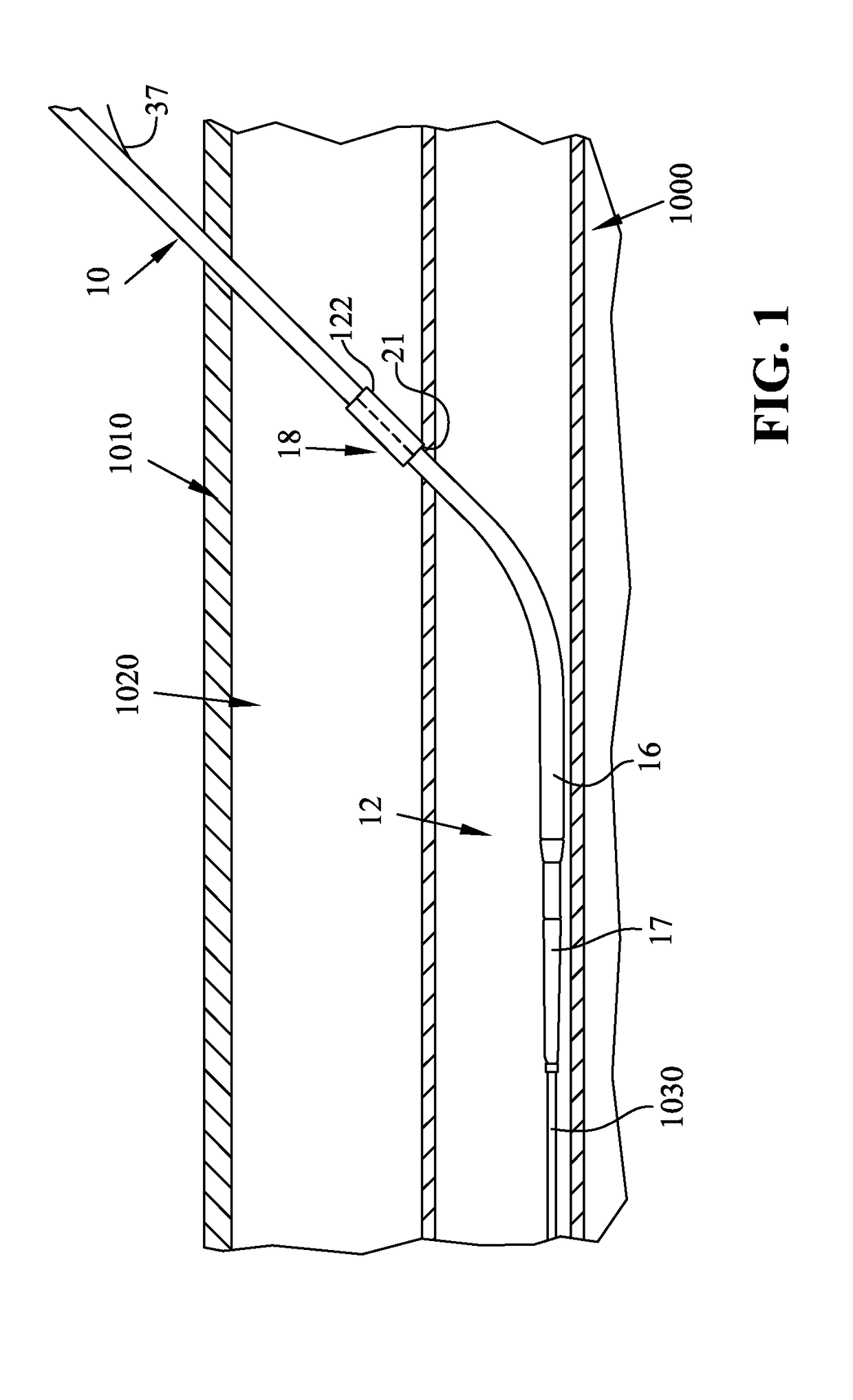
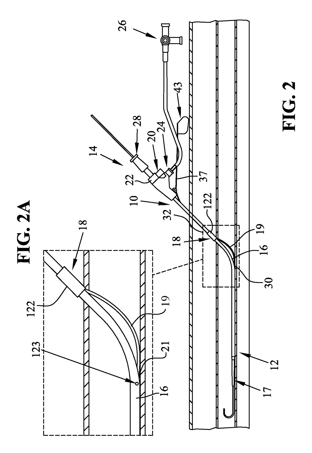
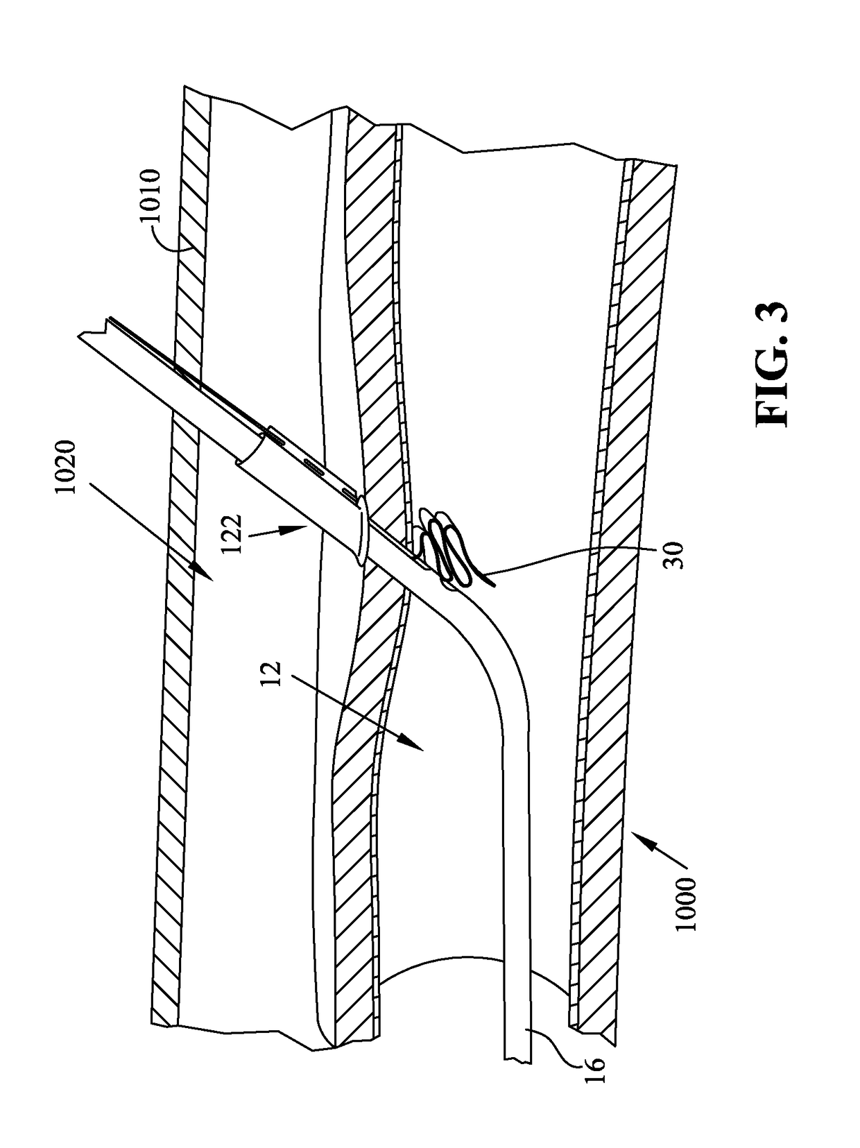
[0019]The disclosures of U.S. application Ser. Nos. 11 / 180,379, 10 / 863,703, 10 / 166,399, 11 / 879,426, 11 / 546,079, and 60 / 297,060 are incorporated herein by reference. The present disclosure is related to an apparatus and a method for sealing a puncture in a tubular tissue structure, such as a blood vessel, or in the wall of a body cavity, with submucosal tissue, another extracellular matrix-derived tissue, or a synthetic bioabsorbable material capable of supporting the growth of endogenous connective tissue in vivo resulting in remodeling of endogenous connective tissue at the puncture site and in formation of a static seal. The apparatus and method of the present disclosure can be used to seal a puncture in a tubular tissue structure, such as a blood vessel, or in the wall of a body cavity, that has been created intentionally or unintentionally during a surgical procedure or nonsurgically (e.g., during an accident). Punctures made intentionally include vascular punctures made in vari...


PUM


Abstract

An apparatus and a method for sealing a puncture in a tubular tissue structure or the wall of a body cavity are provided. The apparatus and method include a bioabsorbable member that is partially external to and partially internal to an introducer sheath before and during placement thereof.

Description

CLAIM FOR PRIORITY[0001]The present application claims priority to U.S. Provisional Application 61 / 061,823 filed Jun. 16, 2008, the disclosure of which is incorporated herein by reference.FIELD OF THE DISCLOSURE[0002]The present disclosure relates to an apparatus and a method for sealing a puncture in a tubular tissue structure or the wall of a body cavity. More particularly, the present disclosure is directed to sealing a puncture site with submucosal tissue or another extracellular matrix-derived tissue capable of remodeling endogenous connective tissue or with a synthetic bioabsorbable material.BACKGROUND AND SUMMARY[0003]The control of bleeding during and after surgery is important to the success of the procedure. The control of blood loss is of particular concern if the surgical procedure is performed directly upon or involves the patient's arteries and veins. Well over one million surgical procedures are performed annually which involve the insertion and removal of catheters i...

Claims


Application Information

Patent Timeline
Patent Type & Authority Patents(United States)
IPC IPC(8): A61B17/03
CPCA61B17/0057A61B2017/00659A61B2017/00004
Inventor MORRIS, EDWARD J.DENARDO, ANDREW J.
Owner MORRIS INNOVATIVE RES
Why Eureka
Try Eureka