
System and method for segmenting vertebrae in digitized images

a technology of digitized images and vertebrae, applied in image enhancement, image analysis, instruments, etc., can solve the problems of inconvenient vertebrae column segmentation in mr images, less methods for vertebrae column segmentation in recent literature, and inability to meet the needs of mri cad applications, etc., to achieve rapid and reliable results

Active Publication Date: 2012-05-08
SIEMENS HEALTHCARE GMBH
View PDF9 Cites 12 Cited by
  • Summary
  • Abstract
  • Description
  • Claims
  • Application Information

AI Technical Summary

Benefits of technology

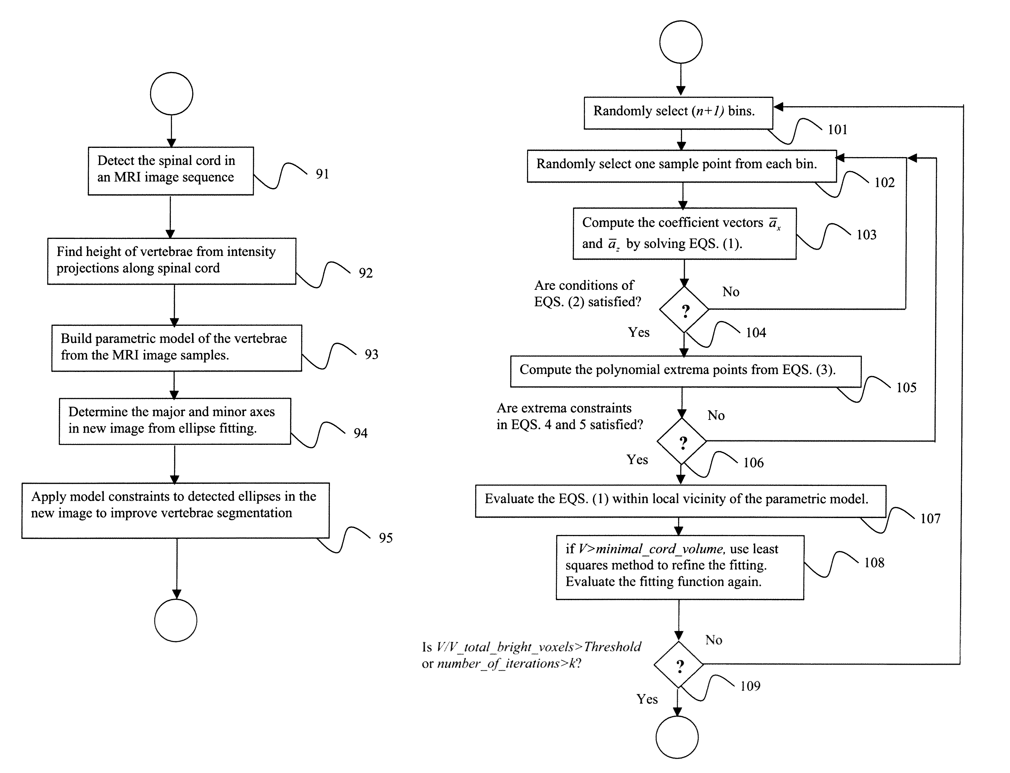
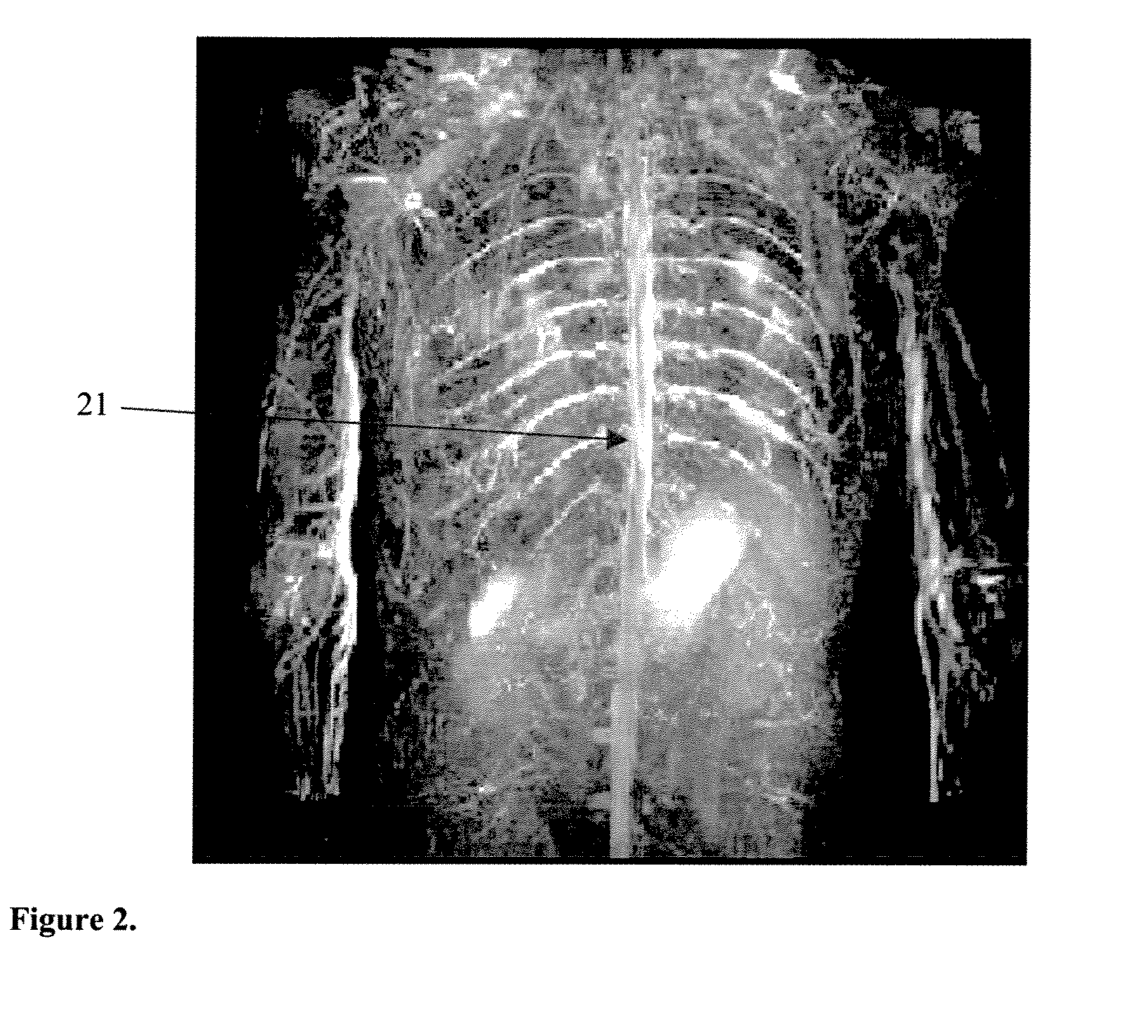
[0005]Exemplary embodiments of the invention as described herein generally include methods and systems for automatically detecting the spinal cord in T2-weighted STIR images, segmenting vertebrae column and vertebrae body based on a given parametric representation of the spinal cord, and modeling naturally periodic structures using the domain knowledge of their periodicity. A method according to an embodiment of the invention involves constructing a model based on geometric primitives from purely anatomical knowledge of organ shape and rough variability limits. The basic intensity range of primary ‘simple’ objects is derived from expert knowledge of image formation for certain tissue types. A method according to an embodiment of the invention can be used for segmentation, characterization and feature extraction, abnormality detection, alignment of anatomical structures or lesions, spinal cord examination, metastasis screening, multi-modal registration, and curved MPR visualization. A 3D-polynomial model via RANSAC (random sample consensus algorithm) provides a fully automatic rapid and reliable estimate of the spinal centerline position and segmentation of the spinal cord.

Problems solved by technology

Spine metastases detection is an important and time-consuming task.
Although MRI, including T1, T2 HASTE (half-fourier acquisition single-shot turbo spinecho) and T2-weighted Short Inversion Time Inversion Recovery (STIR) sequences, is becoming a popular screening modality, development of MRI CAD applications is more complicated than that of CT because of intensity in-homogeneities and higher noise level.
The few methods for vertebrae column segmentation in MR images available in recent literature, such as normalized cuts, are not suitable in the case of severe metastases where dissimilarity between healthy and affected vertebrae could be much greater than between vertebrae and other tissue.

Method used


Image

  • System and method for segmenting vertebrae in digitized images
  • System and method for segmenting vertebrae in digitized images
  • System and method for segmenting vertebrae in digitized images

Examples


Embodiment Construction

[0056]Exemplary embodiments of the invention as described herein generally include systems and methods for detection and segmentation of vertebrae structures in the spine. Accordingly, while the invention is susceptible to various modifications and alternative forms, specific embodiments thereof are shown by way of example in the drawings and will herein be described in detail. It should be understood, however, that there is no intent to limit the invention to the particular forms disclosed, but on the contrary, the invention is to cover all modifications, equivalents, and alternatives falling within the spirit and scope of the invention.

[0057]As used herein, the term “image” refers to multi-dimensional data composed of discrete image elements (e.g., pixels for 2-D images and voxels for 3-D images). The image may be, for example, a medical image of a subject collected by computer tomography, magnetic resonance imaging, ultrasound, or any other medical imaging system known to one of ...


PUM


Abstract

A method for segmenting vertebrae in digitized images includes providing a plurality of digitized whole-body images, detecting and segmenting a spinal cord using 3D polynomial spinal model in each of the plurality of images, finding a height of each vertebrae in each image from intensity projections along the spinal cord, and building a parametric model of a vertebrae from the plurality of images. The method further includes providing a new digitized whole-body image including a spinal cord, fitting an ellipse to each vertebrae of the spinal cord to find the major and minor axes, and applying constraints to the major and minor axes in the new image based on the parametric model to segment the vertebrae.

Description

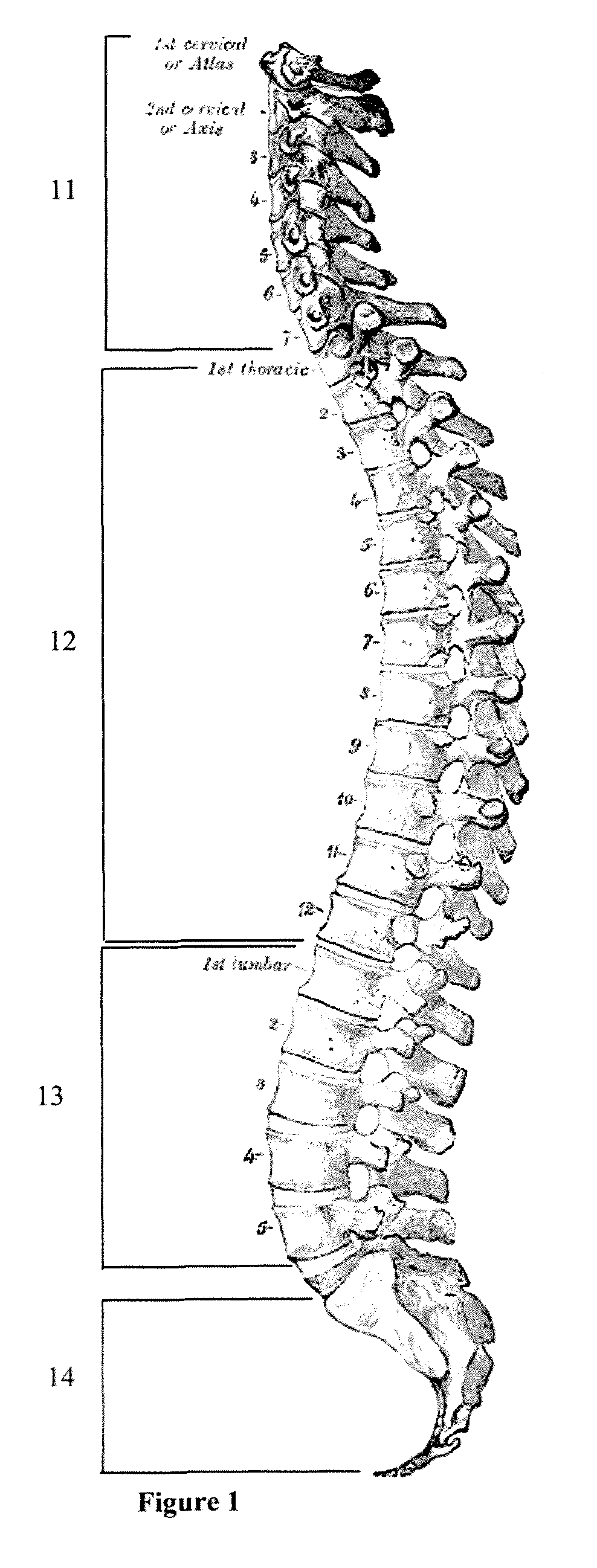
CROSS REFERENCE TO RELATED UNITED STATES APPLICATIONS[0001]This application claims priority from “Parametric model based vertebrae segmentation”, U.S. Provisional Application No. 60 / 838,048 of Liu, et al, filed Aug. 16, 2006, “RANSAC-based Algorithm for Spinal Cord Detection”, U.S. Provisional Application No. 60 / 839,291 of Jerebko et al., filed Aug. 22, 2006, and “Band-pass filtering for segmentation and characterization of structures”, U.S. Provisional Application No. 60 / 839,280 of Anna Jerebko, filed Aug. 22, 2006, the contents of all of which are herein incorporated by reference.TECHNICAL FIELD[0002]This disclosure is directed to the detection and segmentation of vertebrae structures in the spine.DISCUSSION OF THE RELATED ART[0003]Many anatomical structures in human body exhibit natural repetitiveness. For example, vertebrae of the spinal column have heights of certain range, and inter-vertebrae spaces and disks have a certain periodicity. Another example is the colon surface, wh...

Claims


Application Information

Patent Timeline
Patent Type & Authority Patents(United States)
IPC IPC(8): G06K9/00
CPCG06T7/0083G06T7/0089G06T2207/10088G06T2207/20116G06T2207/20168G06T2207/30012G06T2207/30096G06T7/12G06T7/149
Inventor JEREBKO, ANNALIU, JIAMINZHOU, XIANG
Owner SIEMENS HEALTHCARE GMBH
Learn More