
Doppler transceiver and probe for use in minimally invasive procedures

a transceiver and probe technology, applied in the field of minimally invasive procedures, can solve the problems of obliterating the vessel at risk of recurrent bleeding, not being able to visualize the subsurface vessel during laparoscopic procedures, and not being able to determine whether the gi endoscopist is a good candidate for surgery,

Active Publication Date: 2012-06-26
VASCULAR TECH
View PDF27 Cites 7 Cited by
  • Summary
  • Abstract
  • Description
  • Claims
  • Application Information

AI Technical Summary

Benefits of technology

[0017]Still another embodiment of the present invention provides such a system wherein at least one channel of the endoscopic or laparoscopic Doppler probe is coupled to a vacuum source, and whereby a vacuum may be applied to a target, steadying the endoscopic or laparoscopic Doppler probe proximate to the target.

Problems solved by technology

Currently, the GI endoscopist has no way of ascertaining whether an injection or coagulation therapy applied to an ulcer base has adequately occluded or obliterated the vessel at risk for recurrent bleeding.
In addition, subsurface vessels are not readily visualized during laparoscopic procedures.
Tactile sensation, which aids surgeons in the identification and evaluation of vessels in open surgical procedures, is not often available to the surgeon during laparoscopic surgery.
Meticulous dissection to identify a vessel, while necessary to avoid inadvertent injury to the vessel, adds significant time to the surgical procedure.
Other fields employing minimally invasive techniques such as arthroscopy, bronchoscopy, laryngoscopy, mediatinoscopy, thoracoscopy, cystoscopy, ureteroscopy, hysteroscopy, neuroendoscopy, robotic-assisted surgical procedures, and surgical fields in which laparoscopic surgery is utilized, present a similar inability to palpate tissue surfaces to assess blood flow.

Method used


Image

  • Doppler transceiver and probe for use in minimally invasive procedures
  • Doppler transceiver and probe for use in minimally invasive procedures
  • Doppler transceiver and probe for use in minimally invasive procedures

Examples


Embodiment Construction

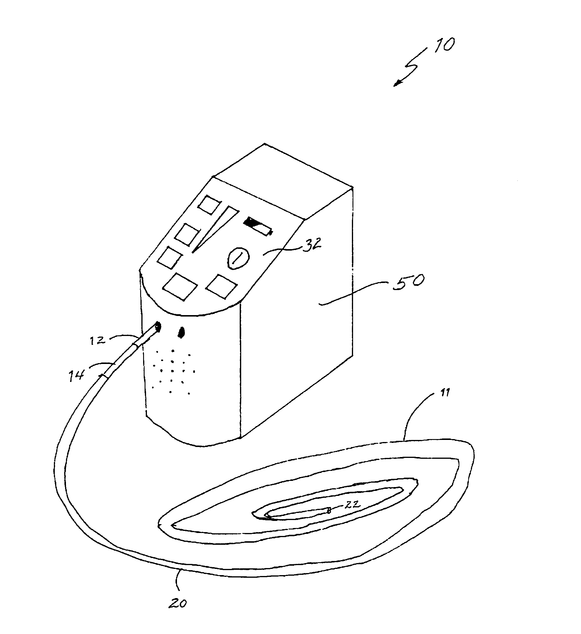
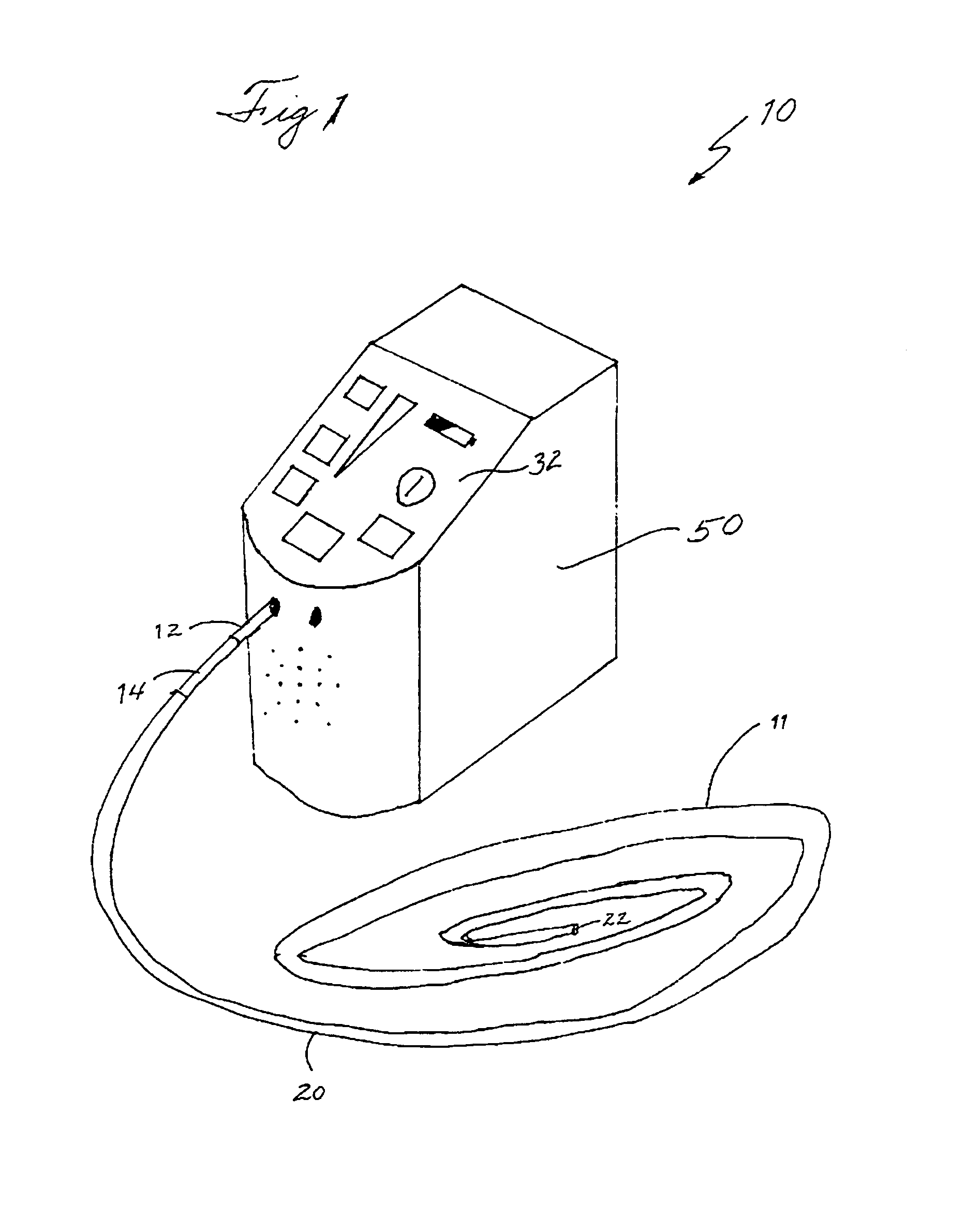
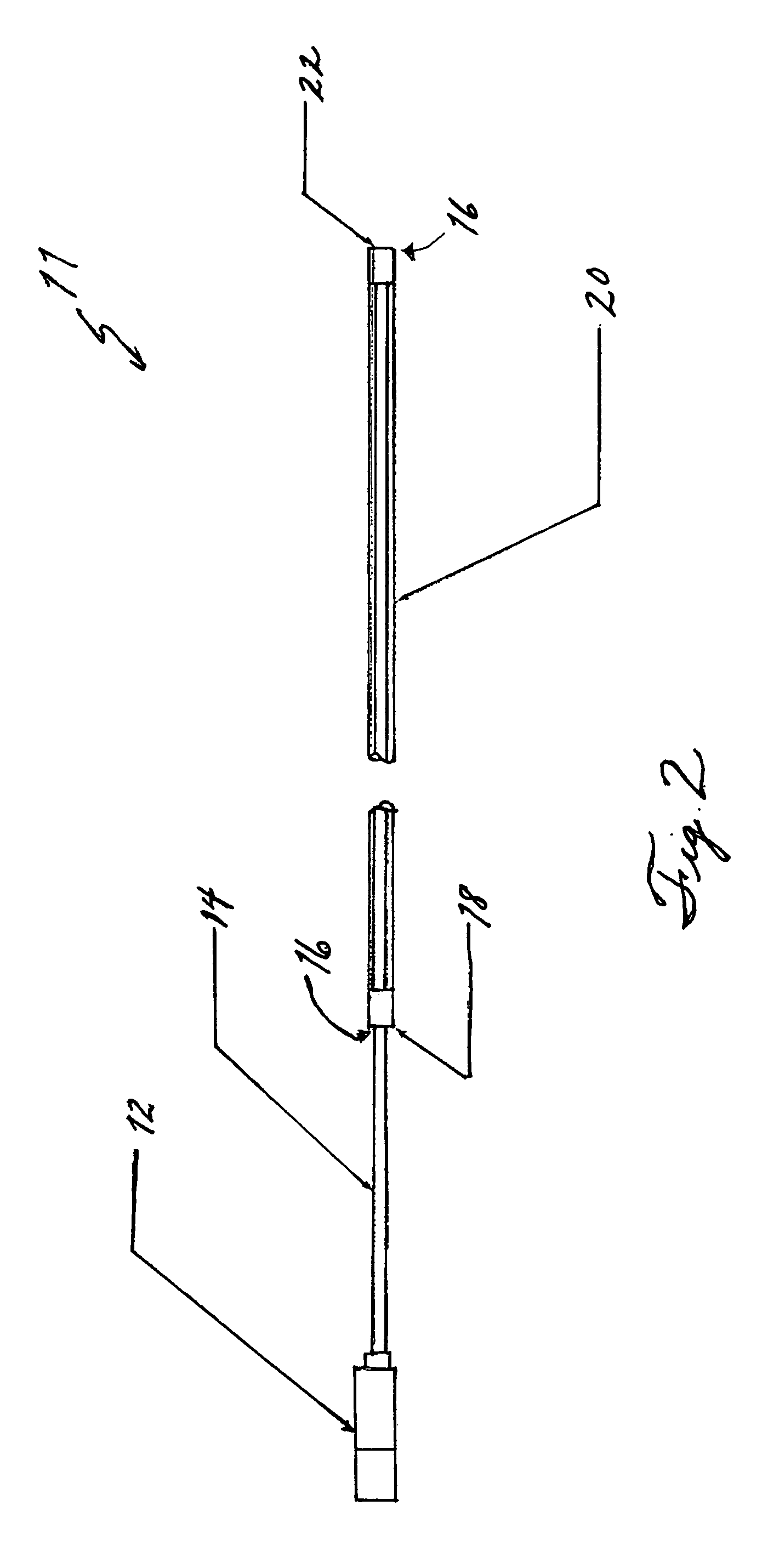
[0050]One embodiment of the present invention provides a non-imaging Doppler system (i.e. one without an ultrasonic images of tissues, but not excluding other visual cues or displays) for use in minimally invasive surgical or medical techniques, such as endoscopy or laparoscopy providing a portable audio blood flow detector, which allows for the identification and assessment of critical blood vessels. One embodiment of the present invention, illustrated in FIG. 1 provides a disposable Doppler probe 11, which can be inserted through the working channel of an endoscope, a laparoscopic port, or other such suitable entry point, and a portable transceiver unit 50, which transmits the Doppler signal in an auditory fashion. A varying audible signal is produced when the crystal 22 of probe 11 is placed upon a vessel within which there is blood flow. The frequency (i.e., pitch) of the signal is proportional to the blood velocity within the vessel. Distinctive tonal patterns are produced whic...


PUM


Abstract

A system is disclosed for the detection of blood flow. The system provides, a portable transceiver unit having a signal generator, a signal comparator, and an audio output whereby a user receives an audio signal indicating differences in Doppler signal, and a Doppler probe coupled to the portable transceiver. The Doppler probe including a piezoelectric crystal disposed at a first end of the Doppler probe; at least one wire transmitting signals between the transceiver unit to the piezoelectric crystal; a rigid or flexible sheath disposed around at least part of the at least one wire and the piezoelectric crystal; and a transceiver unit coupler disposed at a second end of the Doppler probe. The system may also have a quantitative blood flow meter or signal strength meter.

Description

RELATED APPLICATIONS[0001]This application claims the benefit of U.S. Provisional Application No. 60 / 529,779, filed Dec. 16, 2003. This application is herein incorporated in its entirety by reference.FIELD OF THE INVENTION[0002]The invention relates to devices for use in minimally invasive procedures, and more particularly, to an endoscopic or laparoscopic device providing portable blood flow detection.BACKGROUND OF THE INVENTION[0003]There is often a need to identify blood vessels and assess blood flow status in identified vessels during minimally invasive medical procedures. In gastroenterologic endoscopy, examples of such instances include the identification of vessels in peptic ulcer bases, which guides subsequent management of these lesions, identification of vessels at risk prior to polypectomy, and identification of aberrant vessels at risk for bleeding prior to sphincterotomy. Other specialties would yield additional examples of such minimally invasive techniques and procedu...

Claims


Application Information

Patent Timeline
Patent Type & Authority Patents(United States)
IPC IPC(8): A61B8/14
CPCA61B8/06A61B8/12A61B8/488
Inventor DOUGLAS, GARYREGAN, DAVIDSRIVASTAVA, NILENDUSUCHDEV, RACHANA
Owner VASCULAR TECH
Learn More