Tissue classification in medical images

a technology of tissue classification and medical images, applied in the field of medical imaging, can solve the problems of patient not meeting the periodic screening schedule, patient may not be screened as frequently as recommended, and imaging protocols for other organs may be similarly uncomfortabl

Active Publication Date: 2012-06-26
GENERAL ELECTRIC CO
View PDF3 Cites 129 Cited by
  • Summary
  • Abstract
  • Description
  • Claims
  • Application Information

AI Technical Summary

Problems solved by technology

These processes are very uncomfortable to the patient and may result in the patient not complying with the periodic screening schedule recommended for the patient.
As a result, the patient may not be screened as frequently as recommended.
Likewise, the imaging protocols for other organs may be similarly uncomfortable and may discourage or prevent patient compliance with the appropriate examination schedule.

Method used


Image

  • Tissue classification in medical images
  • Tissue classification in medical images
  • Tissue classification in medical images

Examples


Embodiment Construction

[0013]The present techniques are directed to the identification of different anatomical and / or pathological structures in medical images. The technique may be useful for distinguishing between certain types of tissues in different organs. For example, the present disclosure may be useful in distinguishing between lung nodules and healthy lung tissue. Likewise, the techniques may be useful for distinguishing between colon polyps and healthy colon tissue or for identifying other tissue types associated with other organs. In one embodiment, shape-based descriptors are used classify elements of an image (such as voxels in a three-dimensional representation) as different tissue types of interest. In this manner, different structural elements within an image may be labeled in accordance with the type of tissue they represent.

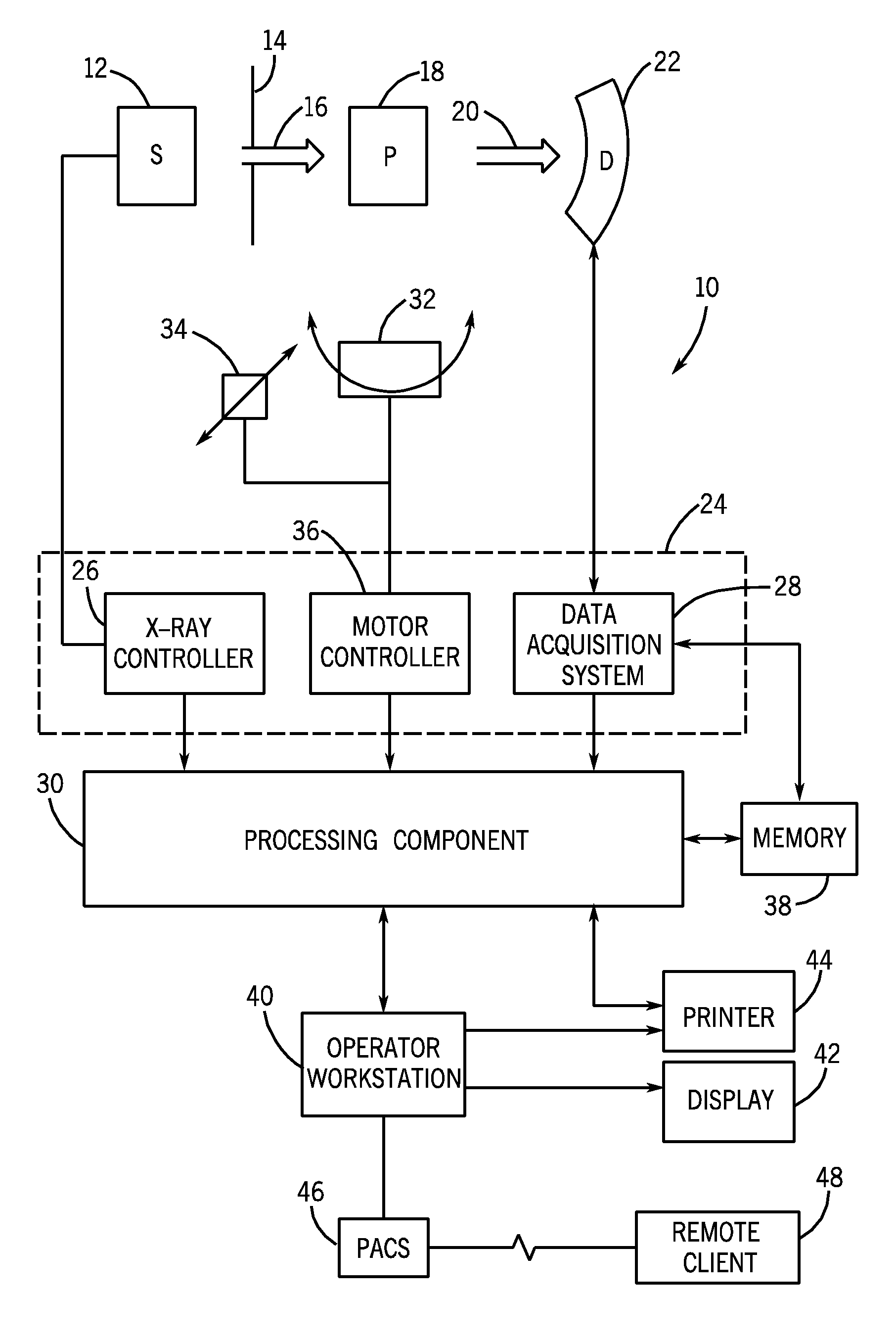
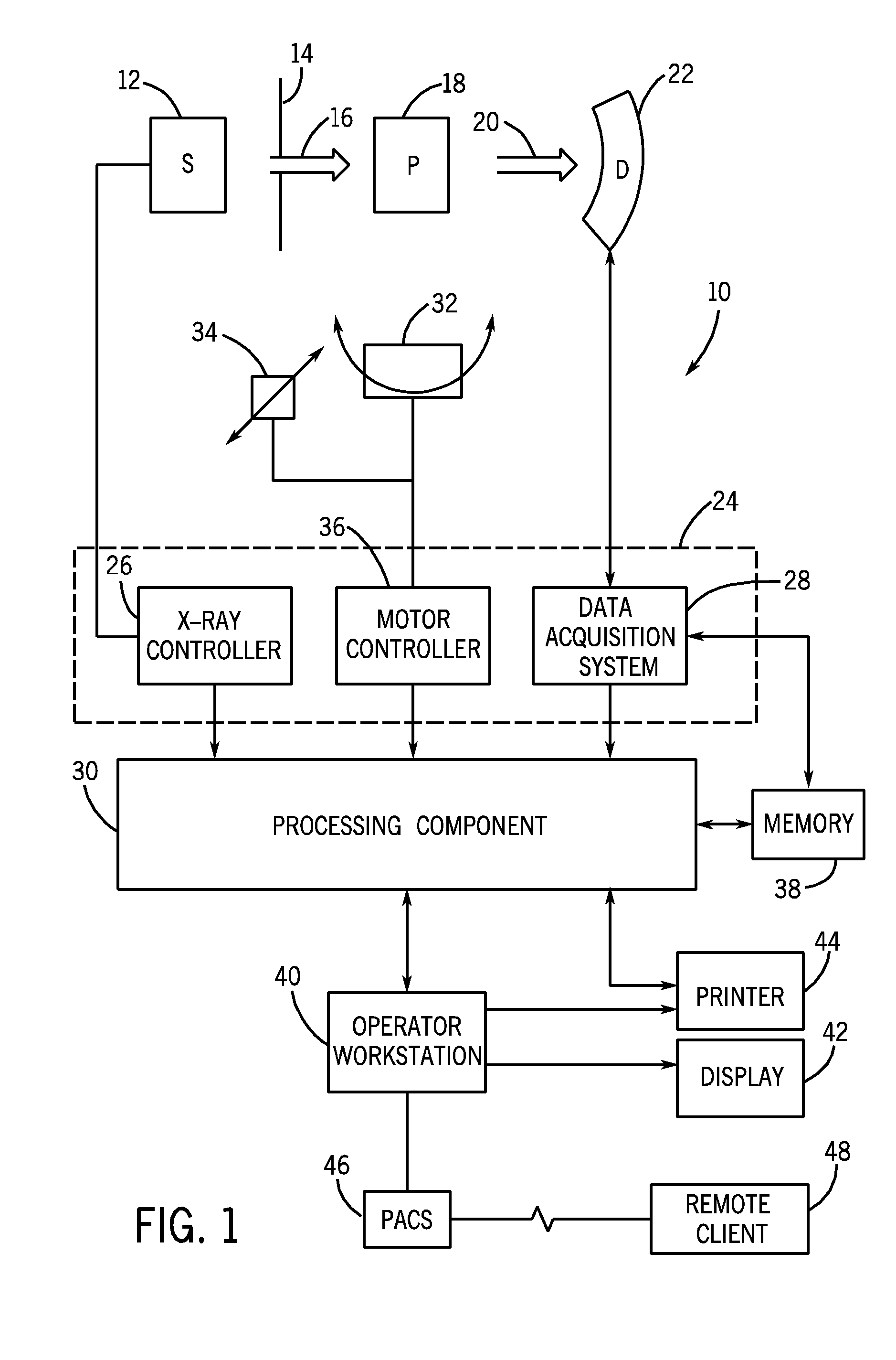
[0014]With this in mind, an example of a computer tomography (CT) imaging system that may be used to acquire images processed in accordance with the present technique...


PUM


Abstract

The present disclosure relates to the use of probabilistic models to classify elements of a medical image into different tissue types. The models may be based upon geometric abstractions of the different tissue types being classified. In addition, the present disclosure teaches the derivation and modification of models for tissue classification based upon the neighboring region of the voxels undergoing classification.

Description

BACKGROUND[0001]The invention relates generally to medical imaging and, in particular, to the identification of different anatomical and / or pathological structures in medical images.[0002]Non-invasive medical imaging technologies allow a caregiver to obtain and view images of the internal structures and organs of a patient without performing surgery or other invasive procedures. In particular, technologies such as X-ray radiography, computed tomography (CT), tomosynthesis, magnetic resonance imaging (MRI), ultrasound, C-arm angiography, positron emission tomography (PET), and single positron emission computed tomography (SPECT) use various physical principles to create two-dimensional and / or three-dimensional representations of the interior of the human body. For example, in those imaging modalities utilizing X-rays, the principle of operation typically involves assessing the differing attenuation of the X-rays by the patient's body at one or more angles. Based upon the differential...

Claims


Application Information

Patent Timeline
Patent Type & Authority Patents(United States)
IPC IPC(8): G06K9/00
CPCA61B6/032A61B6/037G06T7/0014G06T7/0081A61B6/025G06T2207/10081G06T2207/30032G06T7/11
Inventor MENDONCA, PAULO RICARDOSIROHEY, SAADBHOTIKA, RAHULZHAO, FEI
Owner GENERAL ELECTRIC CO
Why Eureka
Try Eureka