Method and apparatus for animal positioning in imaging systems

a positioning method and imaging system technology, applied in the field of invivo animal imaging, can solve the problems of insufficiently providing reproducible positioning of the animal's body, no common platform for imaging animals in multiple systems, and increasing the difficulty of detection of trends over tim

Inactive Publication Date: 2013-01-01
RGT UNIV OF CALIFORNIA
View PDF11 Cites 12 Cited by
  • Summary
  • Abstract
  • Description
  • Claims
  • Application Information

AI Technical Summary

Problems solved by technology

Hence, there is currently no common platform for imaging animals in multiple systems without moving the animals to separate, proprietary stages.
When an animal is used only for a single data point in a test, trends over time become more difficult to detect, as there are frequently individual differences between the animal subjects.
Current positioning systems, such as taping an animal in place or holding its head with toothbars and ear plugs, do not adequately provide such reproducible positioning of the animal's body.
Some imaging devices, such as CT, are subject to considerable artifacts in the resulting images if any part of the animal extends outside the field of view.
Since three dimensional objects, such as small animals, can be placed inside an imaging device in innumerable orientations, registration presents a difficult problem.
These markers, however, may move relative to animals and create inaccuracies in the software image registration.
Software registration is also limited in that only small changes in orientation can be corrected for.
Software registration can be expensive, inaccurate, and time consuming, but is frequently used in small animal imaging for lack of an effective alternative.
Additionally, there may be insufficient data available in one or more images for software methods to properly operate, as in the case when only a spherical tumor and nothing else is visible.
The relative positioning of the fiducial to the animal, however, cannot typically be determined with sufficient accuracy when the orientation of the subject is changed slightly between sessions or devices, so hardware registration is typically not possible with multiple imaging sessions in small animals.
Another problem with current imaging systems is that animals are exposed to pathogens.
Currently, the imaging systems used with these animals do not offer any type of pathogen barrier to shield the animals from pathogens in the open air.
Safe levels of injected anesthetics typically last only 30-50 minutes, and may not be suitable for longer experiments, or for experiments where two or more imaging systems are used to image the animal and exactly the same positioning is desired.
Injected anesthetics also suffer from a variable depth of anesthesia over time, which may affect the biological processes under investigation.
Without heating, the effects of hypothermia will result in physiological stress or even death to the animals, which is likely to adversely effect uptake and metabolism of injected compounds used for examining biological functions or disease processes.
Hypothermia-induced changes are typically not desirable.
Currently, few systems offer any heating options, and there is not an integrated system available to ensure the animals are kept at normal physiological temperatures throughout the whole imaging experiment process.
If the animal is cold and peripheral blood supply is restricted to maintain core body temperature, there may be little or no uptake into subcutaneous tumors, thus compromising the intended investigation.
However, this method delivers little heat, due to the low heat capacity of gasses, and when used for extended times can lead to dehydration of the animals.
The creation of disease models in small animals is often a time consuming and expensive process.
The complex nature of creating these animal disease models often requires weeks or months of preparation and analysis.
Therefore, the loss of even a single animal can be quite substantial.

Method used


Image

  • Method and apparatus for animal positioning in imaging systems
  • Method and apparatus for animal positioning in imaging systems
  • Method and apparatus for animal positioning in imaging systems

Examples


Embodiment Construction

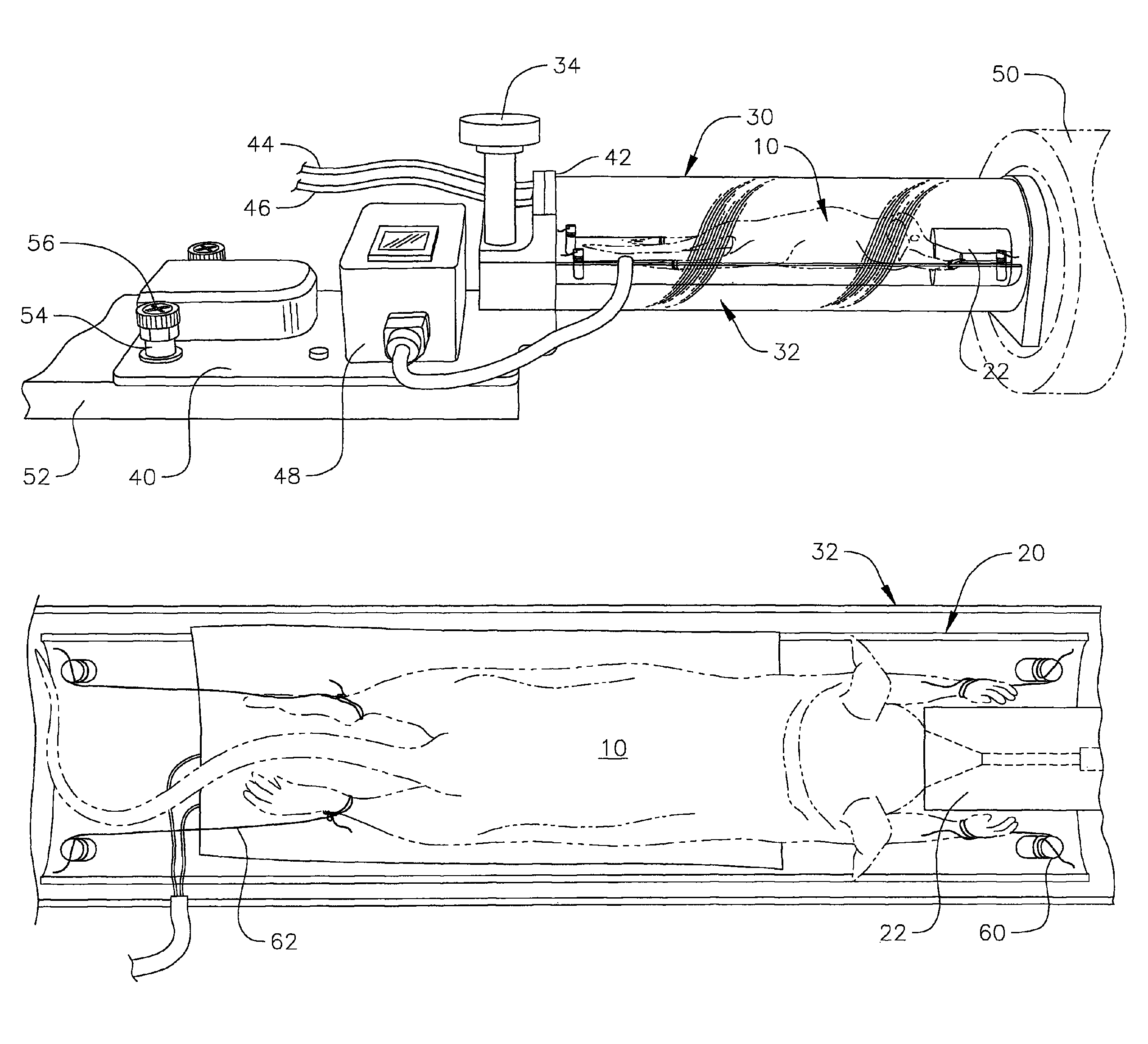
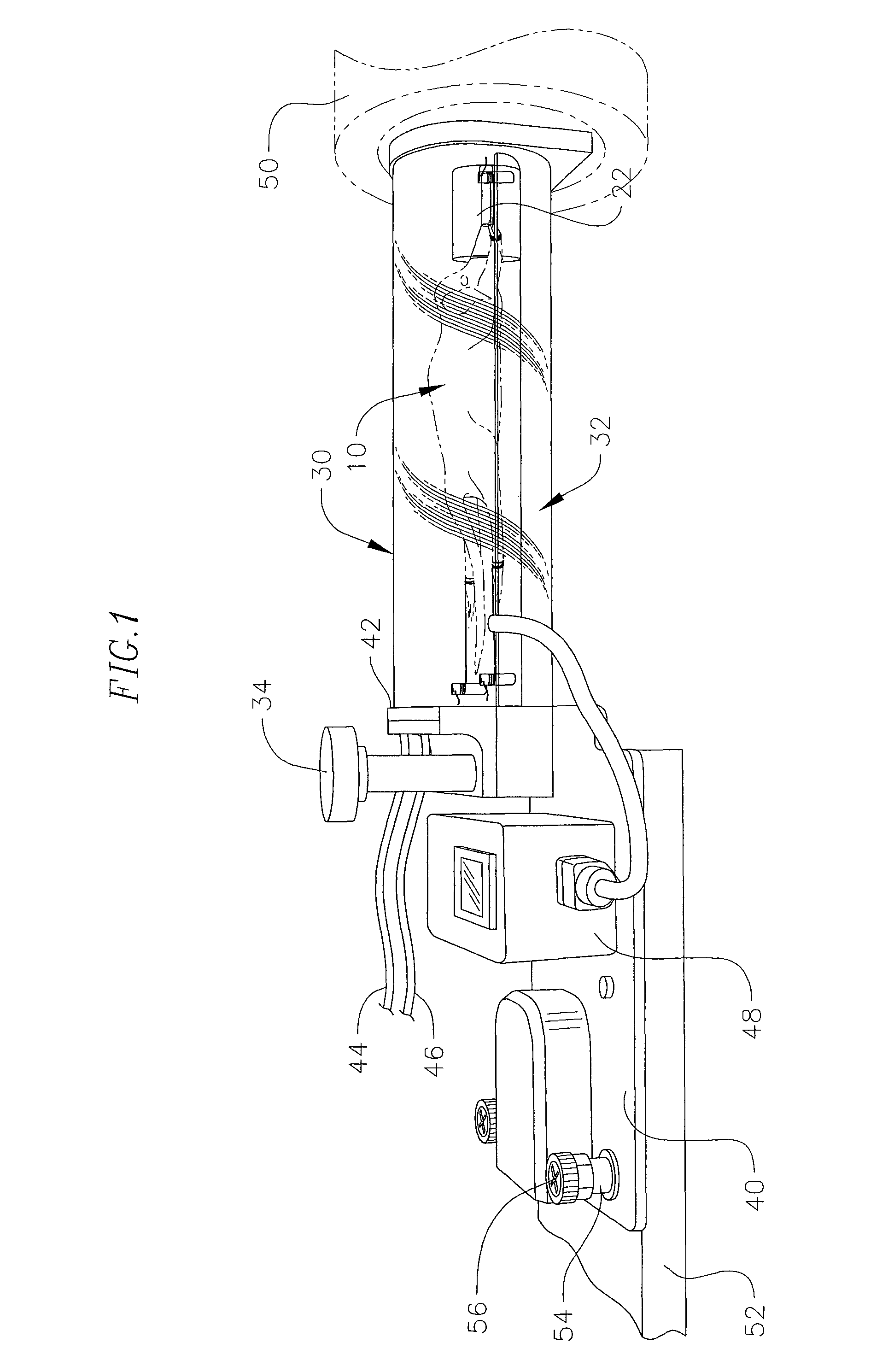
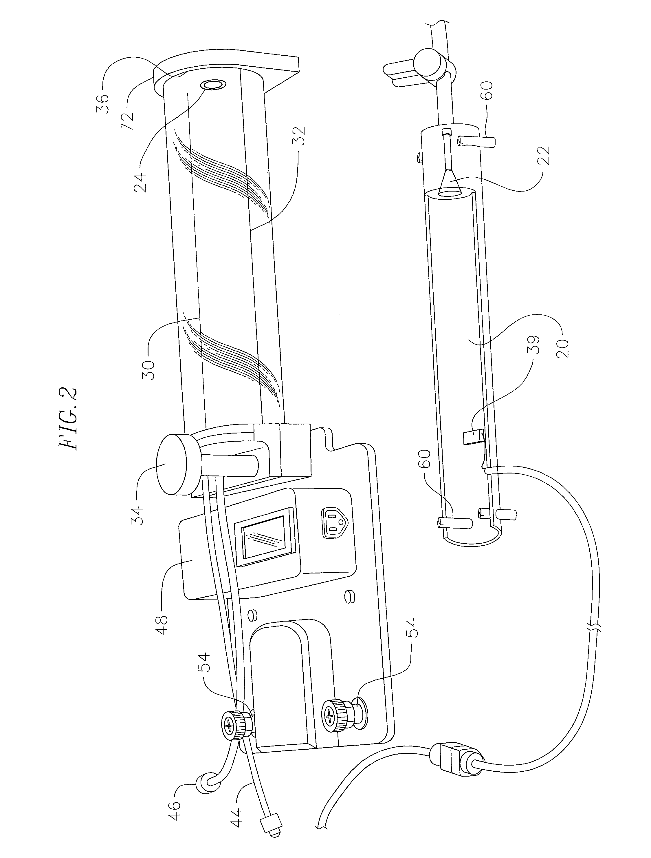
[0030]One embodiment of an apparatus for imaging an animal is shown in FIG. 1. An animal 10 is supported on a bed 20 that sits within a chamber 30, 32. In this embodiment, both the chamber and the bed are acrylic, but it is within the scope of the invention to construct the chamber and bed out of any material suitable for housing and supporting the animal for imaging within the particular imaging device.

[0031]The chamber 30, 32 is fixed to a first mounting surface 40 that also includes an anesthetic delivery system 42 and a heating element control 48. The first mounting surface 40 is fixed to a second mounting surface 52 associated with a first imaging device 50. In this embodiment, the first mounting surface 40 is a metallic plate coupled to one edge of the chamber 30, 32, but it is also within the scope of the invention for the first mounting surface 40 to be any other material compatible with the imaging device 50 or a mountable surface of the bed 20 or chamber 30 itself that wil...


PUM


Abstract

An apparatus for imaging an animal includes a first mounting surface, a bed sized to support the animal and releasably secured to or integral with the first mounting surface. The apparatus also includes a plurality of straps, each having a first end in a fixed position relative to the bed and a second end for tightening around a limb of the animal. A method for in-vivo imaging of an animal includes providing an animal that has limbs, providing a first mounting surface, and providing a bed removably secured to or integral with the mounting surface and sized to support the animal as well as being coupled to a plurality of straps. The method also includes placing the animal on the bed between the plurality of straps and tightening at least two of the plurality of straps around at least two of the limbs such that the animal is substantially secured in place relative to the bed.

Description

ACKNOWLEDGEMENT OF GOVERNMENT SUPPORT[0001]This invention was made with Government support of Grant No. R24 CA 92865, awarded by the National Institute of Health, and DE-FC03-02ER63420, awarded by the Department of Energy. The Government has certain rights in the invention.FIELD OF THE INVENTION[0002]The present invention relates to in-vivo animal imaging, namely a method and apparatus for positioning and imaging small animals for in-vivo imaging.BACKGROUND OF THE INVENTION[0003]In-vivo imaging of small animals to investigate biological functions, particularly those related to cancer, has become commonplace in the past few years. The advent of imaging systems such as the Concorde MicroPET, MicroSPECT, ImTek MicroCAT, MR, Xenogen IVIS optical imaging systems, and others specifically designed to image small animals, has resulted in a substantial increase in research of animal models of disease. Currently, each of these systems is designed and built independently. As such, each manufac...

Claims


Application Information

Patent Timeline
Patent Type & Authority Patents(United States)
IPC IPC(8): A01K15/04
CPCA61B6/508A61B6/045A61B6/0421A61B5/0555A61B5/4821A61B6/03A61B2503/40A61B5/055
Inventor HADJIOANNOU, ARION-XENOFONSTOUT, DAVID B.SILVERMAN, ROBERT W.
Owner RGT UNIV OF CALIFORNIA
Why Eureka
Try Eureka