Simultaneous multiple method out-patient uterus biopsy device and method

a uterus and multiple method technology, applied in the field of system and method for performing a uterus biopsy, can solve the problems of lack of brush or other biopsy sampling device at the distal end, and achieve the effects of improving sampling precision and patient comfort, improving control, and increasing comfort for patients

Active Publication Date: 2013-01-08
MALANOWSKA STEGA ZANETTA +1
View PDF5 Cites 19 Cited by
  • Summary
  • Abstract
  • Description
  • Claims
  • Application Information

AI Technical Summary

Benefits of technology

[0031]In accordance with another embodiment, the plastic tube and twisted wire are contained within a sheath of shorter length than the twisted wire, such that the sheath can be moved along the plastic tube to the atraumatic bulb on the distal end of the twisted wire, thereby covering the brush during insertion and removal after tissue collection. Before insertion, the sheath can be moved into position over the distal end of the twisted wire to protect the brush during insertion. Having the brush covered during insertion also increases comfort for the patient and protects the brush from collecting tissue from unintended areas. The sheath is moved back toward the proximal end of the twisted wire after the device has been inserted to the proper collection depth, exposing the brush and allowing for collection of a tissue sample. The sheath may be moved to completely uncover the brush or may be moved in gradients to uncover portions of the brush. This allows the practitioner to adjust the effective collection area of the brush based on the anatomy of the patient. The plastic tube covering the wire is scored in centimeter gradations along the plastic tube with markings indicating the exact length of the brush inserted into the uterus, starting from the distal tip of the brush to the proximal end of the plastic tube. This allows the clinician to know how deeply the brush is inserted into the uterus. The sheath is approximately the same length as the plastic tube and in position to cover the brush bristles prior to insertion. The sheath may be formed of a clear material such that the gradations on the plastic tube may be viewed through the sheath. The ability to measure insertion depth increases the certainty that the tissue sample collected is from the correct area, After a tissue sample is collected from the proper area, while the tissue sampling device remains inserted, the sheath can be moved back along the distal end of the twisted wire to cover the brush bristles before removing the brush. This allows for the tissue sample to be protected on the brush within the sheath during removal.
[0032]Additionally, the gradations along the flexible tube allow the practitioner to measure the length of bristles exposed. As the practitioner pulls the sheath from its insertion position towards the handle, the further the sheath is pulled the more bristles are exposed. The gradations (ruler) provide a visual confirmation of this measurement and allow the practitioner to be precise in exposing only a certain length of the brush bristles. This measurement allows the practitioner to have better control of where the tissue is sampled and allows the practitioner to adjust the length of brush based on patient specific parameters; such as uterine size measured during previous tests inferred based on patient history. Control of brush exposure increases sampling precision and patient comfort.
[0033]By combining two or more methods into one device, the invention eliminates pain, discomfort, and inconvenience, e.g., a second procedure to obtain an adequate and accurate specimen. The multiple methods of specimen collection, e.g., disruption by physical means, and suction, used together, allows a gentler application of the individual methods, e.g. a gentle disruption and gentle suction applied simultaneously can replace a vigorous disruption, e.g. D&C, and a powerful suction. The combination of multiple gentler methods in one device is safer and more effective than any method alone. On the other hand, to the extent either of the multiple methods is acceptably used at the levels used when employed individually, the apparatus and method according to the present invention has the capacity to produce improved results. The simultaneous combination of multiple methods provides a consistently more satisfactory specimen and more accurate diagnosis.
[0034]The invention can be used in a doctor's office and does not need analgesics or an operating room. It is a safer, faster and less expensive improvement over current methods.

Problems solved by technology

A vacuum biopsy sampling device, such as the known Pipelle endometrial suction curette produces a vacuum and draws it into the sheath by a similar principle, but lacks the brush or other biopsy sampling device at its distal end.

Method used


Image

  • Simultaneous multiple method out-patient uterus biopsy device and method
  • Simultaneous multiple method out-patient uterus biopsy device and method
  • Simultaneous multiple method out-patient uterus biopsy device and method

Examples


Embodiment Construction

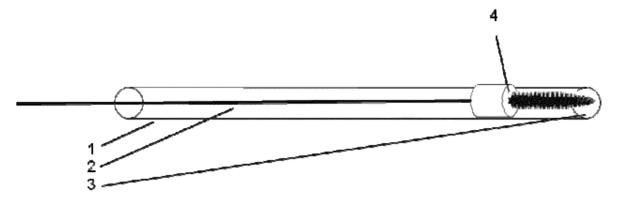
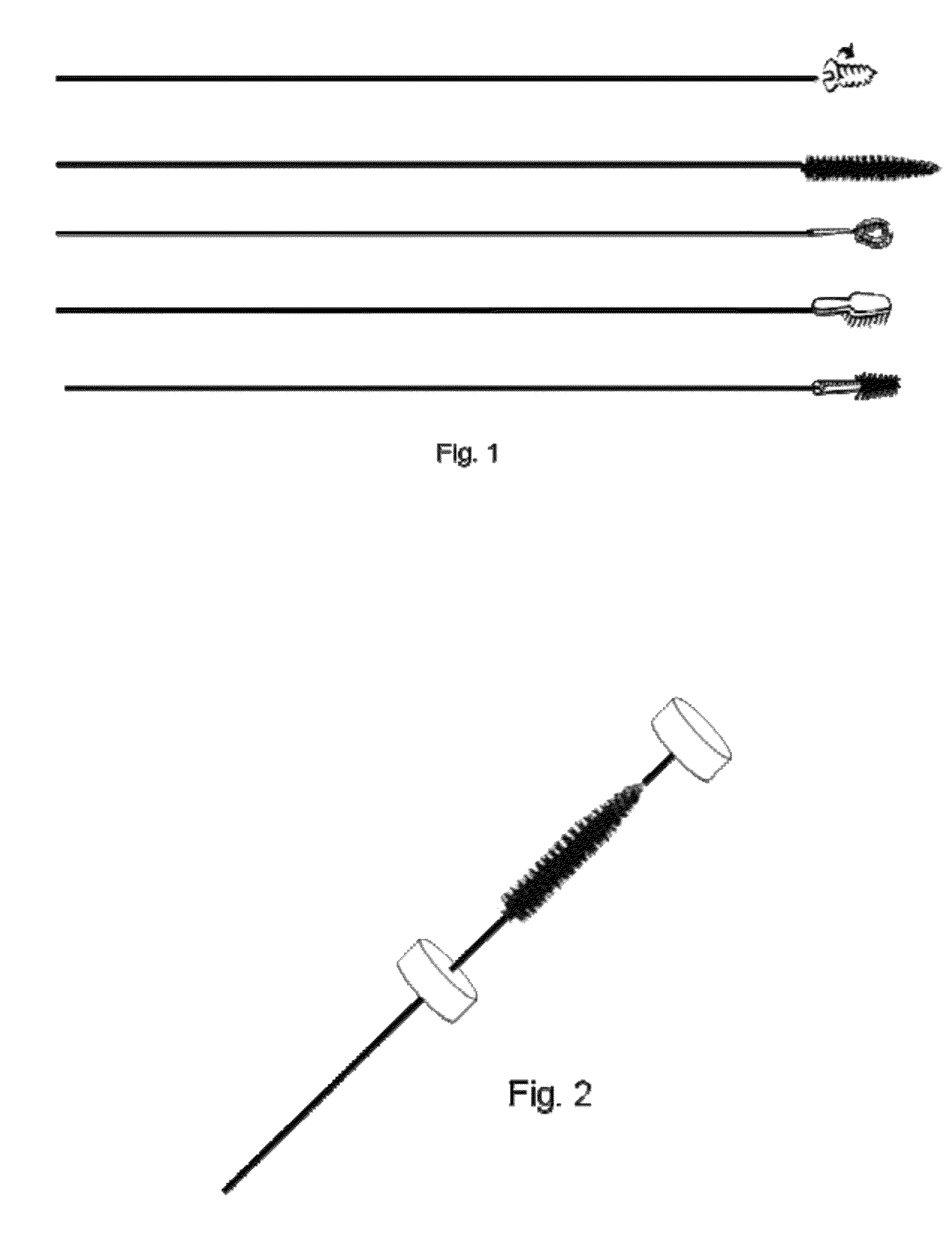
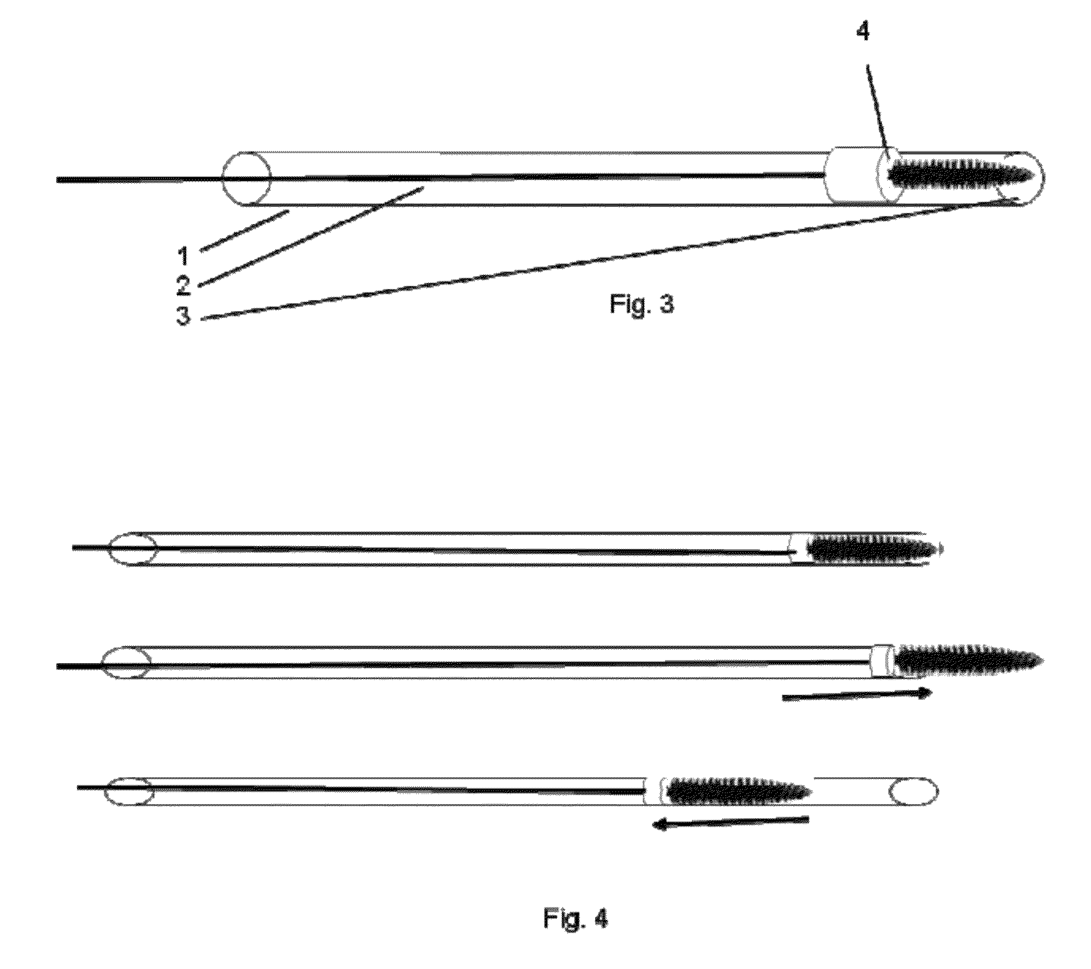
[0059]A preferred embodiment of the present invention consists of an outer thin walled tube (A) of variable diameter and length. In general dimensions are approximately 1.5 mm outside diameter and 1.4 mm inside diameter; length is between 20-50 cm. This tube may be a clear, malleable plastic tube, such as polyethylene. The inner obturator is preferably formed from a thin wire equivalent of approximately 0.1-0.2 mm diameter, having sufficient mechanical properties to convey the forces for extension and retraction during use. At one end, the one that enters the uterus, a disruptive enhancement is attached to the end. For example, the disruptive enhancement may have a screw like pattern mechanical attachment, a thin wire or multiple small semi-rigid projections that contact the uterus and loosen and collect the desired specimen. Alternatively, the disruptive enhancement may also include an enzymatic applicator and liquid based digestive enzymes that can be injected through the obturato...


PUM


Abstract

A biopsy device, comprising a flexible coaxial structure, comprising an obturator within a sheath, the obturator being adapted to be displaced with respect to the sheath along the coaxial axis by a force applied at a proximal end; a disruptor, at a distal end of the obturator, adapted to disrupt a tissue surface to free cells therefrom, having a first position covered within the sheath and a second position freely extending beyond the sheath; an element, having a fixed position on the obturator, and a close clearance with respect town inner surface of the sheath, such that a retraction of the obturator within the sheath causes an associated change in pressure in a space between the obturator distal to the element and the sheath, to thereby draw fluid proximate to an end of the sheath within the sheath.

Description

FIELD OF THE INVENTION[0001]The present invention provides a system and method for performing a biopsy of the uterus. More particularly, it is a mechanical device that performs a mechanical disruption of the uterus, especially the endometrium, and simultaneously aspirates the residue of the disruption.BACKGROUND OF THE INVENTION[0002]Current biopsy devices and methods do not consistently provide an adequate sample to diagnose uterine abnormalities. For instance, if a cancer exists on the inside of the uterus, i.e. the endometrium, current devices and methods may miss the cancer and preclude an accurate diagnosis. To ensure an adequate and accurate specimen currently often requires a painful and dangerous series of procedures.[0003]For example, the Cook Medical Tao Brush™ I.U.M.C. Endometrial Sampler, and the Pipelle endometrial biopsy device (See, Sierecki A R, Gudipudi D K, Montemarano N, Del Priore G., “Comparison of endometrial aspiration biopsy techniques: specimen adequacy.”J R...

Claims


Application Information

Patent Timeline
Patent Type & Authority Patents(United States)
IPC IPC(8): A61B5/00
CPCA61B10/0291A61B2010/0216A61B10/0283A61B2010/0208A61M25/09A61M2025/09191
Inventor MALANOWSKA-STEGA, ZANETTADELPRIORE, GIUSEPPE
Owner MALANOWSKA STEGA ZANETTA
Why Eureka
Try Eureka