
Method and computed tomography scanner for carrying out an angiographic examination

a computed tomography and angiographic technology, applied in the field of methods and/or computed tomography scanners for carrying out angiographic examinations, can solve the problems of inability to examine in a conventional fashion, inability to achieve good results in general only in the process, so as to reduce the dose of x-ray applied to the patient, reduce the workload of examination, and reduce the effect of patient movemen

Active Publication Date: 2013-05-21
SIEMENS HEALTHCARE GMBH
View PDF28 Cites 6 Cited by
  • Summary
  • Abstract
  • Description
  • Claims
  • Application Information

AI Technical Summary

Benefits of technology

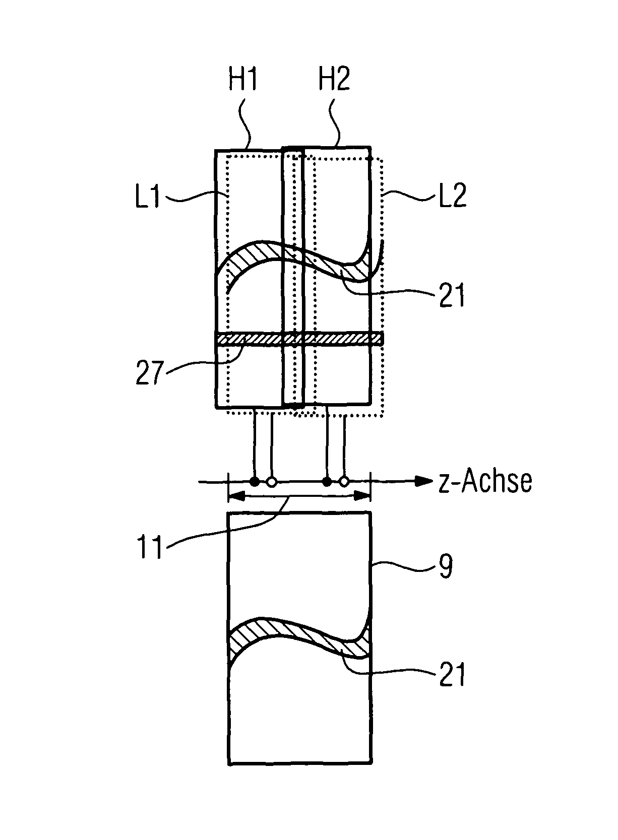
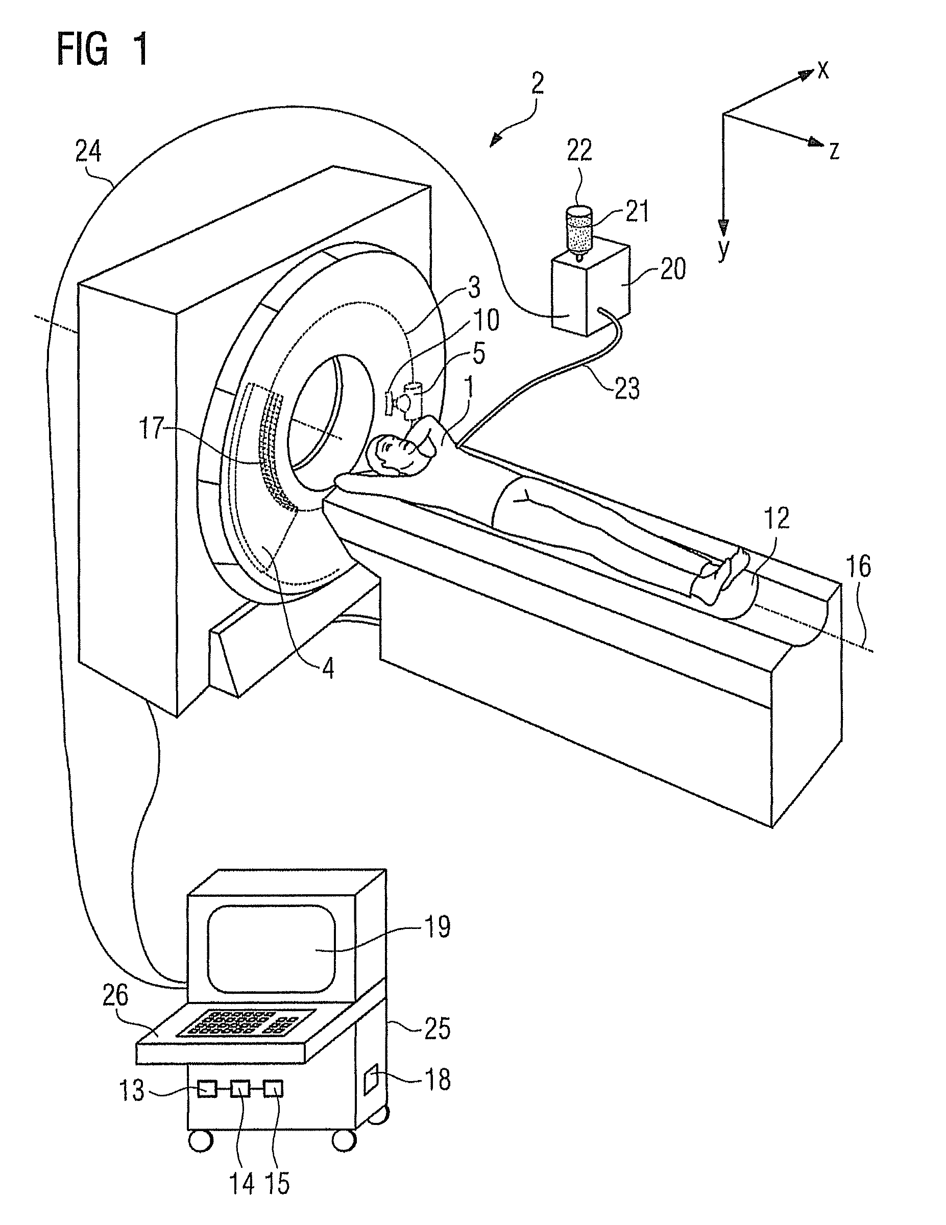
The patent is about using computed tomography scanners for applications that were previously the reserve of other modalities, such as C-arm systems with flat-panel detectors. The inventors have found that multi-row detector computed tomography scanners with small pixel sizes can provide high spatial resolution with large z-coverage, making them ideal for angiographic examinations comparable to subtraction angiography with a C-arm system. Furthermore, the projection data can be acquired within a certain z-axis section, and the measurement region can be repositioned using a continuous or oscillatory motion of the patient table or a flying focus. This allows for a wider field of view and faster imaging.

Problems solved by technology

However, vessels and organs, which do not differ substantially in terms of their attenuation properties from the surrounding soft tissue, cannot be examined in a conventional fashion as a result of the lack of contrast.
A significant disadvantage of this method emerges from the fact that the projection images used for the subtraction were recorded at different times. A patient movement that took place between the recording times, as already caused by breathing, must be compensated for by registering the two projection images before the actual subtraction.
Sufficiently good results can in general only be achieved in the process if the registration is carried out on the basis of computationally expensive affine image transformations.
However, despite image registration, many situations do not allow mathematically completely accurate estimates of the patient movement, which leads to the formation of a certain amount of shadowing in the region of erroneously registered image structures in the resulting projection image.
Even without generating a native slice image, the CTA examinations are disadvantageous in that the patient is exposed to an increased X-ray dose compared to an angiographic examination using a C-arm system as a result of the multiplicity of projections from different angular directions required for the image reconstruction.

Method used


Image

  • Method and computed tomography scanner for carrying out an angiographic examination
  • Method and computed tomography scanner for carrying out an angiographic examination
  • Method and computed tomography scanner for carrying out an angiographic examination

Examples


Embodiment Construction

[0035]Various example embodiments will now be described more fully with reference to the accompanying drawings in which only some example embodiments are shown. Specific structural and functional details disclosed herein are merely representative for purposes of describing example embodiments. The present invention, however, may be embodied in many alternate forms and should not be construed as limited to only the example embodiments set forth herein.

[0036]Accordingly, while example embodiments of the invention are capable of various modifications and alternative forms, embodiments thereof are shown by way of example in the drawings and will herein be described in detail. It should be understood, however, that there is no intent to limit example embodiments of the present invention to the particular forms disclosed. On the contrary, example embodiments are to cover all modifications, equivalents, and alternatives falling within the scope of the invention. Like numbers refer to like ...


PUM


Abstract

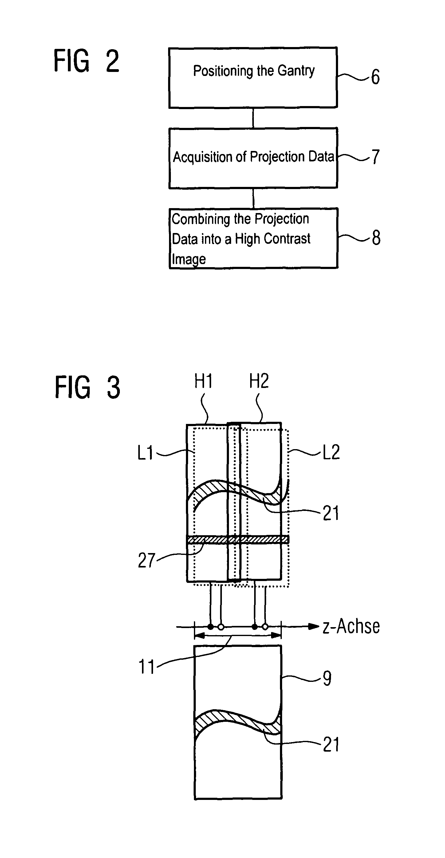
A method and a computed tomography scanner are disclosed for carrying out an angiographic examination of a patient, wherein the utilized computed tomography scanner includes at least one recording system mounted on a gantry such that it can rotate about a z-axis. Projection data is acquired from at least one prescribed angular position of the gantry for at least two different energies of X-ray radiation. The projection data is subsequently combined to form a resulting projection image by evaluating the projection data corresponding to the respective angular position, in which projection image at least one substance, which should be displayed selectively, is imaged with a high image contrast compared to the respective individual projection data. This procedure extends the field of application of the computed tomography scanner to projection-based angiography examinations, which were previously restricted to C-arm systems. 3D image reconstruction methods and projection methods can be carried out on opposite sides and with great flexibility during an examination, without the need for an additional modality. By using a multispectral technique, it is possible to contrast agent. The projection data at dispense with recording a native projection data record without the different energies are moreover acquired with no or little time offset, and so a computationally expensive and error-prone registration of the data records can be dispensed with.

Description

PRIORITY STATEMENT[0001]The present application hereby claims priority under 35 U.S.C. §119 on German patent application number DE 10 2010 027 227.2 filed Jul. 15, 2010, the entire contents of which are hereby incorporated herein by reference.FIELD[0002]At least one embodiment of the invention generally relates to a method and / or a computed tomography scanner for carrying out an angiographic examination.BACKGROUND[0003]It is well-known that X-ray scanners are used in medicine to obtain information about the interior of a patient for the purpose of diagnosis and / or therapy. In the process, the different attenuation properties of the various substances with respect to X-ray radiation generated by the X-ray scanner are utilized for providing contrast in the image. Compared to soft tissue, bone tissue has very different attenuation properties. As a result of the high image contrast connected therewith, bone structures can be analyzed in a simple fashion. However, vessels and organs, whi...

Claims


Application Information

Patent Timeline
Patent Type & Authority Patents(United States)
IPC IPC(8): A61B6/00G06K9/00
CPCA61B6/032A61B6/507A61B6/504A61B6/482A61B6/4035A61B6/405A61B6/4241A61B6/027A61B6/4021
Inventor FLOHR, THOMASGRASRUCK, MICHAELSCHMIDT, BERNHARDSEDLMAIR, MARTIN
Owner SIEMENS HEALTHCARE GMBH
Learn More