Apparatus and method for widefield functional imaging (WiFI) using integrated structured illumination and laser speckle imaging

a widefield functional imaging and structured illumination technology, applied in the field of medical imaging using light, can solve the problems of only providing relative changes in hemodynamic parameters, incomplete picture of underlying mechanisms, and the absolute quantification of the magnitude and origin of cellular and molecular events remains a significant challenge, and achieves the effect of reducing motion artifacts or discomfort for patients

Active Publication Date: 2013-08-13
MODULATED IMAGING
View PDF6 Cites 57 Cited by
  • Summary
  • Abstract
  • Description
  • Claims
  • Application Information

AI Technical Summary

Benefits of technology

[0012]WiFI provides researchers with a quantitative tool to study disease progression and therapeutic response with 1) a high degree of fidelity and spatial localization, and 2) sufficient spatiotemporal resolution and probe volume to study events on length scales that have broad biologic and clinical relevance (i.e., mm-cm). With WiFI the ambiguity that exists in planar imaging modalities (between molecular reporter depth and signal strength) will be overcome, resulting in absolute measurements of signal and more accurate comparisons of multiple experimental conditions. The knowledge of both local metabolic activity and molecular reporter dynamics will result in an improved understanding of cell-vascular coupling phenomena. With absolute quantification of local oxygen saturation and blood flow, researchers will be able to draw comparisons among data collected in serial measurement sessions on a single patient and among patients measured at different sites worldwide. Furthermore, with absolute quantification of tissue parameters, we envision the possibility of WiFI-based epidemiologic studies to facilitate development of physiologically meaningful quantitative metrics of tissue function (i.e., “normal” vs. “abnormal” blood flow).
[0016]A fast, clinic-friendly imaging instrument (WiFI Instrument 3) is fabricated for therapy guidance and wound healing monitoring. Instrument 3 is a clinic-friendly “snapshot” hyperspectral (500-1000 nm) system, capable of broadband spatial-frequency-domain imaging on a sub-second timescale. The device enables mapping of the spatial distributions of hemoglobin, lipid, water, and tissue scattering in layered tissue systems. This lightweight system is mounted on an articulating arm to allow arbitrary positioning for a variety of clinical applications, including flap and diabetic wound monitoring, melanoma studies, and port-wine stain imaging. The key component of this system is a custom holographic computed tomographic imaging spectrometer (CTIS). In combination with the 2K×4K Dalsa 11M04 camera, the 7-order filter provides ˜5 nm spectral resolution of absorption and scattering across the entire spectral range from 500-1000 nm, all with only three phase projection images. This allows snapshot clinical measurements and multiple chromophore map extraction before, during, and after therapies with minimal motion artifacts or discomfort to the patient. The projection subsystem is comprised of a separate DMD light engine from Apogen geared toward lightweight construction (no filter wheels, magnesium exoskeleton, and fiber light guide tungsten source delivery), and is designed for integration with the CTIS / Dalsa imaging arm. The Apogen light engine and Dalsa Pantera 11M04 camera are synchronized via the same underlying platform developed for WiFI Instruments 1 and 2. We constructed this system initially with a liquid crystal tunable filter (LCTF), later replaced with the CTIS upon delivery of the device. A graphics processor unit (GPU) provides acceleration of the CTIS tomographic reconstruction code. While acquisition with the proposed system is less than 1 s, the CTIS reconstruction step is currently limited to more than 1 min / image for a total of more than 3 min computational time. While this delay in feedback is acceptable for longitudinal studies of chronic disease progression and therapeutic response, it is incompatible with applications geared at informing a physician while monitoring an acute therapy, such as port-wine stain treatment, and resection of cancerous tissues in brain, melanoma, and breast cancer surgeries. We utilize the programmable, massively data-parallel nature of GPUs to solve the CTIS expectation-maximization (EM) problem (a naturally-parallel algorithm). This is implemented using the CUDA programming model by nVidia, an abstracted set of floating-point libraries aimed at general purpose GPU computation (GPGPU). Preliminary reconstructions of CTIS data have yielded long reconstruction times (1 minute per image). A work station with GPUs will reduce the reconstruction time of the CTIS to allow near real-time (1 fps) quantitative hyper-spectral imaging.
[0028]The apparatus comprises a laser, and a LCOS spatial light modulator to permit laser projection as a motion-free system by preventing speckle dephasing in the integrated device itself.
[0037]The apparatus comprises a source, a detector, and dual filter wheels in front of the source and detector to allow a flexible combination of multispectral reflectance and fluorescence measurements, where filter wheel alignment is maintained and stray light rejected to maximize fluorescence contrast, where the source, detector, and dual filter wheels are mutually synchronized with each other.
[0043]The integrated SI and LSI devices comprise a holographic computed tomographic imaging spectrometer (CTIS) with a camera capable of spectral resolution of absorption and scattering across a spectral range from 500-1000 nm with only three phase projection images to allow real time clinical measurements and multiple chromophore map extraction before, during, and after therapies with minimal motion artifacts or discomfort to a patient.

Problems solved by technology

Despite these tremendous advances in molecular imaging, absolute quantification of the magnitude and origin of cellular and molecular events remains a significant challenge.
Local changes in intrinsic signals have been attributed to an increase or decrease in local neurovascular activity, but separation of these signal dynamics into basis components such as oxy / deoxygenation of blood, changes in blood volume, and optical scattering, has not been performed, resulting in an incomplete picture of the underlying mechanisms.
Technologic advances such as combined reflectance / fluorescence imaging, multi-parameter full-field imaging, and laminar optical tomography enable improved separation of the signals to study important parameters such as local tissue metabolic dynamics; however, these technologies currently can provide only relative changes in hemodynamic parameters and do so without consideration of optical scattering effects on extracted tissue parameters.

Method used


Image

  • Apparatus and method for widefield functional imaging (WiFI) using integrated structured illumination and laser speckle imaging
  • Apparatus and method for widefield functional imaging (WiFI) using integrated structured illumination and laser speckle imaging
  • Apparatus and method for widefield functional imaging (WiFI) using integrated structured illumination and laser speckle imaging

Examples


Embodiment Construction

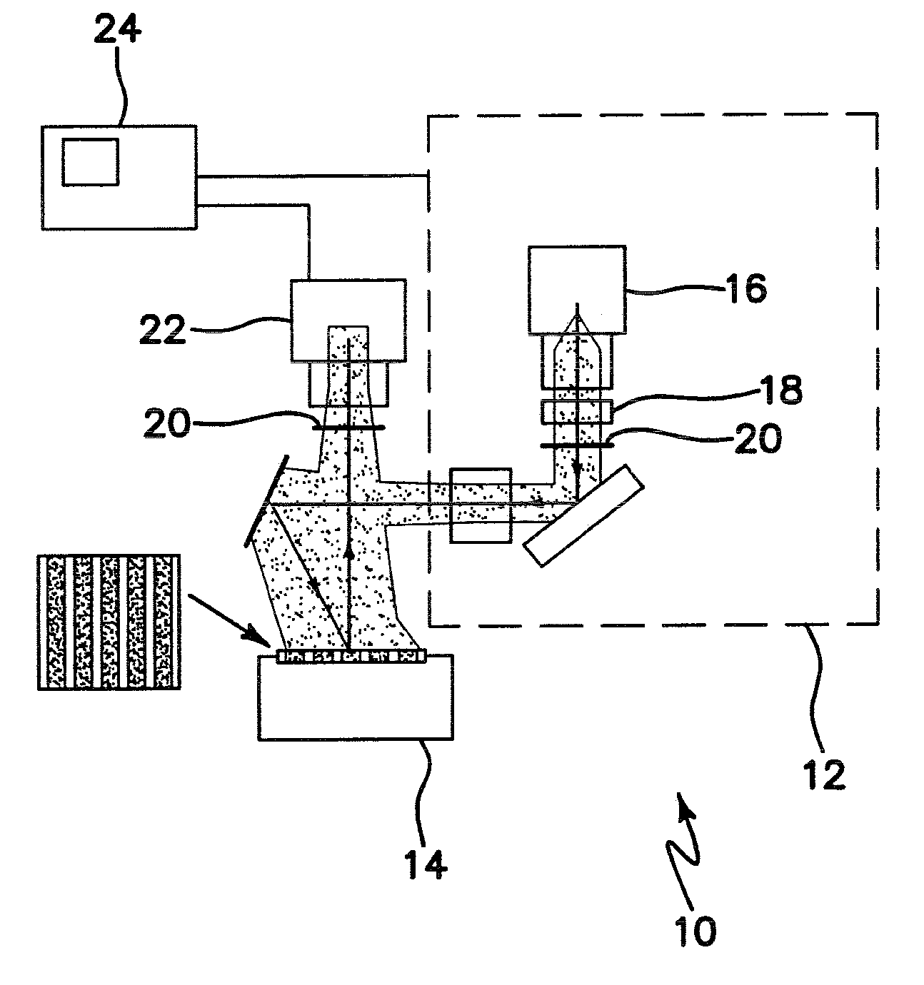
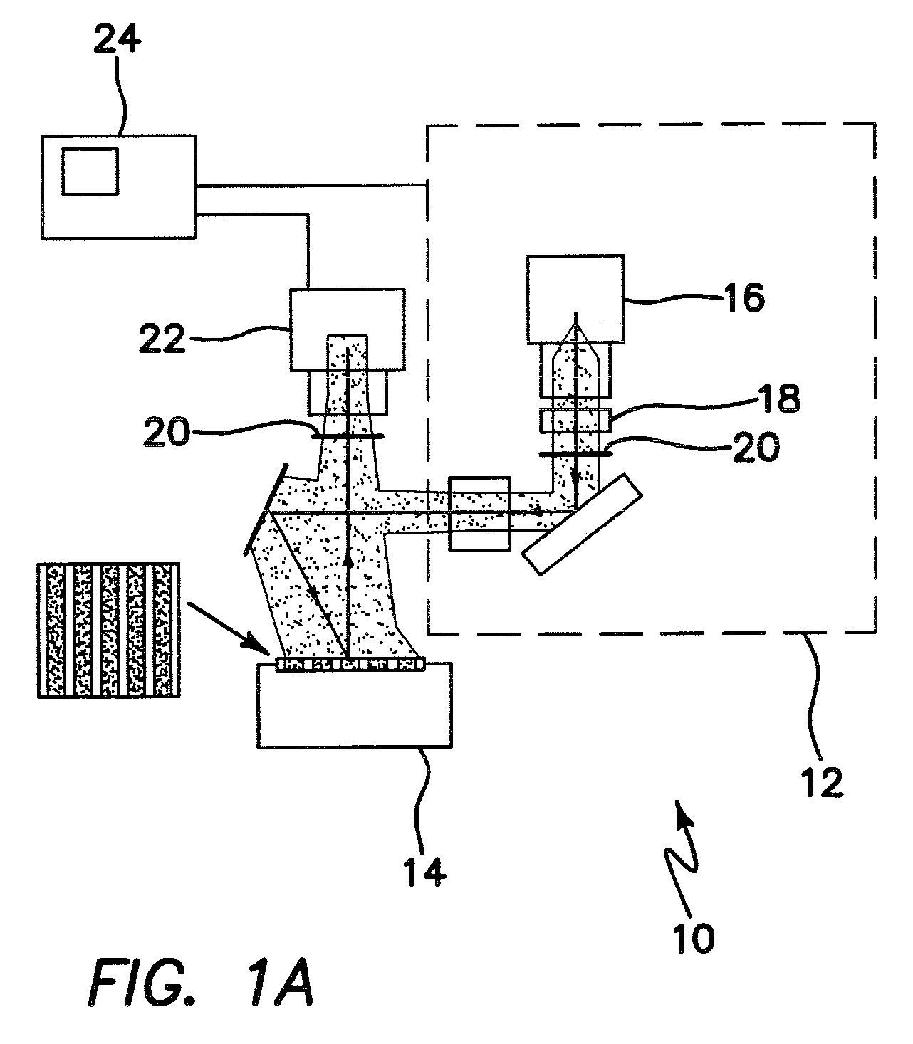
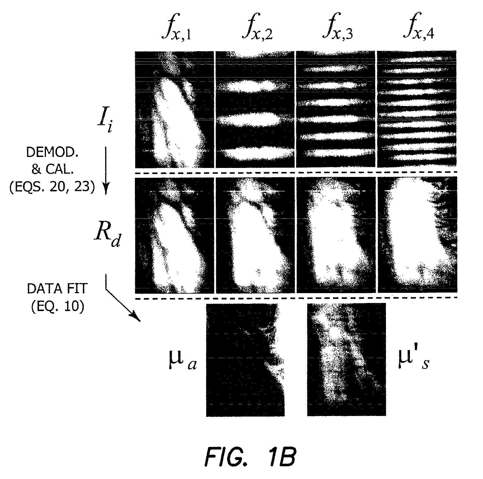
[0064]Quantitative characterization of tissue structure and function across spatial scales is one of the most challenging problems in medical imaging. Field of view, depth of interrogation, and resolution are critical features that dramatically impact image quality and information content. Optical methods can potentially provide a single platform for imaging biological tissues with resolution and depth sensitivity from microns to centimeters, limited by fundamental light-tissue interactions.

[0065]The broad advantage of the wide-field functional imaging (WIFI) core is that it is an integrated imaging platform capable of quantitative subsurface metabolic imaging across spatial scales. WiFI simultaneously measures tissue blood flow, biochemical composition (i.e. oxy- and deoxy-hemoglobin, water and lipid content), and molecular fluorescence in turbid tissues. It possesses sufficient spatio-temporal resolution to study both fast (i.e., ms timescale) and localized (i.e., tens of μm to mm...


PUM


Abstract

An apparatus for wide-field functional imaging (WiFI) of tissue includes a spatially modulated reflectance / fluorescence imaging (SI) device capable of quantitative subsurface imaging across spatial scales, and a laser speckle imaging (LSI) device capable of quantitative subsurface imaging across spatial scales using integrated with the (SI) device. The SI device and LSI device are capable of independently providing quantitative measurement of tissue functional status.

Description

RELATED APPLICATIONS[0001]The present application is related to U.S. Provisional Patent Application Ser. No. 60 / 985,872, filed on Nov. 6, 2007, which is incorporated herein by reference and to which priority is claimed pursuant to 35 USC 119.GOVERNMENT RIGHTS[0002]This invention was made with government support under grant RR001192 awarded by the National Institutes of Health. The government has certain rights in the invention.BACKGROUND OF THE INVENTION[0003]1. Field of the Invention[0004]The invention relates to the field of medical imaging using light.[0005]2. Description of the Prior Art[0006]Due to its relatively low cost and ease of implementation, optical imaging is an attractive technology to study intrinsic signals associated with endogenous chromophores as well as targeted exogenous probes. With the explosive growth in available molecular reporter strategies for studying fundamental biophysical processes, there has been a paradigm shift in research efforts from ex vivo des...

Claims


Application Information

Patent Timeline
Patent Type & Authority Patents(United States)
IPC IPC(8): A61B6/00
CPCA61B5/0073G01N21/4795G01N21/6456A61B5/4094
Inventor DURKIN, ANTHONY J.CUCCIA, DAVIDTROMBERG, BRUCE J.MAZHAR, AMAANCHOI, BERNARD
Owner MODULATED IMAGING
Why Eureka
Try Eureka