APPARATUS AND METHOD FOR WIDEFIELD FUNCTIONAL IMAGING (WiFI) USING INTEGRATED STRUCTURED ILLUMINATION AND LASER SPECKLE IMAGING

a widefield functional imaging and structured illumination technology, applied in the field of medical imaging using light, can solve the problems of only providing relative changes in hemodynamic parameters, incomplete picture of underlying mechanisms, and the absolute quantification of the magnitude and origin of cellular and molecular events remains a significant challenge, and achieves the effect of reducing motion artifacts or discomfort for patients

Active Publication Date: 2009-05-07
MODULATED IMAGING
View PDF6 Cites 118 Cited by
  • Summary
  • Abstract
  • Description
  • Claims
  • Application Information

AI Technical Summary

Benefits of technology

[0042]The integrated SI and LSI devices comprise a holographic computed tomographic imaging spectrometer (CTIS) with a camera capable of spectral resolution of absorption and scattering across a spectral range from 500-1

Problems solved by technology

Despite these tremendous advances in molecular imaging, absolute quantification of the magnitude and origin of cellular and molecular events remains a significant challenge.
Local changes in intrinsic signals have been attributed to an increase or decrease in local neurovascular activity, but separation of these signal dynamics into basis components such as oxy/deoxygenation of blood, changes in blood volume, and optical scattering, has not been performed, resulting in an incompl

Method used


Image

  • APPARATUS AND METHOD FOR WIDEFIELD FUNCTIONAL IMAGING (WiFI) USING INTEGRATED STRUCTURED ILLUMINATION AND LASER SPECKLE IMAGING
  • APPARATUS AND METHOD FOR WIDEFIELD FUNCTIONAL IMAGING (WiFI) USING INTEGRATED STRUCTURED ILLUMINATION AND LASER SPECKLE IMAGING
  • APPARATUS AND METHOD FOR WIDEFIELD FUNCTIONAL IMAGING (WiFI) USING INTEGRATED STRUCTURED ILLUMINATION AND LASER SPECKLE IMAGING

Examples


Embodiment Construction

[0062]Quantitative characterization of tissue structure and function across spatial scales is one of the most challenging problems in medical imaging. Field of view, depth of interrogation, and resolution are critical features that dramatically impact image quality and information content. Optical methods can potentially provide a single platform for imaging biological tissues with resolution and depth sensitivity from microns to centimeters, limited by fundamental light-tissue interactions.

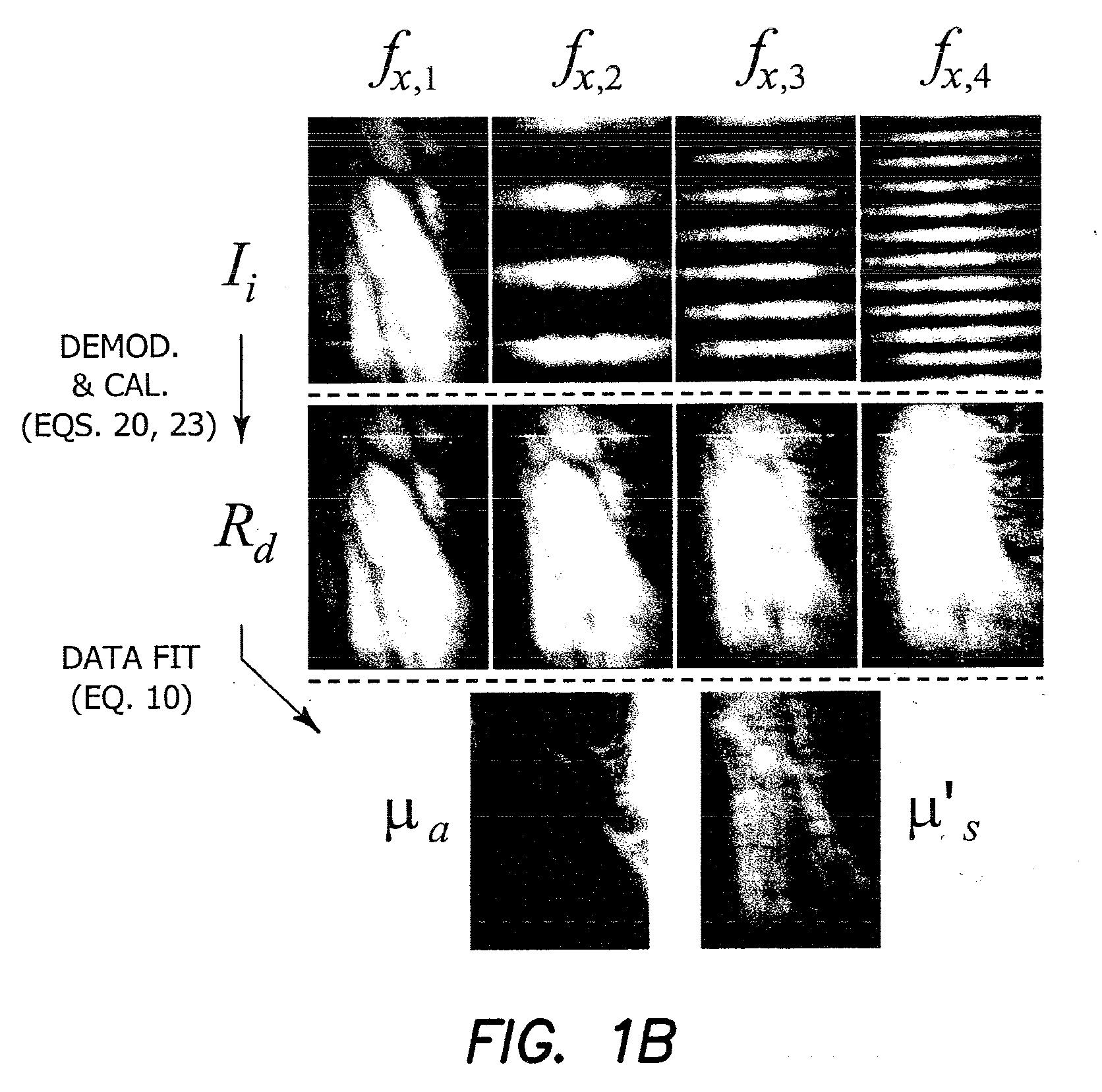
[0063]The broad advantage of the wide-field functional imaging (WIFI) core is that it is an integrated imaging platform capable of quantitative subsurface metabolic imaging across spatial scales. WiFI simultaneously measures tissue blood flow, biochemical composition (i.e. oxy- and deoxy-hemoglobin, water and lipid content), and molecular fluorescence in turbid tissues. It possesses sufficient spatio-temporal resolution to study both fast (i.e., ms timescale) and localized (i.e., tens of μm to mm...


PUM


Abstract

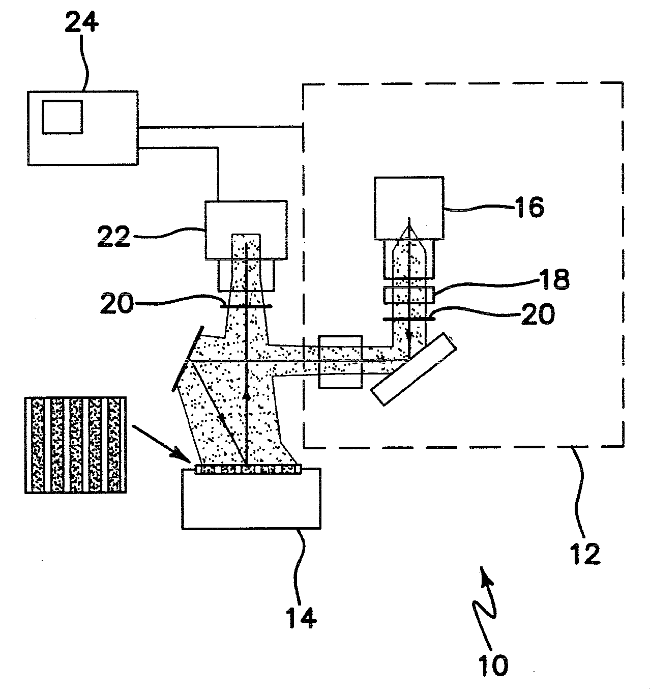
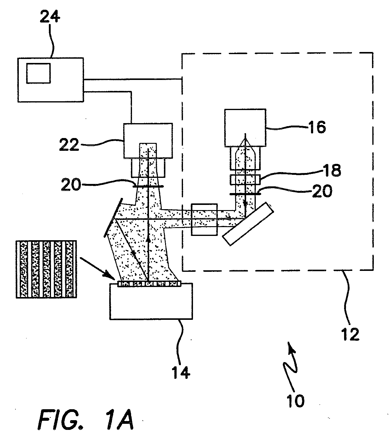
An apparatus for wide-field functional imaging (WIFI) of tissue includes a spatially modulated reflectance/fluorescence imaging (SI) device capable of quantitative subsurface imaging across spatial scales, and a laser speckle imaging (LSI) device capable of quantitative subsurface imaging across spatial scales using integrated with the (SI) device. The SI device and LSI device are capable of independently providing quantitative measurement of tissue functional status.

Description

RELATED APPLICATIONS[0001]The present application is related to U.S. Provisional Patent Application Ser. No. 60 / 985,872, filed on Nov. 6, 2007, which is incorporated herein by reference and to which priority is claimed pursuant to 35 USC 119.BACKGROUND OF THE INVENTION[0002]1. Field of the Invention[0003]The invention relates to the field of medical imaging using light.[0004]2. Description of the Prior Art[0005]Due to its relatively low cost and ease of implementation, optical imaging is an attractive technology to study intrinsic signals associated with endogenous chromophores as well as targeted exogenous probes. With the explosive growth in available molecular reporter strategies for studying fundamental biophysical processes, there has been a paradigm shift in research efforts from ex vivo destructive evaluation to in vivo analysis, allowing for characterization of dynamic biological processes and for each animal to serve as its own control. Despite these tremendous advances in ...

Claims


Application Information

Patent Timeline
IPC IPC(8): A61B6/00
CPCA61B5/0073G01N21/6456G01N21/4795A61B5/4094
Inventor DURKIN, ANTHONY J.CUCCIA, DAVIDTROMBERG, BRUCE J.MAZHAR, AMAANCHOI, BERNARD
Owner MODULATED IMAGING
Why Eureka
Try Eureka